Implications
Ultra-low storage temperature alters (i) the long-range order of collagen packing in fibrils, resulting in fibril rupture at higher stresses, and (ii) the composition of extra-fibrillar matrix, resulting in an increase in the interaction energy between fibrils via collagen-bound proteoglycans. Consequently, the tissue strength, stiffness and fracture toughness increase.
Introduction
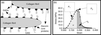
This study is concerned with the effects of frozen storage temperature on the elasticity of tendons, which are biological examples of fibre-reinforced composites comprising highly paralleled collagen fibrils embedded in the hydrated proteoglycan (PG)-rich extracellular matrix (ECM; Hukins and Aspden, Reference Hukins and Aspden1985; Figure 1a). The basic mechanism of reinforcement in tendons addresses the transfer of stress generated in the deforming PG-rich matrix, in response to an external load, to the collagen fibrils (Goh et al., Reference Goh, Meakin, Aspden and Hukins2005). Regulating this mechanism involves the interactions of PGs on the fibrils with those in the surrounding matrix and between adjacent fibrils (Goh et al., Reference Goh, Meakin, Aspden and Hukins2007). The stress transfer mechanism is fundamental to understanding the biomechanics of load transmission, from muscle to bone by tendons, in many studies. These studies may be found in biomedical science, for example, addressing the biomarkers of ageing (Derwin and Soslowsky, Reference Derwin and Soslowsky1999), injury and repair (Lin et al., Reference Lin, Cardenas and Soslowsky2004), and meat science, for example, investigating the mechanical response in relation to the quality of the attachment of muscle to bone from livestock (Moussa et al., Reference Moussa, Babilé, Fernandez and Remignon2007; Gondret et al., Reference Gondret, Hernandez, Remignon and Combes2009). Unfortunately, experimental designs in these studies often involve long sample preparation time. To minimise biological degradation, tendons are stored by freezing, usually at around −20°C (Moussa et al., Reference Moussa, Babilé, Fernandez and Remignon2007; Goh et al., Reference Goh, Holmes, Lu, Richardson, Kadler, Purslow and Wess2008; Gondret et al., Reference Gondret, Hernandez, Remignon and Combes2009) or at ultra-low temperatures, for example, −80°C (Giannini et al., Reference Giannini, Buda, Di Caprio, Agati, Bigi, De Pasquale and Ruggeri2008), until the day of testing. Although the processes that regulate the formation of ice crystallites and the disruption of cells and ECM organisation are well-known (Muldrew and McGann, Reference Muldrew and McGann1990), how frozen storage temperature affects the basic mechanisms that regulate the ECM elasticity, leading to changes in the mechanical properties, is not clear.
(a) Extracellular matrix of tendons comprising of collagen fibrils embedded in a hydrated PG-rich matrix. Decorin core proteins, with glycosaminoglycans (GAGs) attached, are depicted on the surface of collagen fibrils. (b) A stress-strain curve (M20) illustrating the areas under the curve corresponding to the strain energy density to maximum stress (u) and to fracture (up); arrows P1 and P2 indicate the point of inflexion and the maximum stress (σ) point, respectively.

We have investigated the influence of frozen storage temperatures on the mechanical properties of tendons from the tail of a small murine model (C57BL6 mouse). Here, we are concerned with the maximum stress, σ, on the stress-strain curve, stiffness (E), strain (ε) at σ, strain energy densities up to ε (u) and from ε until complete rupture (up) (Goh et al., Reference Goh, Meakin, Aspden and Hukins2005). Since the genome of the C57BL6 mouse has been sequenced, it has been regarded as an ideal murine model in the context of integrative biology study; it is widely used for laboratory experiments to illuminate basic mechanisms that could address similar outcomes in other mammals (Laissue et al., Reference Laissue, L’Hôte, Serres and Vaiman2009). In particular, tissues from the murine model have been used extensively to evaluate biomechanical properties to study systemic changes caused by injury and during healing (Lin et al., Reference Lin, Cardenas and Soslowsky2004), ageing of ECM (Goh et al., Reference Goh, Holmes, Lu, Richardson, Kadler, Purslow and Wess2008) and disruption of the signal pathway for regulating ECM in genetically engineered mice that produced alterations in collagen (Derwin and Soslowsky, Reference Derwin and Soslowsky1999) and PGs (Derwin and Soslowsky, Reference Derwin and Soslowsky1999). Application of the principles of fibre-reinforced composites to connective tissues such as tendons has yielded insights on the mechanisms of elastic and plastic stress transfers in collagen fibrils and on fibril–fibril interactions across the hydrated PG-rich matrix within ECM for regulating tissue elasticity (Goh et al., Reference Goh, Meakin, Aspden and Hukins2005). Here, these insights will be used to speculate on the sensitivity of these mechanisms to low storage temperatures.
Material and methods
Sample preparation
Tendons, of about 200 μm thick, were dissected from the tail of a 16-month-old mouse (C57BL6) which was killed in accordance with the procedure laid down by the National Research Council for the care and use of laboratory animals. The tendons were transected and divided equally into three groups consisting of ten samples per group. One group was designated as control and was tested immediately. Samples from the second (M20) and third (M80) groups were immediately placed in freezers operating at −20°C (MDF-U5411, Sanyo, Osaka, Japan) and −80°C (MDF-U73V, Sanyo), respectively. After 24 h, the frozen samples were thawed at room temperature for analysis.
Mechanical tests
A small-scale horizontal tensile rig, designed and built in-house, was used for mechanical tests (Goh et al., Reference Goh, Holmes, Lu, Richardson, Kadler, Purslow and Wess2008). Each sample was mounted onto the rig, secured by the grips at its ends and hydrated by submerging in phosphate buffered saline, pH 7.2, at room temperature. The rig was mounted onto an inverted microscope (DM-IL, Leica Microsystems GmbH, Wetzlar, Germany) for sample observation during the test. A displacement rate of 0.067 mm/s was applied to stretch the sample until the tissue ruptured (Goh et al., Reference Goh, Holmes, Lu, Richardson, Kadler, Purslow and Wess2008). The force generated in the sample was recorded by a load cell (UF-1, Pioden Controls Ltd) coupled to one grip; the sample extension corresponded to the relative displacement of the grips, which was recorded by a linear variable displacement transducer (CDP-M, Tokyo Sokki Kenkyujo, Tokyo, Japan).
Determination of mechanical properties
Stress-strain plots were obtained from the load-extension data. Here, stress is defined as the ratio of the load to the nominal cross-sectional area of the sample; strain is the ratio of the change in the sample length to the length described by the grip-to-grip distance. By fitting an appropriate polynomial equation to the data points on the plot, from stress = 0 to σ, the point of inflexion was identified (Figure 1b). The gradient at this point parameterised E (Goh et al., Reference Goh, Holmes, Lu, Richardson, Kadler, Purslow and Wess2008).
From the area under the stress-strain curve (Figure 1b), the strain energy density (u) up to σ was evaluated to determine the energy needed to cause tissue failure; in this region the energy absorbed is dominated by fibril recruitment and fibril yielding (Goh et al., Reference Goh, Holmes, Lu, Richardson, Kadler, Purslow and Wess2008). We have also determined the strain energy density up, from ε until when the tissue ruptured completely, to account for the energy absorbed by the dominant failure modes, namely fibril pull-out by frictional sliding at the fibril–PG matrix interface, as well as PG matrix rupture (Sikoryn and Hukins, Reference Sikoryn and Hukins1988).
Statistical analyses
Representative (mean ± s.e.) values of the mechanical parameters, that is, σ, E, ε, εy and u, for each treatment were determined. Paired sample t-test was used to analyse for differences between the control v. each treatment group at α = 0.05. Differences due to the treatment were considered significant if the P-value was < 0.05.
Results
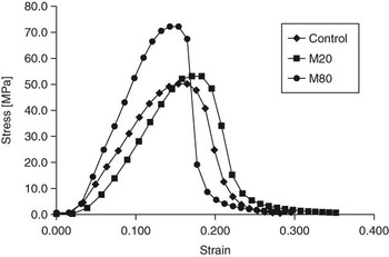
Figure 2 shows stress-strain plots derived from the control, M20 and M80 groups. In general, during the loading stage, all samples featured a toe region followed by a rapid increase in the rate of change of stress with respect to strain; during tissue failure, that is beyond the strain at maximum stress, the gradients decreased rapidly until rupture. Of the three groups, the M80 group yielded the largest gradient during the loading stage and during tissue failure.
Plot of stress v. strain from control and treated samples. Data points represent a typical experiment from one sample each.

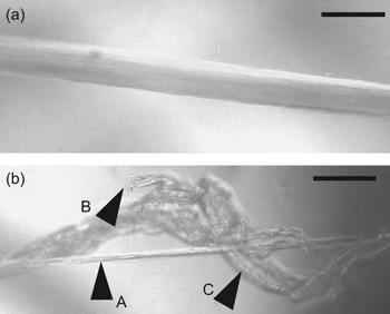
Examination of the microstructure of tendons from the control, M20 and M80 groups revealed wavy collagen fibres (which are one hierarchy above collagen fibrils) that exhibited the usual structural changes, that is de-crimping and straightening (Figure 3a), during loading. All the three groups responded with similar structural changes during mechanical loading. Immediately after σ, the tendon began to fail (Figure 3b). Here, a thin section bridging the bulk of tissue was observed (arrow A). Rupture and pull-out of collagen fibres were also observed; the ends of these structures are indicated by arrow B. At the lower hierarchical level, this could implicate collagen fibril rupture and pull-out. Longitudinal splits in the tendon were also observed, implicating the rupture of the PG matrix (arrow C).
Microscopic examination of tendon samples (a) during initial loading, (b) post-peak stress, σ. The images in part (a) and (b) were taken from a sample belonging to the M20 (samples treated at −20°C) group. Horizontal (scale) bar has a length of 100 μm. Arrow A indicates a thin section bridging the bulk of tissue. Arrow B indicates the ends of the fibres. Arrow C indicates longitudinal splits in the tendon.

The results of σ, E, ε, up and u for the control, M20 and M80 groups are listed in Table 1. In all the cases considered here, no significant difference was observed between the control and M20 groups. No significant difference was observed between the control and M80 groups for ε and up. However, the M80 group yielded magnitudes of σ, E and u, which were 1.4, 1.5 and 1.6 times significantly higher, respectively, than the control group.
Mechanical properties of tail tendons from the C57BL6 mouse

M20 = samples treated at −20°C; M80 = samples treated at −80°C.
Values are mean ± s.e. (n = 10).
*Significant difference of treatment (with respect to the control) at P < 0.05.
Discussion
Collagen fibrils feature a crystalline arrangement of collagen molecules. This crystalline arrangement is characterised by a long-range order of radially packed (side-to-side) collagen molecules and an ordered array of axially staggered collagen molecules along the fibril axis (Laing et al., Reference Laing, Cameron and Wess2003). The long-range order characterises the crystalline system in which portions deep within the fibril, that is, far from the fibril–PG matrix interface, exhibit similar strain response; strain energy will not concentrate at the interface but will eventually be absorbed by molecules at greater depths from the interface. Decorin PGs associated with the collagen fibrils consist of a core protein, bound to collagen, with a side chain known as glycosaminoglycans (GAGs; Figure 1; Screen et al., Reference Screen, Chhaya, Greenwald and Bader2006). The non-steric interactions between GAGs, from the respective decorin PGs on adjacent fibrils, is implicated for regulating the stress transfer to collagen fibrils as the fibrils slide relative to one another when the tissue is loaded (Screen et al., Reference Screen, Chhaya, Greenwald and Bader2006; Goh et al., Reference Goh, Meakin, Aspden and Hukins2007). According to the elasticity of materials (Sun, Reference Sun2009), it follows that E and σ may be determined by equilibrating the external mechanical load to the responses of the intermolecular forces of the tendons. Thus,


where U 1 and U 2 are the sum of cohesive energy per bond, at equilibrium between the biomacromolecules, within a volume, V (Sun, Reference Sun2009). Although E and σ have similar dimensions, the molecular interactions involved in these parameters are not the same. Equation
(1)is valid whether the tendon is undergoing elastic or plastic deformation, which is determined by the forces of interactions between the GAGs of decorin PGs on adjacent collagen fibrils. Equation
(2)is only valid when the tendon is experiencing plasticity, which could involve the plastic deformation of fibrils and the breaking of covalent bonds between collagen molecules in the fibril. Thereafter, the broken bonds could inhibit the motion of collagen molecules around the dislocations, leading to a strengthened tissue as energy of the remaining bonds increases and the bond length contracts (Sun, Reference Sun2009).
It follows that any increase in E is regulated by the van der Waals (VDW) forces between GAGs from collagen-bound PGs, at adjacent fibrils. The interactions are significant for as long as the fibrils are in close proximity to one another. During freezing, as water withdraws from the water compartment, beginning in the PG matrix (Bevilacqua et al., Reference Bevilacqua, Zaritzky and Calvelo1979), and precipitates into ice crystallites, the ion concentration increases in the non-water compartment (Muldrew and McGann, Reference Muldrew and McGann1990), creating a concentration gradient across the unfrozen zone. As cations (e.g. Na+) diffuse to the anionic charge of the GAGs, the increase in the concentration of cations around the GAGs increases the attractive forces between the GAGs. This effect may be appreciable at −80°C but not at −20°C. Moreover, the changes occurring in the tendons from the M80 group may not be completely reversible during thawing; in this case, a greater GAG–GAG interaction increases the resistance for fibril–fibril sliding when the tissue is stretched so that the tissue becomes stiffer than unfrozen ones. To understand how this could have happened, a molecular model was developed to study GAG–GAG interactions (following from Redaelli et al., Reference Redaelli, Vesentini, Soncini, Vena, Mantero and Montevecchi2003). Mobile cations, namely Na+, were designated in closed proximity to each group to model a hypertonic PG matrix environment; the number of Na+ (N) was varied to investigate the effects on the VDW energy, which was evaluated as a function of the distance between the PG molecules, δ. The model was solved by molecular mechanics using software Hyperchem (Version 7.5, Hypercube Inc., Florida, USA). It was found that VDW energy, which decreased as δ increased, was affected by N. In particular, VDW energy increased by about 1.2 times as N increased twofold, but the profile of the energy-δ curves was not affected. Further details can be found in supplementary material. Thus, we concluded that the overall effect of freezing led to a localisation and densification of charge and energy. Hence, E has to be increased as predicted by equation (1).
Any increase in σ could be attributed to changes in the packing of collagen molecules in the fibril. Low storage temperature could alter the long-range order of radial packing of collagen molecules, which could be assessed by the diffuse-scattering region from X-ray diffraction patterns of these tissues. Changes to the diffuse region occur during freezing; tissues frozen at −150°C exhibit reduced diffuse regions suggesting that the long range order is increased (Laing et al., Reference Laing, Cameron and Wess2003). Whether the effect is reversible upon thawing is not clear; Hickey and Hukins (Reference Hickey and Hukins1979) had shown that thawed tissues (frozen at −35°C) exhibited smaller diffuse regions as compared with control samples. An increase in the long-range order implies that the fine structure, that is, the crystalline arrangement of collagen molecules, of the fibril would have to be disturbed to a very much greater depth. More molecules would be involved in absorbing the energy needed to cause fibril rupture; thus more energy is absorbed by the tissue. As pointed out earlier in this section, stresses will not concentrate at regions near to the fibril surface, an important factor determining the failure of fibrils. Instead, stresses will be distributed deeper within the fibril, thus making the fibril less susceptible to fracture so that a higher u, and consequently higher σ as predicted by equation (2), would be needed to rupture the tendon.
We note that up is not affected by low storage temperatures. This suggests that low storage temperature did not have a significant effect on the molecular interaction at the fibril–PG interface and in the bulk of the PG matrix. We also note that the extensibility of the tendon, which is parameterised by ε, was not sensitive to low storage temperatures. We recalled that X-ray diffraction patterns of stretched tendons revealed that the tissue strain would always be larger than the strain within the collagen fibrils (Puxkandl et al., Reference Puxkandl, Zizak, Paris, Keckes, Tesch, Bernstorff, Purslow and Fratzl2002). It is possible that the changes affecting the strain in individual fibrils would have negligible influence on the overall strain at maximum stress in the tissue stored at sub-zero temperature.
The purpose of this paragraph is to discuss the effects of cooling on the size of the ice crystallites formed in the tendons. According to Bevilacqua et al. (Reference Bevilacqua, Zaritzky and Calvelo1979), a combination of the effects of solute concentration (due to an increase in viscosity) and thermal gradients (due to latent heat release and heat lost from the unfrozen zone), in the region between the crystallites, could lead to super-saturation and super-cooling. Theoretically, the cooling rate of the tissue is proportional to the square of the moving speed of the freezing front, u 2, while the temperature gradient at the solid-liquid interface (G) is proportional to u (Miyawaki et al., Reference Miyawaki, Abe and Yano1992). Here, G = Tm − Ts/x where x is the length of the frozen crystallite and Ts and Tm are the operating temperature of the freezer and the temperature at the surface of the crystallite, respectively. Empirically, the thickness (dp) of ice crystallites is related to u and G; this relationship can be represented as

Tiller and Rutter (Reference Tiller and Rutter1956). It follows that the cooling rate is related to dp in equation
(3). Thus, slow freezing rates cause ice to grow in extracellular locations, resulting in large crystallites and maximum dislocation of water; rapid cooling produces small ice crystallites in both cells and ECM (Bevilacqua et al., Reference Bevilacqua, Zaritzky and Calvelo1979). In particular, slow cooling rates correspond to wider spread of sizes; fast cooling rates correspond to narrower spread of sizes and the crystallite growth is more organised (Pardo et al., Reference Pardo, Suess and Niranjan2002). Consider a model of the crystallite in the tendon. On the basis of the results of Bevilacqua et al. (Reference Bevilacqua, Zaritzky and Calvelo1979) and Chevalier et al. (Reference Chevalier, Le Bail and Ghoul2000), using an order of magnitude estimate, we find x∼100 μm. Owing to convection in the freezers, we may approximate Ts∼Tf, = 0°C, the freezing temperature of water. With these values, equation
(3)was evaluated for the upper limit (0.41 mm/s; Miyawaki et al., Reference Miyawaki, Abe and Yano1992) and lower limit (0.06 mm/s; Chevalier et al., Reference Chevalier, Le Bail and Ghoul2000) of u, that is, corresponding to four combinations, for each case of Tm = −20°C and −80°C. By determining the ratio dp | −20°C/dp | −80°C, this simple analysis predicts that the ice crystallites formed at the higher freezer temperature are bigger than those formed at the lower freezer temperature by 1.16 to 3.46 times. In this case, the more organised smaller crystallites are dispersed uniformly within the interfibrillar matrix, raising the ion concentration around the GAGs. This argument complements an earlier suggestion that the increase in GAG interactions, arising from a localisation and densification of charge and energy around the GAGs associated with collagen fibrils, yields a higher E.
In conclusion, ultra-low storage temperature affects only σ, E and u in tail tendons from the C57BL6 mouse, leading to tissues with larger σ, E and u. The results of this study imply that care is required when considering the method for storing and preserving tendons in order to minimise changes to the elasticity of the tissue.
Acknowledgements
CY was supported by an NTU research scholarship. This study was supported by grants from the EU Framework V, and the Merlion-France 2007 programme (from the French Ministere des Affaires Etrangeres et Europeennes).