Acute rheumatic fever is an inflammatory disease that develops as a result of immunological response against infection of group A β haemolytic streptococcus. Although the incidence of rheumatic heart disease has been declining in developed countries, it is still one of the most important causes of acquired heart disease in young children and adults in developing countries.Reference Demirbağ, Sade, Aydın, Bozkurt and Acartürk 1 Involvement of the heart valves is the most important long-term effect of acute rheumatic fever, closely associated with morbidity and mortality.Reference Carapetis, Steer, Mulholland and Weber 2 Heart valve involvement has been observed in 60% of patients with acute rheumatic fever, and the most commonly affected valves are the mitral and aortic valves.Reference Carapetis, Steer, Mulholland and Weber 2 Although the mechanism of valvular damage is not clear, the role of inflammatory cytokines in the pathophysiology of acute rheumatic fever is well known. Furthermore, activation of complements, accumulation of lymphocytes, and excess release of adhesion molecules are present in the heart tissue.Reference Guilherme, Cyry and Demarchi 3 , Reference Settin, Abdel-Hady, El-baz and Saber 4
Red blood cell distribution width is associated with inflammatory markers in chronic inflammation.Reference Förhécz, Gombos, Borgulya, Pozsonyi, Prohászka and Jánoskuti 5 , Reference Güngör, Ozcan and Ozpamuk Karadeniz 6 It has been found that this parameter causes the progression of diseases characterised by chronic inflammation and even increases in many cardiovascular diseases such as heart failure, acute myocardial infarction, and coronary artery disease.Reference Güngör, Ozcan and Ozpamuk Karadeniz 6 – Reference Tonelli, Sacks, Arnold, Moye, Davis and Pfeffer 9 In addition, because of platelet activation, which has a very important role in inflammation, it has been observed to secrete mediators such as chemokines and cytokines.Reference Bath and Butterworth 10 Mean platelet volume and platelet distribution width are platelet activation markers, which are associated with serious inflammation.Reference Herve, Humbert and Sitbon 11 , Reference Pitchford and Page 12
The aims of the present study were to compare red blood cell distribution width, mean platelet volume, and platelet distribution width values in children with rheumatic heart disease with healthy children and to demonstrate the evaluation of these three parameters in combination in clinical follow-up of patients at long term.
Materials and methods
Patients who were admitted to the Pediatric Cardiology Unit between April, 2012 and December, 2015 diagnosed with rheumatic heart disease by transthoracic echocardiography were retrospectively recorded. A total of 151 patients who were diagnosed with rheumatic carditis according to the pathological valvular insufficiency criteria via transthoracic echocardiography or had been diagnosed with acute carditis and treated properly for 6 months were included. All the patients had secondary prophylaxis with antibiotics. Transthoracic echocardiography was performed on all patients by an experienced paediatric cardiologist. The demographic characteristics of the patients were obtained from patient information system. In addition, a control group was included consisting of 148, healthy, age- and sex-matched children with normal transthoracic echocardiographic findings.
Complete blood count including white blood cell count, platelet count, red blood cell distribution width, mean platelet volume, and platelet distribution width, erythrocyte sedimentation rate, and C-reactive protein levels of all patients and control subjects were recorded. Transthoracic echocardiography was performed via Vivid S5 Pro Ultrasound System (GE Medical Systems, Horten, Norway), using 3 and 6 MHz transducers in two-dimensional, M-mode, and colored Doppler visualising modes. Cardiac valves were evaluated for annular and chordal pathologies and regurgitation, prolapses, and presence of vegetation. Mitral and aortic valve regurgitations were assessed by colored Doppler echocardiography in the apical and parasternal long-axis windows. Pathological valve insufficiency was considered for the mitral valve as continuation of regurgitation during systole and for the aortic valve as continuation of regurgitation during diastole at least in two windows as a jet of mosaic colour 1 cm in length with a peak velocity of 2.5 m/second.Reference Bisno, Butchart and Ganguly 13
Exclusion criteria for both patient and control groups included the following: having any infectious or inflammatory diseases or using anti-inflammatory or anticoagulant drugs in the last 1 month, having anaemia, thrombocytopaenia, or haematological problems. Local Ethics Committee approval was obtained for the study protocol.
Statistical analysis
Data are summarised as means and their standard deviations, medians, and minimum and maximum values.
Control of numerical data was performed using the “Shapiro–Wilk Normality Test”. The “Mann–Whitney U-Test” was used for red blood cell distribution width, and the “Independent t-test” was used for the other parameters. “Spearman’s Correlation Analysis” was used to determine the relationships between parameters.
After evaluation of red blood cell distribution width, platelet distribution width, mean platelet volume, and C-reactive protein levels one by one (univariate) between subgroups of patients and the control group to evaluate the effects of them together, analyses of multiple variants were performed (multivariate); p<0.05 was considered statistically significant.
Results
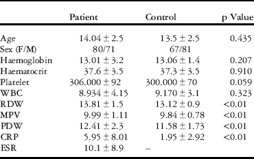
In this study, 151 patients (80 male and 71 female) who were diagnosed with rheumatic heart disease and 148 age- and sex-matched healthy children were included. The mean age of the patients was 14.04±2.5 (with a range from 7 to 18) years. Mitral regurgitation existed in 52 patients and aortic regurgitation in 33 of them, and both were present in 66 patients. Demographic and laboratory characteristics of the patient and control groups are shown in Table 1. Red blood cell distribution width, platelet distribution width, mean platelet volume, and C-reactive protein levels of the patient group were significantly higher when compared with the control group (p<0.01); however, there were no significant differences in values of haemoglobin, haematocrit, platelet count, and white blood cell count between the groups (p>0.05).
Demographic and laboratory characteristics of the patient and control groups.

CRP=C-reactive protein; ESR=erythrocyte sedimentation rate; F=female; M=male; MPV=mean platelet volume; PDW=platelet distribution width; RDW=red blood cell distribution width; WBC=white blood cell count
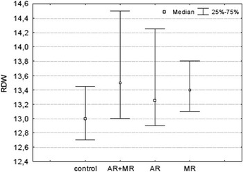
The red blood cell distribution width levels according to valve involvement between the groups are shown in Figure 1. The difference of red blood cell distribution width level was statistically significant between the two valve involvement groups and the control group (p=0.002). When C-reactive protein and platelet distribution width were added to the evaluation of red blood cell distribution width, the value of statistical model was increased (p=0.001). The red blood cell distribution width alone had no impact on the difference between the patients with aortic regurgitation and the controls (p=0.334), but when added C-reactive protein, platelet distribution width and mean platelet volume to the model, it was found to be statistically significant (p=0.001).
Red blood cell distribution width (RDW) levels according to rheumatic valvular involvement. AR=aorta regurgitation; MR=mitral regurgitation.

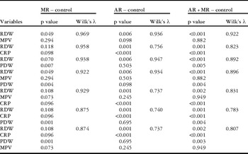
It was also observed that red blood cell distribution width differed between patients with mitral regurgitation and controls (p=0.007). Moreover, red blood cell distribution width was still significant when evaluated together with the effects of the haemogram parameters, platelet distribution width, and mean platelet volume (p=0.049), but when C-reactive protein was added to the model significance was lost (p=0.108) (Table 2).
Comparison of subgroups by valvular involvement with the control group when red blood cell distribution width (RDW), platelet distribution width (PDW), mean platelet volume (MPV), and C-reactive protein (CRP) are included.

AR=aorta regurgitation; MR=mitral regurgitation
Red blood cell distribution width was found to have a significantly positive correlation with both C-reactive protein (r=0.271, p=0.035) and sedimentation (r=0.308, p=0.006). The red blood cell distribution width and platelet distribution width values were higher in patients with involvement of both valves, but this increase was not statistically significant (p>0.05).
Discussion
This is the first study in children with chronic rheumatic valvular heart disease to evaluate the combination of red blood cell distribution width, platelet distribution width, and mean platelet volume parameters, and all of them were significantly increased. Our study also identified that there is a positive correlation between inflammatory markers and red blood cell distribution width in patients with chronic rheumatic carditis.
Rheumatic fever is an autoimmune disease that develops after group A streptococcus infection due to abnormal cellular and hormonal responses in susceptible individuals. Although major histocompatibility antigens, tissue-specific antigens, and antibody formation have been considered significant risk factors in the pathogenesis of this disease, the exact developing mechanism of the acute rheumatic fever is still undefined. The clinical severity of the disease is shaped not only by abnormal immune response but also by genetic predisposition of the host, virulence of the organism that infects, and environmental conditions. Rheumatic valvular disease is the most serious complication of acute rheumatic fever and is in turn one of the most common causes of acquired heart disease in developing countries.Reference Demirbağ, Sade, Aydın, Bozkurt and Acartürk 1 Cytokines secreted by activated lymphocytes and macrophages, tumour necrosis factor, and interleukins play an important role in the pathogenesis of rheumatic heart disease. M protein and anti-carbohydrate antibodies of group A streptococcus cause damage to the valvular endothelium in susceptible individuals, resulting in increased release of vascular cell adhesion molecule 1 and other adhesion molecules. Subsequently, the interaction between VCAM-1 and activated lymphocytes stimulates cluster of differentiation 4 and cluster of differentiation 8 T cells. The inflammatory reaction that is triggered by the activation of the complement system as a consequence of T cell infiltration and autoreactivity causes a progressive and permanent damage to the valves.Reference Bisno, Butchart and Ganguly 13 , Reference Stollerman 14
Mean platelet volume and platelet distribution width levels that demonstrate the variability in size of platelets can be used as markers of platelet activation. Moreover, it has been shown that these two markers reflect the severity of inflammation and also increase in myocardial infarction and chronic pulmonary diseases.Reference Bath and Butterworth 10 , Reference Endler, Klimesch and Sunder-Plassmann 15 , Reference Arıca, Ozer, Arıca, Karakus, Celik and Gunesacar 16 The mechanism of increase in mean platelet volume and platelet distribution width is not understood exactly until now. It has been emphasised that inflammatory cytokines induce the production of large and reactive platelets and these circulating platelets have a shorter life span.Reference Endler, Klimesch and Sunder-Plassmann 15
Ozdemir et alReference Ozdemir, Karadeniz and Doksoz 17 compared the levels of mean platelet volume and platelet distribution width in patients with acute rheumatic fever before and after the treatment in a study and they found no significant differences. Mean platelet volume levels in patients with acute rheumatic fever and controls were also compared in a study by Sert et alReference Sert, Aypar and Odabas 18 ; the result showed lower levels in patients. The results of these two studies are conflicting in terms of mean platelet volume and platelet distribution width levels. In our study, on the other hand, mean platelet volume and platelet distribution width levels were significantly higher in patients with chronic rheumatic heart disease compared with the control group. Ozdemir et al did not detect any changes in these parameters before and after treatment, which can be explained as continuation of the inflammation process after the treatment. In another study, on the contrary, the level of mean platelet volume was found to be lower in acute rheumatic fever and in chronic inflammatory diseases, whereas in myocardial infarction and pulmonary diseases the marker was found to be increased.Reference Bath and Butterworth 10 , Reference Endler, Klimesch and Sunder-Plassmann 15 , Reference Arıca, Ozer, Arıca, Karakus, Celik and Gunesacar 16 In our study, high C-reactive protein levels have been accepted as an indicator of chronic inflammation in patients, and simultaneous elevation in the value of mean platelet volume and platelet distribution width has been concerned due to the chronic inflammation as well.
Red blood cell distribution width, reflecting the variability in size of red blood cells in circulation, increases in some heart diseases regardless of the haemoglobin level and is considered as a new marker for predicting mortality.Reference Förhécz, Gombos, Borgulya, Pozsonyi, Prohászka and Jánoskuti 5 , Reference Güngör, Ozcan and Ozpamuk Karadeniz 6 – Reference Isik, Uyarel and Tanboga 8 , Reference Patel, Semba and Ferrucci 19 Oxidative stress and chronic inflammation shorten the life span of erythrocytes and increase the number of immature and different-sized erythrocytes in circulation.Reference Felker, Allen and Pocock 20 Therefore, in this study, increased red blood cell distribution width in patients when compared with the control group suggests that red blood cell distribution width is a marker of the underlying chronic inflammation, which increases the risk of cardiovascular disease. Similarly, C-reactive protein is one of the well-known inflammatory markers. C-reactive protein level was found to be higher in patients with chronic rheumatic heart disease compared with the control group, and this increase was accepted as an indicator of the continuing inflammation.Reference Gölbasi, Uçar and Keles 21 , Reference Chiu-Braga, Hayashi, Schafranski and Messias-Reason 22 Some studies show a strong correlation between red blood cell distribution width and C-reactive protein levels, and some suggest that red blood cell distribution width can be used in determining the risk of mortality.Reference Perlstein, Weuve, Pfeffer and Beckman 23 In addition, some studies in patients with coronary artery disease underline the elevation in red blood cell distribution width level as associated with C-reactive protein level, which may be an independent predictor of mortality.Reference Lappé, Horne and Shah 24 , Reference Ma, Li and Li 25 Furthermore, Tonelli et alReference Tonelli, Sacks, Arnold, Moye, Davis and Pfeffer 9 found an independent relationship between red blood cell distribution width and death and/or the risk of cardiovascular events in patients with asymptomatic heart failure who had myocardial infarction previously. Akboğa et alReference Akboğa, Abacı and Canpolat 26 in a study including patients with rheumatic mitral valvular stenosis found that the red blood cell distribution width levels were significantly increased in parallel with the severity of valvular stenosis, which was also correlated with C-reactive protein levels. In our study, similarly, the red blood cell distribution width level in patients with chronic rheumatic heart disease was significantly higher compared with the control group. Furthermore, there was a positive correlation between C-reactive protein and red blood cell distribution width levels as in the literature.
On the basis of the data, it is assumed that evaluating the red blood cell distribution width and platelet markers together would be more effective in follow up patients with acute rheumatic fever having valvular involvement. Meanwhile, the red blood cell distribution width levels in patients with mitral regurgitation and both mitral and aortic regurgitation were significantly higher compared with the control group, and this significance increased with the addition of platelet distribution width and mean platelet volume to the comparison. Even when red blood cell distribution width level alone was not different in patients with aortic regurgitation compared with the control group, the results were found to be statistically significant when red blood cell distribution width together with platelet distribution width, mean platelet volume, and C-reactive protein were interpreted. Thus, as noted in many other studies previously, our results support the role of chronic inflammation in the pathophysiology of chronic rheumatic heart disease and the association of red blood cell distribution width, platelet distribution width, and mean platelet volume with this inflammation.Reference Endler, Klimesch and Sunder-Plassmann 15 , Reference Perlstein, Weuve, Pfeffer and Beckman 23 , Reference Lappé, Horne and Shah 24 , Reference Akboğa, Abacı and Canpolat 26
Consequently, we have been assuming that parameters such as red blood cell distribution width, platelet distribution width, and mean platelet volume that can be determined by complete blood count can be used to predict valvular involvement in the follow-up of patients with chronic rheumatic carditis. Furthermore, red blood cell distribution width can be used as a risk-determining marker in children with rheumatic heart disease, as it has a positive correlation with C-reactive protein, which is a chronic inflammatory marker. Nevertheless, distinguishing congenital mitral regurgitation and patients with quiet carditis may sometimes be a bit difficult. Owing to this fact, it has been assumed that the use of markers such as red blood cell distribution width, mean platelet volume, and platelet distribution width, which demonstrate the process of chronic inflammation and can be easily examined anywhere, will help in overcoming this distinction in near future; however, in order to evaluate the relationship, more comprehensive prospective studies are required.
The retrospective design of the study is a major limitation. Besides inflammation, parameters known to affect red blood cell distribution width such as erythropoietin, iron, folic acid, and fibrinogen levels were not taken into consideration; however, patients with anaemia or any haematological problems were not included in the study.
Acknowledgements
None.
Financial Support
This research received no specific grant from any funding agency or from commercial or not-for-profit sectors.
Conflicts of Interest
None.
Ethical Standards
The authors assert that all procedures contributing to this work comply with the ethical standards of the relevant national guidelines on human experimentation (please name) and with the Helsinki Declaration of 1975, as revised in 2008, and has been approved by the institutional committees of Mersin University.