Introduction
The nervus intermedius is a branch of the facial nerve with both parasympathetic and sensory components. Wrisberg named it after its intermediate location between the motor component of the facial nerve and the superior vestibular nerve. The nervus intermedius is rarely clinically significant. Nervus intermedius neuralgia is characterised by severe pain deep in the ear that may spread to the ear canal, outer ear, mastoid or eye regions. Hitselberger's sign is anaesthesia of the concha bowl and posterior external auditory canal resulting from a space-occupying lesion of the cerebellopontine angle that causes compression of the nervus intermedius.Reference Guinto and Guinto1
Tumour of the nervus intermedius is also quite rare. Because of its location, this tumour involves the internal auditory canal and is therefore often incorrectly diagnosed as vestibular schwannoma. Meningioma of the nervus intermedius has not been described. We performed a literature search with PubMed using the following keywords: nervus intermedius, nervus intermedius meningioma, cerebellopontine angle tumour and internal auditory canal tumour. The following case describes the clinical presentation, audiological and imaging findings, and the intra-operative and post-operative course of a patient with nervus intermedius meningioma.
Case report
A 48-year-old male presented to our clinic with a 5-year history of progressive right-sided hearing loss. He had developed associated dizziness and unilateral tinnitus a few years after first noting the hearing loss. His dizziness had increased in frequency and now occurred daily, with episodes lasting 2–3 hours each. There had not been any otalgia or otorrhoea. Eardrum, tuning fork, and cranial nerve test results were normal; there was no facial twitching or droop.
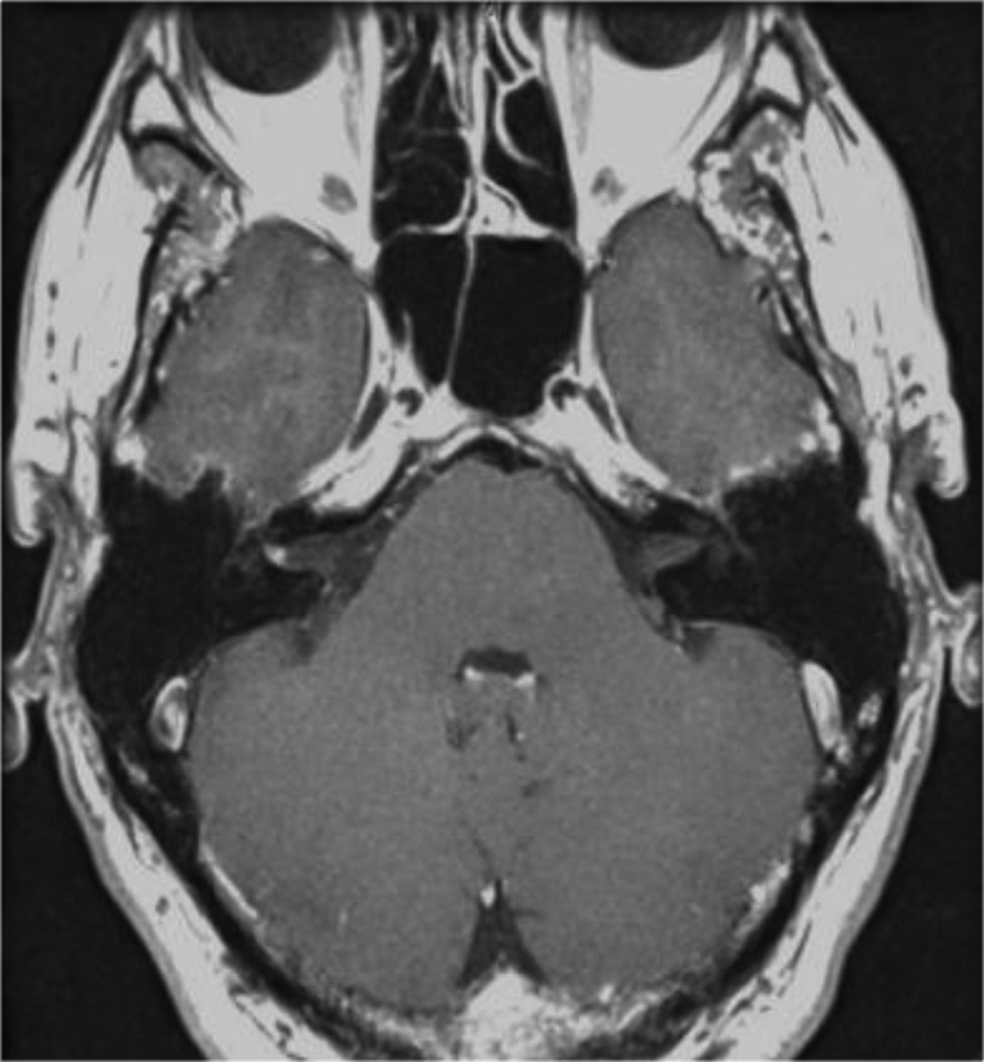
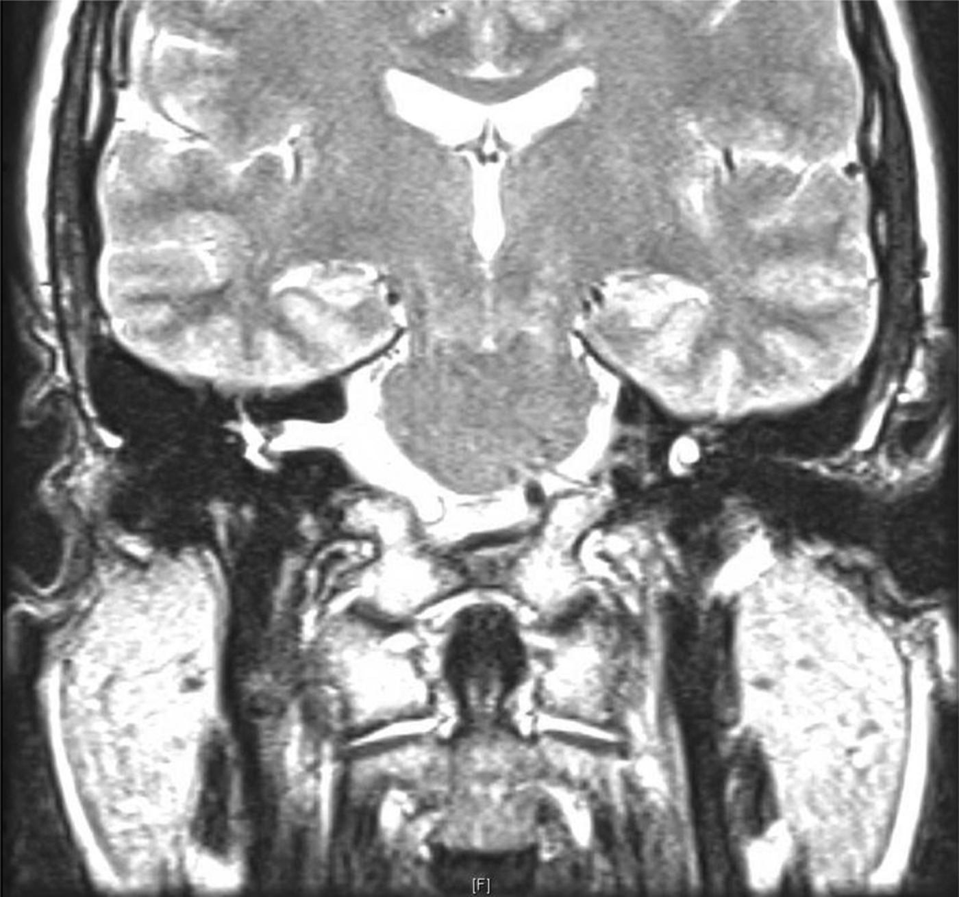
The pure tone average for the right ear was 58 dB compared with 10 dB for the left (Figure 1). Magnetic resonance imaging (MRI) showed a 2 mm enhancing mass impacted in the right fundus superiorly (Figures 2 and 3).
Pre-operative audiogram for the (a) right and (b) left ears, showing flat, moderate, right sensorineural hearing loss. Δ = right ear air unmasked; [=right ear bone unmasked; × = left ear air unmasked; >= left ear bone unmasked

Focused axial magnetic resonance image of the right internal auditory canal, showing an enhancing lesion laterally at the fundus which appears to arise from the superior vestibular nerve.
Focused coronal magnetic resonance image of the right internal auditory canal, showing an enhancing lesion laterally at the fundus which appears to arise from the superior vestibular nerve.
We suspected a vestibular schwannoma; however, we requested that facial nerve electroneuronography was carried out because of concern for a facial schwannoma given the tumour's proximity to the labyrinthine segment of the facial nerve. We treated the patient symptomatically with a low salt diet, Dyazide® and meclizine.
Electroneuronography showed a 71 per cent response on the right side, demonstrating less dys-synchrony of the facial nerve motor fibres than we expected for a tumour primarily involving the facial nerve.
Despite the addition of clonazepam, the patient's dizziness did not satisfactorily improve. The options of observation, radiosurgery and tumour removal were discussed with the patient, who opted for the latter. Given the tumour location, level of hearing loss, and small potential for a facial schwannoma, removal was carried out via a translabyrinthine craniotomy.
Intra-operatively, we found that the tumour was neither a superior vestibular nerve schwannoma nor a facial nerve tumour, but had an attachment to another, smaller nerve. It was arising from the nervus intermedius (Figure 4), and our intra-operative impression was a nervus intermedius schwannoma. Gross total tumour removal was achieved with anatomical and electrophysiological preservation of the motor branch of the facial nerve. The eustachian tube and middle ear were packed, and the craniotomy defect was filled with abdominal fat and secured with a titanium cranioplasty plate.
Intra-operative translabyrinthine view of the mass and its relation to the superior vestibular and facial nerves.
The patient's post-operative course was uneventful, and he was discharged home 2 days after surgery. His House–Brackmann score upon awakening revealed grade II (slight) nerve damage, and he was given facial nerve exercises and underwent a slow steroid taper. His dizziness bouts and balance have since improved.
Histological sections of the resected tumour confirmed a well-differentiated World Health Organization grade I meningioma, measuring 0.3 × 0.2 × 0.1 cm (Figures 5 and 6). Staining of vimentin indicated diffuse tumour immunoreactivity. Patch epithelial membrane antigen expression was also present. S-100 and neurofilament immunostaining highlighted the nervus intermedius but were not expressed in the tumour.
The tumour consists of bland meningothelial cells arranged in lobules diagnostic of meningioma. (H&E; ×300)
An S-100 immunostain highlights peripheral nerve elements but the tumour lacks S-100 expression, as is typical for meningioma but not for schwannoma. (S-100; ×30)
Discussion
The nerve of Wrisberg is relatively obscurely located between the motor component of the facial nerve and the superior vestibular nerve. Parasympathetic fibres within the nervus intermedius originate in the superior salivary nucleus. They travel along the facial nerve to their target organs, and the nasal and oral cavities, via the greater petrosal and chorda tympani nerves. Afferent sensory information that travels within the nervus intermedius originates from diffuse locations in the head and neck, including the nasopharynx and the external acoustic meatus. The nervus intermedius also relays special sensory taste information from the anterior tongue, hard palate and floor of the mouth to the solitary nucleus. Though it typically becomes relevant clinically from a neuropathy standpoint (either nervus intermedius neuralgia or Hitselberger's sign), it is occasionally the origin of neoplastic pathology.Reference Guinto and Guinto1
Pathology is limited in nervus intermedius tumour; only schwannomas have been described to date. A variety of different approaches to resection have been utilised. Fuentes and Uziel first described surgical resection of a schwannoma arising from the nervus intermedius at the genu with extension into the internal auditory canal in 1983.Reference Fuentes and Uziel2 This required a suboccipital approach followed by a second surgery via a subpetrosal approach for complete resection.
Mowry et al. described 16 patients with facial nerve schwannomas presumed to be vestibular schwannomas prior to surgery.Reference Mowry, Hansen and Gantz3 Two of these patients’ tumours arose from the nervus intermedius. Both patients underwent excision via the middle cranial fossa approach. The motor branch was preserved in both cases. One patient had delayed onset facial weakness of House–Brackmann grade III severity, but this improved to House–Brackmann grade II prior to the patient being lost to follow up. The other patient had transient House–Brackmann grade II nerve damage post-operatively that subsequently improved to normal facial function.
• Nervus intermedius tumour is indistinguishable from vestibular or facial motor schwannoma on magnetic resonance imaging (MRI)
• This has implications regarding potential facial nerve outcomes
• Current literature suggests better facial nerve prognosis with removal of nervus intermedius tumour versus motor branch of facial nerve tumour
• Nervus intermedius meningioma has not been reported previously and facial nerve prognosis after resection is unknown
• A translabyrinthine approach can be used for nervus intermedius meningioma resection with favourable facial nerve outcome
• Advances in MRI will improve pre-operative evaluation of nervus intermedius
Sherman et al. reported 10 patients with facial schwannomas, 1 of which had a nervus intermedius schwannoma.Reference Sherman, Dagnew, Pensak, van Loveren and Tew4 They did not specify the approach used to resect this tumour; however, they noted that the patient had no injury to the motor branch but did suffer a mild sensorineural hearing loss due to the operation.
Rizer et al. described a patient with mixed hearing loss who was diagnosed with otosclerosis.Reference Rizer, Guthikonda, Lippy and Schuring5 This patient was scheduled to undergo stapedectomy only to experience sudden hearing loss prior to surgery. The MRI scans revealed a cerebellopontine angle mass, and at surgery the patient was found to have a schwannoma at the junction of the motor branch and the nervus intermedius.
Diagnosis of neoplasm from the nervus intermedius typically occurs in the intra-operative setting. However, advances in MRI technology in terms of higher power fields have recently been applied to the cerebellopontine angle with the goal of identifying the nervus intermedius. Currently, the normal nerve can be identified in 60 per cent of cases.Reference Burmeister, Baltzer, Dietzel, Krumbein, Bitter and Schrott-Fischer6 Whether this increased resolution will translate to cerebellopontine angle and internal auditory canal neoplasms remains to be seen. Nevertheless, the nervus intermedius can be identified and distinguished from the motor branch of the facial nerve using intra-operative facial nerve monitoring. However, distinguishing between the two nerves requires careful attention as both may result in stimulation of the facial nerve monitor. Still, monitoring has been shown to be helpful in both locating the nervus intermedius and protecting the motor branch.Reference Ashram, Jackler, Pitts and Yingling7
Conclusion
Nervus intermedius tumour is indistinguishable from vestibular or facial motor schwannoma on MRI. This has implications regarding potential facial nerve outcomes as vestibular and nervus intermedius schwannomas carry a potential for anatomical facial nerve preservation, whereas a tumour arising from the motor branch of the VIIth cranial nerve does not. Facial nerve schwannoma can be pre-operatively suspected in cases of clinical facial nerve weakness or facial nerve dys-synchrony on electroneuronography in the setting of a relatively small tumour. Current literature suggests better facial nerve prognosis with removal of a nervus intermedius tumour compared with removal of the motor branch of a facial nerve tumour. However, there may still be some low-grade facial nerve paresis and mild sensorineural hearing loss. This varies with the surgical approach. In our case, a translabyrinthine approach was used because of poor pre-operative hearing and the lateral location of the tumour. More reports of meningioma of the nervus intermedius are needed to determine if a different complications profile exists for these cases. This may have implications for the preferred surgical approach too.
Advances in MRI may improve our ability to evaluate the nervus intermedius pre-operatively. For now, the otologist should continue to be mindful of the nervus intermedius when considering the diagnosis of vestibular schwannoma, and when dissecting in the cerebellopontine angle where facial nerve monitoring may give false security about the location of the motor branch of the facial nerve due to nervus intermedius stimulation.