Introduction
Lack of appropriate vaccines, drug resistance in malaria parasites, and insecticide resistance in Anopheles vectors has hindered the global control of malaria. One of the newest strategies in malaria control focuses on protection using vaccines and interruption of the transmission cycle of malaria (UNDP/World Bank/World Health Organization, 2000). Transmission-blocking vaccines (TBVS) are based on either sexual stage of parasite antigens or mosquito mid-gut antigens that serve as receptors for parasite ligands in the mid-gut of mosquitoes (Lavazec and Bourgouin, Reference Lavazec and Bourgouin2008). The general idea about TBVs is that the antibodies against sexual stage parasite antigens or mosquito mid-gut receptors taken up by mosquitoes during blood feeding mask receptors.
The mid-gut is the first cellular barrier that the parasite must overcome to be transmitted to a new vertebrate host; therefore, recognizing these molecular sites which are involved in mosquito mid-gut and parasite interaction is the most important part for developing a TBV. The mosquito mid-gut is composed of one layer of epithelial cells, which on the apical side are folded into numerous microvilli. The apical side of the microvillus is covered with the glycocalyx composed of diverse glycoproteins. Mid-gut glycoproteins have been shown to play a role in the establishment of parasite infections in the mosquito body (Dinglasan et al., Reference Dinglasan, Kalume, Kanzok, Ghosh, Muratova, Pandey and Jacobs-Lorena2007). Lectins, such as jakalin, adhere to the microvillar surface (Rudin and Hecker, Reference Rudin and Hecker1989), suggesting the presence of abundant carbohydrate molecules at this site. It has been shown that mid-gut glycoproteins are mostly O-glycoproteins (mucin and mucin-like proteins) and experiments are underway to characterize these glycoproteins (Dinglasan et al., Reference Dinglasan, Fields, Shahabuddin, Azad and Sacci2003). Several lines of evidence support the presence of mucins on mosquito epithelial surfaces (Shen et al., Reference Shen, Dimopoulos, Kafatos and Jacobs-Lorena1999; Dinglasan et al., Reference Dinglasan, Fields, Shahabuddin, Azad and Sacci2003; Injera et al., Reference Injera, Kabiru, Gicheru, Githure and Beier2013). The carbohydrate structures found on mucins offer a vast array of potential binding sites for pathogenic organisms (Dinglasan et al., Reference Dinglasan, Fields, Shahabuddin, Azad and Sacci2003). Mucins have a protective function in epithelium against proteolytic enzymes (Wang and Granados, Reference Wang and Granados1997). While it is well accepted that mucins are involved in many biological processes, from cell signalling (Carraway et al., Reference Carraway, Ramsauer, Haq and Carothers Carraway2003) and signal transduction (Singh and Hollingsworth, Reference Singh and Hollingsworth2006) to immune responses (Sheehan et al., Reference Sheehan, Kesimer and Pickles2006), as well as host−pathogen interactions, our understanding of how mucin molecules manipulate these complex processes at the molecular level is still in its infancy (Hang and Bertozzi, Reference Hang and Bertozzi2005). Considering the adhesive nature of mucin-type O-linked glycan, they can serve as discrete ligands for cell receptors. AgMuc1 (mucin of Anopheles gambiae) (Shen et al., Reference Shen, Dimopoulos, Kafatos and Jacobs-Lorena1999) and AeIMUC1 (mucin of Aedes aegypti) (Morlais et al., Reference Morlais, Mori, Schneider and Severson2003) were described as the genes encoding the respective intestinal mucin glycoproteins. It has been shown that AelMUC1 is associated to a locus that affects Plasmodium gallinaceum invasion of the mid-gut (Morlais et al., Reference Morlais, Mori, Schneider and Severson2003). Immunization of BALB/c mice with AgMuc1/IL-12 cDNA has been shown to kill mosquitoes when they feed on these mice (Injera et al., Reference Injera, Kabiru, Gicheru, Githure and Beier2013), and adhesion of the white blood cells to the surface of the mosquito mid-gut epithelium could be due to the expression of mucin after blood feeding (Injera et al., Reference Injera, Kabiru, Gicheru, Githure and Beier2013).
Anopheles stephensi Liston (Diptera: Culicidae) is the major vector of human malaria in the Middle East and South Asia regions. Based on egg-float ridge numbers, there are three biological forms in A. stephensi including type, intermediate and mysorensis forms (Sweet and Rao, Reference Sweet and Rao1937; Subbarao et al., Reference Subbarao, Vasantha, Adak, Sharma and Curtis1987). The type form is an efficient vector in urban areas whereas the intermediate form is active in semi-urban/rural areas, and mysorensis is a rural species with a lower vector capacity (Subbarao et al., Reference Subbarao, Vasantha, Adak, Sharma and Curtis1987). In other studies, it has been shown that the length polymorphism of the mucin gene in the mid-gut of Anopheles gambiae can affect the vectorial capacity of this vector (Shen et al., Reference Shen, Dimopoulos, Kafatos and Jacobs-Lorena1999). The main objective of our study was the characterization and molecular analysis of the mucin gene from A. stephensi. Our study shows that this mucin is a surface-associated protein of the mid-gut and possesses a length polymorphism between these two strains of A. stephensi used in this study.
Materials and methods
Biological material
Mosquito samples
The experimental work was performed using a colony of the Kazerun strain of A. stephensi reared at the insectary of Tehran University of Medical Sciences, School of Public Health that has been in existence for more than 30 years, and has been renewed many times using field-caught mosquitoes. This strain originated from Kazerun, Fars Province, south Iran. The Liston strain of A. stephensi came from a colony reared since 1988 at the insectary of the Laboratory of Parasitology of University of Camerino in Italy. Anopheles stephensi Liston is a major malaria vector with a geographical range from the Middle East through the Indian subcontinent and China. The mosquitoes were maintained in standard conditions, 70 ± 10% relative humidity at 27°C on a 12 h:12 h light:dark cycle.
RNA extraction and cDNA synthesis
Total RNA was extracted using Ambion's RNAqueous™-Micro Kit according to the manufacturer's instruction. Mosquito mid-guts were dissected in sterile PBS 1X under a binocular microscope and stored at −70°C until the time of using. A single dissected mid-gut of A. stephensi was used for each RNA extraction. Complementary DNA synthesis was conducted using the Revert Aid TM First Strand cDNA Synthesis Kit (Fermentas, Canada) with oligo (dT) primers (20 pmol/µl) according to the manufacturer's instructions, and used as a template in PCR. The integrity of cDNA was checked with the A. stephensi housekeeping gene by amplification of 118 bp housekeeping S7 gene using the primers S7F: 5′-ATGGCTATGGTGTTCGGTTC-3′ and S7R: 5′-TGATGATCGGCCTTCTTGTTG-3′.
Primer design and rapid amplification of cDNA ends (RACE-PCR)
To identify the entire sequence of the mucin gene of A. stephensi in both strains, we used RACE-PCR. For primary amplification, we chose an expressed sequence tag (EST) obtained from the mid-gut tissue of the adult female A. stephensi mosquito (Patil et al., Reference Patil, Atanur, Dhotre, Anantharam, Mahajan, Walujkar, Chandode, Kulkarni, Ghate, Srivastav, Dayananda, Gupta, Bhagwat, Joshi, Mourya, Patole and Shouche2009). This sequence has the most similarity to A. gambiae mucin gene sequences obtained from NCBI with GenBank accession number: AF046924.1. These A. stephensi EST sequences have been submitted at the EST DataBase (http://mycompdb.bioinfo-portal.cdac.in/cgi-bin/est/index.cgi) (Patil et al., Reference Patil, Atanur, Dhotre, Anantharam, Mahajan, Walujkar, Chandode, Kulkarni, Ghate, Srivastav, Dayananda, Gupta, Bhagwat, Joshi, Mourya, Patole and Shouche2009). The DNA fragments were amplified using specific primers designed manually based on these A. stephensi EST sequences as follows: MUCS1: ACCGCTGCATCTGGTCAAGCT and MUCS2: GCCAACTCCACATCAGCACTGCT.
To establish the unknown regions of 5′ and 3′ flanking sequences of the aforementioned EST, we carried out 5′ and 3′RACE-PCR. 5′ RACE-PCR was applied based on a new method published in the NIH protocol with modifications (Shi and Jarvis, Reference Shi and Jarvis2006). We used MUCS2 primer for the first strand cDNA synthesis. The ‘ligation adaptor’ was generated by mixing two commercial oligonucleotides, termed ‘sense adaptor’ (SA) and ‘antisense adaptor’ (ASA) together at final concentrations of 10 μm each. The sequences of the AS and ASA primers are available in the protocol. Based on this protocol, the mixture was heated to 100°C, then allowed to slowly cool down to room temperature. The resulting adaptor was fused to the cDNA using T4 DNA ligase. For the first round of amplification, we used the 5MUC1 primer and long adaptor (Universal Primer Mix, UPM long) while for nested PCR, we used the 5MUC2 primer and a short adaptor (Universal Primer Mix, UPM short). Sequences of specific gene primers which were designed based on the amplified EST are as follows: 5MUC1: CTCAACACTTGCGGACGT and 5MUC2: GATTCAGGTGCGACGGTAGT, and the sequences of UPM long and UPM short are available in the protocol.
The first round of amplification by Touchdown PCR was performed on a Corbet thermal cycler (Corbet, model: CG1-96, AUS), with the following profile: 5 min at 96°C for initial denaturation, five cycles each at three different annealing temperatures (70, 63 and 56°C), and then 30 cycles with an annealing temperature of 51°C. Each PCR cycle included a 30-s denaturation step at 96°C, a 30-s annealing step at the temperatures mentioned above, and two extension steps (15 s at 72°C, followed by 45 s at 74°C), which was designed to progressively melt any secondary structures that might have formed during the preceding annealing step. Nested secondary PCRs were performed using 1 μl of the primary PCR product. The secondary PCR cycling conditions included an initial 3 min denaturation step at 96°C, followed by 30 cycles of 30 s denaturation at 96°C, 30 s annealing at 63°C and ascending extension times and temperatures, as described above.
3′ RACE-PCR was based on the Rapid Amplification of cDNA Ends Kit (Gibco/BRL, Rockville, MD) with modifications. The first strand cDNA synthesis was initiated using oligo (dT) containing Adapter Primer (AP). The target gene was amplified in 3′ RACE-PCR using two primers, one a specific primer (3MUC: AGCAGTGCTGATGTGGAGTTGGC) that anneals to a site located within the cDNA molecule, and the other a universal amplification primer that targets the mRNA of the cDNA, complementary at the 3′ end of the mRNA. Thermo cycle profile was as follows: 3 min at 95°C for initial denaturation step, 30 s at 95°C, 45 s at 57°C and 1 min at 72°C for 30 cycles, and 10 min at 72°C for final extension. All the PCR product fragments were purified and cloned into T/A cloning vector PTZ57R/T (Fermentas, Canada) and then were submitted for DNA sequencing.
Exploring introns in mucin gene from Anopheles stephensi
To discover the possible presence of AsMuc introns, DNA was isolated from a single whole mosquito body. Amplification was conducted using specific forward primer IntF 5′-ATGTTGAAA- GTTCTTCTGGGAC-3′ and reverse primer IntR1 5′-TTAATTGATTAAGATCGACAACAC-3′, corresponding to the specific sequence of mucin cDNA from A. stephensi.
In silico study of molecular analysis and structural protein of AsMuc
The whole sequence of protein AsMuc from the two strains was characterized using several bioinformatics tools. Physical and chemical parameters for AsMuc protein were obtained in the ProtParam tool (http://web.expasy.org/protparam/) and hydrophobicity or hydrophilicity scales in the Portscale tool (http://web.expasy.org/protscale/) of the Expasy sequence analysis prediction server. Hydropathy analysis was done by Kyte-Doolittle plot. Prediction of signal peptide, transmembrane domain, and localization and prediction of glycosylation sites were surveyed in the CBS server prediction (http://www.cbs.dtu.dk/services/) using SingnalP, TMHMM, TargetP and NetOGlyc tools. The GPI anchor site was predicted by big-PI Predictor (http://mendel.imp.ac.at/sat/gpi/gpi_server.html).
Cloning of AsMuc into transfer baculovirus vector
To demonstrate subcellular localization of AsMuc, we used a baculovirus expression system. The full length of the coding domain region of AsMuc (Liston strain) with signal peptide sequence was amplified by PCR using Pfu DNA polymerase (Promega, Madison, WI, USA). Amplification was carried out from cDNA synthesized using oligo (dT). The primer sequences which introduce EcoRI and XhoI restriction sites in the ends of PCR product are as follows: ERmuc GCGAATTCATGTTGAAAGTTCTTCTGGGAC and Xhmuc GCCTCGAGTTAATTGATTAAGATC- GACAACAC. A stop codon was engineered into the reverse primer to ensure correct termination and proper folding (Kutzler and Weiner, Reference Kutzler and Weiner2008). Amplification was performed on the aforementioned Corbet thermal cycler program with initial denaturation at 95°C for 3 min, followed by amplification for 30 s at 95°C, 30 s at 62°C, 30 s at 72°C for 30 cycles, and the final extension at 72°C for 5 min. Digestion of the PCR products with Xho1 and EcoR1 (Promega, Madison, WI, USA) allowed unidirectional insertion of the coding-region DNA into the multiple cloning site (MCS) of baculovirus vector (Invitrogen, USA). The correct DNA sequence and reading frame within the recombinant expression plasmid (pFastBac HTa/AsMuc) were confirmed by automated DNA sequencing using an ABI Prism dye terminator cycle sequencing kit (Gene Fanavaran, IRI). The mucin CDS was subcloned into the EcoRI and XhoI sites of the transfer plasmid pFastBacHTa (Invitrogen). The recombinant pFastBacHTa/AsMuc plasmid was transformed into DH10BAC Escherichia coli competent cells to generate recombinant bacmid by co-transfection following transposition of the pFastBac expression construct into bMON4272 bacmid DNA. To verify the presence of the AsMuc gene in the recombinant bacmid, we used PCR analysis with pUC/M13 forward and reverse primers that hybridize to sites flanking the mini-attTn7 site within the lacZα-complementation. We prepared recombinant bacmid using the procedures provided in the Bac-to-Bac® Baculovirus Expression System manual, Invitrogen.
Cell culture and AsMuc localization
To produce baculovirus expressing AsMuc, Sf9 (Spodoptera frugiperda) insect cells were transfected with recombinant bacmid using a cationic lipid Cellfectin® II Reagent (Life Technologies, USA) as recommended by the supplier. Briefly, Sf9 cells were seeded in a 6-well plate at 80−85% confluency in Grace's Insect Medium (Life Technologies, USA) which supplemented with 10% Fetal Calf Serum (FBS), 100 U/ml penicillin/100 μg/ml streptomycin, and grown at 27°C one day before transfection. Recombinant baculovirus production was monitored daily by visualization of the cytopathic effects (CPE). The culture medium was collected, clarified by centrifugation at 1000 r.p.m., and subjected for multiple rounds of virus propagation.
Immunofluorescent assay
Sf9 cells were cultured in a 6-well plate and infected with recombinant baculovirus in parallel with uninfected Sf9 cells as a negative control. All plates were incubated at 27°C. After 72 h, the cells were washed in phosphate-buffered saline (PBS, pH 7.0) for 5 min and then fixed with paraformaldehyde (4% paraformaldehyde, 100 mm PBS) on ice for 60 min. The cells were washed three times in PBS for 5 min each, permeabilized in methanol for 10 min at −20°C, and allowed to air dry. Then the cells were treated with blocking buffer [1% bovine serum albumin (BSA), 1% fish gelatin in PBS] for 30 min at 37°C. Sf9 cells expressing fusion protein AsMuc were probed with mouse anti-His antibody diluted 1:200 in blocking buffer overnight at 4°C. The cells were washed three times for 5 min each in PBS followed by incubation with FITC-conjugated anti-mouse IgG as a secondary antibody, diluted 1:500 in blocking buffer for 2 h at 37°C. Following rinsing with PBS, the cells were visualized by inverted fluorescence microscopy.
Results
Analysis of the nucleotide and deduced amino-acid sequences, and domain organization
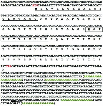
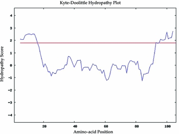
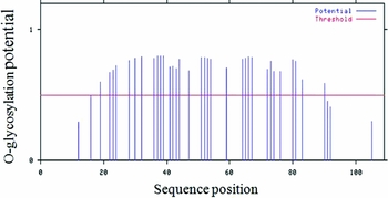
The nucleotide sequences of AsMuc for Liston and Kazerun strains were submitted in GenBank under accession numbers JQ026104.1-JQ026108.1 and JQ228229.1-JQ228232.1, JQ013478.1. The full length of AsMuc CDS from the Liston strain is 330 bp and from the Kazerun strain 291 bp, which includes the signal peptide sequence in N-domain in both strains. The specific primers amplified whole CDS sequences in both strains located from starting translational site to stop codon. Nucleotide sequence analysis revealed that mucin gene in the Kazerun strain in comparison with the Liston strain lacks 39 bp in its central core. This missing sequence encodes one repeat of GQATTTTVAPESP, which might be related to a length polymorphism within the mucin domain in different biological forms. The sequence of the full length of AsMuc cDNA was determined using 3′RACE PCR and 5′RACE PCR techniques. We could define the 3′ end region of AsMuc gene by the Rapid Amplification of cDNA Ends Kit (Gibco) protocol, but the 5′ end of AsMuc gene was not established using this protocol, potentially because of the secondary structure of mRNA or repeated motifs within the regions. We therefore changed our strategy to an alternative method in RACE-PCR using the NIH protocol, which is designed for difficult templates. The 5′ region of AsMuc sequence is a stretch of 76 bp nucleic acids encompassing the 5′-untranslated region (5′-UTR), and including a potential start codon preceded by AAAAC. This motif matches the Drosophila Kozak consensus sequence (C/A)AA(A/C)ATG), suggesting that it represents the codon for initiation of translation (Cavener, Reference Cavener1987). These results showed that there are two possible termination polyadenylation signal sequences (AATAAA) with four starting polyadenylation sites in the 3′R (Fig. 1). The four poly (A) signals are located at the nucleotide acid positions 154, 165, 495 and 510, downstream of the stop codon. These alternative sites may contain regulatory sequences that can regulate the expression of the gene. To explore possible existence of AsMuc introns, the AsMuc gene was amplified using DNA isolated from a single, whole mosquito body. The results showed that AsMuc is an intron-less gene like A. gambiae mucin gene. The cDNA of AsMuc includes a single ORF encoding a protein of 109 amino acids in the Liston strain and 96 aa in the Kazerun strain with estimated molecular weights of 10.6 and 9.35 kD, respectively. The isoelectric point of AsMuc is 4.32 to 4.49 in the two strains, which is as a result of the high acidic amino acid composition of the predicted protein (Fig. 1). Computation of instability index (38.18) classified the protein as a stable protein. A schematic representation of the deduced amino acid sequence of AsMuc is depicted in (Fig. 1). The first 20 amino acids in the N-terminus, predicted as the signal sequence, is followed by a Ser/Thr-rich region serving as a docking site for O-glycosylation (S + T = 35%). Repeated threonine-rich motifs are found in a variety of vertebrate and invertebrate mucins. Indeed, mucins are defined as if the threonine and serine content of the proteins is greater than 20% of the amino acid composition, and forms a cluster sequence(s) within the protein. AsMuc satisfied these criteria. The deduced protein sequences of AsMuc in both strains have two hydrophobic fragments, one at the N-terminus and the other at the C-terminus flanking a central core of repetitive and non-repetitive serine/threonine-rich fragments (Fig. 1). A hydropathic analysis of amino acid sequences showed the deduced proteins have two hydrophobic fragments, one at the N terminus and the other at the C-terminus. Based on other evidence, both hydrophobic domains are required to target AsMuc on the cell surface (Shen et al., Reference Shen, Dimopoulos, Kafatos and Jacobs-Lorena1999). Analysis of the secondary structure of AsMuc protein confirmed the presence of a transmembrane helix in the C-terminal domain. The C-terminus hydrophobic domain, VLPVLLGAYVVLSILI, may contribute to the cell membrane attachment. This hydrophobic domain is comparable to the hydrophobic patch at the C-terminus of mucin gene from A. gambiae (Fig. 2). In the Kazerun strain, one of the tandem repeats consensus motifs is absent (Fig. 1). This variation might be a result of slipped strand DNA replication or unequal recombination within a certain range. BLAST analysis of AsMuc protein demonstrated that the similarity between A. stephensi Liston strain and A. gambiae 4a, r/r pink eye strain is 44% (Evale 5e-21) (Fig. 3), whereas the percentage of similarity with mucin sequences of other mosquito species submitted in GenBank significantly reduced. For example, similarity between A. stephensi mucin and Anopheles funestus mucin is 16% (E value 0.25). A comparison of the predicted amino acid sequence of the A. stephensi (AsMuc) mucin with A. gambiae mucin, and salivary gland mucin from A. funestus (accession number ABI83779.1) and Trypanosoma cruzi (accession number U32447) shows differences in mucin repeated sequences among species. Both AsMuc and AgMuc1 mucin contain TTTTVAP repeats whereas Trypanosome cruzi mucins contain TTTTTTTTKPP repeats and A. funestus contains TTTEEA repeats. NetO glycol prediction server in CBS predicted 33–34 O-glycosylation sites in the AsMuc gene in Liston strain in the threonine-rich region, but not in the hydrophobic terminal domains (Fig. 4). AsMuc has no potential N-glycosylation site. Nuclear magnetic resonance (NMR) analysis of short peptide substrates suggests that the density and glycosylation pattern of acceptor substrates affect the ability of enzymes to modify Thr/Ser residues within mucin domains (Hang and Bertozzi, Reference Hang and Bertozzi2005). N-linked glycosylation takes place at a definite consensus sequence (AsnXaaSer/Thr), but no primary amino acid consensus sequence is observed for mucin-type O-linked glycosylation (Hang and Bertozzi, Reference Hang and Bertozzi2005). GPI anchor prediction by Big-PI showed one potential site for GPI anchor in C-terminal.
Nucleotide and deduced amino acid sequence of the Anopheles stephensi mucin mRNA (AsMuc). The putative signal peptide is underlined. The hydrophobic N-domain and C-domain are underlined. The possible termination polyadenylation signal sequences in the 3′-UTR are wave underlined. The polymorphic region missing in Kazeruni strain is boxed.

Kyte-Doolittle plot of AsMuc, with a window size of 19. Peaks in signal peptide region with scores greater than 2 indicate hydrophobic regions and strong negative peaks indicate hydrophilic regions of proteins.

Alignment of amino acid sequences of mucin between Anopheles gambiae (CAA07489.1) and Anopheles stephensi (AEY77529.1). Alignment was generated by ClustalW program. Identical residues are denoted by “*”, grey boxed, conserved substitutions are denoted by “:”, and semiconserved substitutions are denoted by “.”

Prediction of O-glycosylation sites in Anopheles stephensi mucin. Vertical lines indicate serine and threonine residues. A red line indicates the threshold level. Residues with an O-glycosylation potential exceeding the threshold level are expected to be glycosylated.

Immunological localization of His-AsMuc fusion protein in insect cells
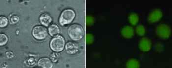
His-AsMuc fusion protein is expressed in Sf9 insect cells via a baculovirus vector. Sf9 cells were infected with recombinant baculovirus that expresses the AsMuc under the control of the viral polyhedron promoter. Localization of the recombinant protein was detected by fluorescence microscopy using a FITC-conjugated antibody (Fig. 5). AsMuc was detected on the cell surface of Sf9 cells expressing recombinant protein, but not on uninfected cells. These results indicate that the mucin is a membrane protein. Cell surface localization of AsMuc was detected at the later stages of infection of Sf9 cells with recombinant virus (72−84 h post infection). This distribution pattern was predicted based on the presence of a putative signal peptide in AsMuc.
Subcellular localization of AsMuc in Sf9 insect cells. Infected Sf9 cells with recombinant virus were incubated with FITC-labelled antibody in insect culture medium at 37°C. Cells were washed and observed under a fluorescence microscope.

Discussion
We have characterized an invertebrate intestinal mucin gene of A. stephensi in Liston and Kazerun strains sharing common features with intestinal mucin of A. gambiae (Shen et al., Reference Shen, Dimopoulos, Kafatos and Jacobs-Lorena1999).
The existence of many glycosylation sites within AsMuc is assumed to be a component of the carbohydrate-rich glycocalyx in the surface of the microvillus. This property suggests a possible common role of AsMuc and AgMuc1 in mid-gut physiology. It has been shown that the AgMuc1 gene is highly expressed in intestinal tissues after blood feeding (Abraham and Jacobs-Lorena, Reference Abraham and Jacobs-Lorena2004), and the presence of mRNA in unfed guts suggests translational regulation of AgMuc1 expression in the mid-gut (Shen et al., Reference Shen, Dimopoulos, Kafatos and Jacobs-Lorena1999). There is growing evidence that the carbohydrates located on the mosquito microvillar membrane might serve as the first receptor−ligand interaction between the malaria parasites and the gut epithelial cells (Dinglasan and Jacobs-Lorena, Reference Dinglasan and Jacobs-Lorena2005). For instance, it has been shown that P. gallinaceum ookinetes could not attach to A. aegypti mid-gut after chemical amendment of carbohydrates located on microvillus (Zieler et al., Reference Zieler, Nawrocki and Shahabuddin1999).
Low similarity of AsMuc with other mosquitoes may display different biological behaviours of this gene in different species. These differences may be related to in-depth evolutionary speciation.
Considering that each mucin has its own more or less conserved tandem repeats, it is probable that the central domain has evolved under selective pressure to keep serine and threonine codons corresponding to the O-glycosylation sites, and that the repeated motifs have arisen through internal successive duplications (Chaturvedi et al., Reference Chaturvedi, Singh and Batra2008).
Shen et al. (Reference Shen, Dimopoulos, Kafatos and Jacobs-Lorena1999) expressed AgMuc1 tagged with a green fluorescent protein (GFP) in insect cells using a baculovirus vector and showed that the protein localized on the cell surface. In our study, surface localization of His-AsMuc in insect cells suggests the presence of AsMuc on the cell surface of mid-gut epithelial cells. Surface localization is dependent on both the N-terminal secretory signal sequence and the C-terminal hydrophobic domains (Shen et al., Reference Shen, Dimopoulos, Kafatos and Jacobs-Lorena1999), and surface localization of AsMuc can be related to the putative signal peptide in the N-terminal and a GPI-anchor site in the C-terminal of protein. The hydropathic analysis of AsMuc shows the hydrophobic feature of N-domain and C-domain in AsMuc protein. All these features can help the surface localization of AsMuc in the mosquito mid-gut and its possible involvement in mosquito−parasite interaction.
In A. gambiae, the length polymorphism in the mucin alleles is associated with genetic localization of this gene and parasite−vector interaction (Shen et al., Reference Shen, Dimopoulos, Kafatos and Jacobs-Lorena1999). A shorter AgMucb1 allele lacking one threonine-serine repetitive unit was detected in the malaria-susceptible A. gambiae 4A r/r strain, whereas another A. gambiae refractory strain tested (L3–5) was found to have the longer AgMuc1 allele (Shen et al., Reference Shen, Dimopoulos, Kafatos and Jacobs-Lorena1999). The allelic polymorphism of the mucin gene observed between the Liston and Kazerun strains in this study may mediate different biological and functional features of molecules at the interaction sites between parasite and mosquito mid-gut and lead to different vector capacities in these strains. Based on our previous study (Oshaghi et al., Reference Oshaghi, Yaaghoobi, Vatandoost, Abaei and Akbarzadeh2006) the Kazerun strain belongs to intermediate form and was responsible for transferring malaria in Fars Frovince in southern Iran in the past (Manouchehri et al., Reference Manouchehri, Javadian, Eshghy and Motabar1976), and sporozoite rate was reported between 0.2 to 0.7% (Manouchehri et al., Reference Manouchehri, Javadian, Eshghy and Motabar1976). The Liston strain belongs to type form. Type form is considered an efficient vector of malaria in urban areas (Subbarao et al., Reference Subbarao, Vasantha, Adak, Sharma and Curtis1987). Compared to the intermediate form, the type form is a more efficient malaria vector with a higher sporozoite rate of 3.6% (Korgoankar et al., Reference Korgaonkar, Kumar, Yadav, Kabadi and Dash2012)−6.7% (Banerjea, Reference Banerjea1930).
It is worth assessing the interaction of ookinetes with expressed AsMuc proteins from different alleles and monitoring the potential infection intensity phenotypes. Many stimulating aspects of AsMuc remain to be explored in future investigations. Characterization of insect intestinal mucins could lead to the development of new strategies for disease and pest control (Rayms-Keller et al., Reference Rayms-Keller, McGaw, Oray, Carlson and Beaty2000).
In the present study, we characterized the AsMuc gene and showed surface localization of protein in eukaryotic cells. This recombinant mucin is a feasible and promising candidate for further surveys as an agent associated with epithelial cell interactions. This work can pave the way for fundamental studies in TBVs strategies.
Acknowledgements
This paper is a part of the results of the first author's dissertation for fulfilment of a PhD in Medical Entomology and Vector Control from the Department of Medical Entomology and Vector Control, School of Public Health, Tehran University of Medical Sciences, Tehran, Iran. This investigation received technical support from the departments of Medical Entomology and Medical Genetics of Tehran University of Medical Sciences, and the School of Biosciences and Biotechnology, Camerino University, Italy, and Influenza Lab Research of Pasteur Institute of Iran. This investigation received financial support from Tehran University of Medical Sciences, with grant number: 5864-27-02-86.