Introduction
Recent studies of the developmental origins of adult disease (DOAD) hypothesis have demonstrated that early life events may contribute to the pathogenesis of many adult diseases, including Alzheimer’s disease (AD) and other neurodegenerative diseases.Reference Barker 1 – Reference Bilbo 4 One system at risk from these early life events is the immune system. The developing immune system is highly sensitive to exogenous insults and can be associated with systematic changes that persist into adulthood.Reference DeWitt, Peden-Adams, Keil and Dietert 5 These persistent effects have been associated with pediatric diseases, and are predictive of later-life disease onset.Reference Dietert and Zelikoff 6 However, little headway has been made to uncover the myriad mechanisms and systemic interactions that underlie the risk of adult disease following an early life insult.
Microglia, the innate immune cells of the central nervous system (CNS), are thought to be potential targets of developmental disruption, subsequently contributing to a variety of adult diseases within the CNS.Reference Harry and Kraft 7 , Reference Streit, Xue, Tischer and Bechmann 8 These neuroimmune cells have a multitude of homeostatic functions in the developing brain, including promoting synaptogenesis and synaptic pruning.Reference Santambrogio, Belyanskaya and Fischer 9 Likewise, they are the first responders to brain injury, pathogens and toxicants through a sensitive, stimuli-dependent response typically involving a phenotypic change to an activated state.Reference Gehrmann, Matsumoto and Kreutzberg 10 , Reference Olah, Biber, Vinet and WGM 11 The role of microglia in neurodegenerative diseases, however, is unclear in the later stages of pathology, and even less is known about their potential contribution to the developmental origins of these diseases. Furthermore, as the exact mechanism of developmental microglial dysfunction in AD is unknown, the methods of analysis are similarly lacking.Reference Walker and Lue 12
The brain undergoes sexually dimorphic development that, in part, is thought to be due to the direct involvement of microglial activation; thus, specifically timed disruption of this tightly regulated signaling may result in sex-dependent susceptibility to AD and other neurodegenerative disorders.Reference Lenz, Nugent, Haliyur and McCarthy 13 As females are at greater risk of developing AD correlation to periods of distinct developmental sexual dimorphism is of particular interest. In this study, we investigated the role of microglia in the etiopathology of AD as a consequence of early toxicant exposure during a critical window of development for microglia, defined by microglia-driven postnatal brain development during this time. The prototypical toxicant utilized during this critical window was lead acetate, a well-defined developmental immunotoxicant and neurotoxicant, at a physiologically relevant dose to mimic an acute, low-level exposure to environmental lead. This time-sensitive dosing paradigm was applied in a genetically predisposed mouse model for AD [triple transgenic murine model for AD (3×TgAD)] to assess the pathological consequences of early toxicant exposure. An advantage to this ‘double-hit’ model is that we could more clearly differentiate increased vulnerability to AD, as evidenced by earlier or more severe pathological markers, consequent to precise postnatal disruption of neuroimmune-relevant development. In addition, the 3×TgAD mouse model was reported to exhibit female bias in amyloid accumulation similar to that in humans, increasing the translational power of the model and interpretation of sex-specific results.Reference Carroll, Rosario and Kreimer 14 We hypothesized that microglia would become activated in a sex-dependent manner following early postnatal lead exposure and that this change in activation would be inversely correlated with earlier or more severe AD pathologies, suggesting an early role for microglia in the pathological progression of AD.
Here we demonstrate the utility of this double-hit DOAD model, specifically in the context of the developing neuroimmune parenchyma and microglia, in detecting early changes involved in the pathophysiology of AD. We report that postnatal toxicant exposure increased AD pathologies in females to a greater extent than in males, and that this may be correlated with early disruption of female microglia functionality.
Materials and methods
Animal handling
All experimental animal handling and dosing was carried out in accordance with procedures approved by the East Carolina University’s Institutional Animal Care and Use Committee (IACUC). Pregnant transgenic dams [3×TgAD; B6; 129-Psen1tm1MpmTg(APPSwe, tauP301L) 1Lfa/Mmjax] were obtained from the seed colony in the East Carolina University’s Department of Comparative Medicine and kept on a 12:12 h light–dark cycle, with access to food and water ad libitum. Litters were culled to six after birth [postnatal day (PND) 1], weaned at PND 21 and housed by sex in groups of no more than three.
Dosing and tissue preparation
Dosing solution concentration of lead acetate (100 ppm in sterile water, at 10 µl/g body weight/ day; Sigma-Aldrich, St. Louis, MO, USA) was prepared weekly, and dosing was performed based on daily body weight. This concentration was determined based on rodent model toxicity and risk assessment recommendations,Reference Moser, Walls and Zoetis 15 , Reference Zoetis and Walls 16 as well as previous reports of long-term potentiation impairment due to microglial activation following exposure to Pb at this concentration.Reference Liu, Liu and Wang 17 From PND 5–15, neonates were dosed with lead acetate (Pb) or vehicle (ctl) once per day using a modified gavage technique.Reference Butchbach, Edwards, Schussler and Burghes 18 No overt signs of lead-induced toxicity, such as changes in body weight, or behavioral abnormalities, such as decreased feeding or huddling, were observed in any of the animals. One mouse per sex, litter and treatment group was euthanized at either PND 50, 90 or 180. These terminal endpoints were chosen based on reports of the 3×TgAD mouse strain displaying amyloid-β immunoreactivity as early as 3 months of age.Reference Oddo, Caccamo, Kitazawa, Tseng and LaFerla 19 Sample sizes included 3–6 mice/sex/age/exposure. Immediately following euthanasia, the brain was carefully removed and hippocampi dissected in ice-cold phosphate buffered saline (PBS). For histochemical analysis, hippocampi were fixed for 24 h in 10% neutral buffered formalin followed by 70% ethyl alcohol before paraffin embedding. For flow cytometric analysis, the remaining brain tissue sans cerebellum was placed in fluorescence-activated cell sorting (FACS) buffer (3% fetal calf serum (FCS), 0.1% sodium azide and 10 mM ethylenediaminetetraacetic acid (EDTA) in PBS) for subsequent homogenization the same day as extraction.
Flow cytometry
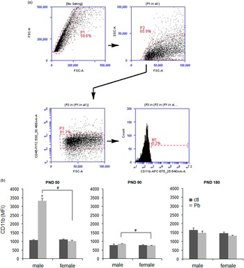
Brain tissue was homogenized in a 15 ml Tenbroeck glass homogenizer and filtered through a 70 μm nylon mesh filter. Myelin removal was accomplished via centrifugation in 30% isotonic Percoll (GE Healthcare, Uppsala, Sweden), and the fatty layer was removed from the top of the cell suspension. Cells were then resuspended in blocking buffer [5% normal mouse serum, 5% normal rat serum, 1% Fcr block (polyclonal anti-mouse IgG CD16/32; eBioscience Inc., San Diego, CA, USA) in FACS buffer] and incubated for 10 min. Cells were passed through a 40 μm nylon mesh filter, centrifuged, resuspended in incubation buffer (10% FBS, PBS) and incubated with the following primary antibodies for 1 h: APC-conjugated CD11b [1:100, rat monoclonal IgG2b kappa (M1/70); Abcam, Cambridge, MA, USA] and FITC-conjugated CD45R [1:100, rat monoclonal IgG2a kappa (C363.16A); eBioscience Inc.]. Following incubation, cells were centrifuged, resuspended in FACS buffer and read using a BD Accuri C6 flow cytometer (BD Biosciences, Franklin Lakes, NJ, USA). A total of 10,000 events were recorded. Color compensation was performed using isotype controls, and quadrants were determined based on fluorescence minus one (FMO) positive controls. Figure 1a depicts the gating schematic used to differentiate resident microglia (CD45lo) from peripheral macrophages (CD45hi). The data were analyzed using the BD Accuri C6 software (BD Biosciences), and are represented as the CD11b mean fluorescence intensity±s.e.m.
Developmental Pb exposure increased CD11b expression in males at postnatal day (PND) 50. There was no observable change in CD11b associated with Pb exposure at PND 90 and 180. (a) Representative flow cytometric plots and gating schematic for discrimination of doublets, live/dead and peripheral macrophages (CD45hi, CD11b+). (b) Bar graphs depicting CD11b expression as a result of early Pb exposure at PND 50, 90 and 180 in males and females. n=3–6 mice/sex/age/exposure. Data are presented as mean±s.e.m. P<0.05 was considered statistically significant compared with age-matched control values. *, Significant between exposure; #, significant between sex; FSC, forward scatter; SSC, side scatter; MFI, mean fluorescence intensity; ctl, control; Pb, lead.

Immunohistochemistry
Formalin-fixed, paraffin-embedded (FFPE) hippocampi were sliced on a rotary microtome at 5 μm and mounted on Superfrost Plus slides (Azer Scientific, Morgantown, PA, USA). In brief, slides were dewaxed in Histo-Clear II (Electron Microscopy Sciences, Halfield, PA, USA), followed by washes in 100 and 95% ethyl alcohol and PBS. Antigen unmasking was accomplished using a heat-mediated citrate buffer, followed by incubation in 0.3% hydrogen peroxide for 30 min. All subsequent staining was performed using Sequenza-Coverplate racks (Thermo Scientific, Waltham, MA, USA). Sections were permeabilized with phosphate buffered saline with Tween 20 (TPBS) and blocked with diluted normal serum (ABC Vectastain; Vector Laboratories, Burlingame, CA, USA). Slides were then incubated with primary antibody, ionized calcium-binding adapter molecule 1 (Iba1, 1:500; Wako Chemicals USA, Inc., Richmond, VA, USA), for 60 min at room temperature or overnight at 4°C. Sections were visualized using ABC Vectastain and DAB kits (Vector Laboratories) and counterstained with Harris’ Alum Hematoxylin. After sequential washing in ethyl alcohol and Histo-Clear, slides were coverslipped with Permount (Fisher Scientific, Fair Lawn, NJ, USA) and cured overnight before visualization.
Slides were visualized using a Leica DM1000 light microscope at 20× magnification with a SPOTTM Idea camera attachment and SPOT Advanced imaging software. Two regions of interest (ROI) per hippocampal section were selected at random, with the viewing frame containing as much tissue as possible, and the percentage of immunopositive microglia/ROI was analyzed using ImageJReference Schneider, Rasband and Eliceiri 20 (NIH, Bethesda, MD, USA). All data are represented as the mean per cent microglia density per ROI±s.e.m.
Immunofluorescence
FMO control optimizations, the use of fluorphores with relatively distinct emission spectra and a sequential double-staining immunofluorescence procedure were utilized to reduce cross-reactivity and spectral bleed-through. Slide dewaxing, rehydration and antigen retrieval was performed in a similar manner to immunohistochemistry. Slides were blocked with 10% donkey serum in TPBS for 30 min at room temperature, followed by a 1-h incubation with primary antibody for amyloid-β (1:1000, rabbit pAb; Abcam). Subsequent incubation with Cy5-conjugated secondary antibody (1:1000, goat anti-rabbit IgG-Cy5; Abcam) was performed and cell nuclei were stained with 41,6-diamidino-2-phenylindole (DAPI) (300 nM; Life Technologies, Eugene, OR, USA). Slides were coverslipped with ProLong Gold antifade reagent (Life Technologies), cured for 24 h and stored upright to protect from light.
Hippocampal sections were visualized with a Zeiss LSM 700 laser scanning confocal microscope with the EC Plan-Neofluar 10×/0.30 M27 objective. Two counting frames per animal (640.17×640.17 μm) were used for ROIs. Amyloid load density was defined as percentage of amyloid-β+immunoreactivity/ROI area, and the two ROIs were averaged per animal. Fluorescence intensity was considered positive over the background fluorescence threshold. Data are represented as mean±s.e.m.
Statistical analysis
All analyses were performed using Statistical Analysis System (SAS Institute, Cary, NC, USA). Two-way analysis of variance for sex and exposure effects was performed for immunohistochemistry and flow cytometry at each age. Individual pairwise comparisons between sex within treatment and between treatments within sex were made with a t-test or a Tukey’s studentized range distribution method. All data in bar charts are represented as mean±s.e.m. P<0.05 was used to define statistical significance.
Results
Developmental Pb exposure increased microglial activation only in males at PND 50
To determine how toxicant exposure during a critical window of development altered microglia and markers of AD, we used two techniques to assess microglial activation: upregulation of activation surface markers with flow cytometry and microglial density with immunohistochemistry.
Flow cytometry was carried out using markers of microglial/macrophage activation, CD45 and CD11b.Reference Sedgwick, Schwender and Imrich 21 – Reference Nikodemova and Watters 24 CD45lo microglia were discriminated from peripheral macrophages (Fig. 1a), and CD11b expression was used to determine activation and general functionality, as CD11b (CR3A) is part of the innate complement system and involved in phagocytosis, migration and chemotaxis.Reference Roy, Fung, Liu and Pahan 25 , Reference Kobayashi, Imagama and Ohgomori 26 Microglial activation was higher in Pb-exposed males than in females at PND 50, with Pb-exposed female microglial responsiveness comparable with ctl mice (Fig. 1b). By PND 90 the sex difference in Pb-exposed mice was still detectable, but there was no change in CD11b expression between ctl and Pb mice of either sex at PND 90 and 180.
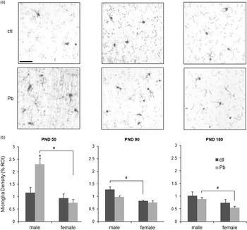
The hippocampus is vulnerable to damage with AD, and associated with amyloid-β deposition and accumulation in the 3×TgAD mouse model.Reference Oddo, Caccamo, Kitazawa, Tseng and LaFerla 19 Therefore, microglial activation within this region of the brain was assessed at three time points (Fig. 2a). To determine microglial abundance and activation, hippocampi were stained with Iba1, a general marker for microglia. As more infiltrating microglia would increase overall density, and activated microglia take up more space due to enlarged somas and thickened processes, microglial density was used as a general maker for neuroprotective microglial presence.Reference Schwarz, Sholar and Bilbo 27 Immunohistochemical evaluation indicated that Pb-exposed male mice had greater microglial density than the ctl males at PND 50, and Pb-exposed females had significantly lower microglial density than Pb-exposed males (Fig. 2b). Female mice had consistently lower microglial density at all time points, with Pb-exposed females differing significantly from Pb-exposed males at the earliest (PND 50) and latest (PND 180) time points.
Developmental Pb exposure increased microglial density in males at postnatal day (PND) 50, as assessed by ionized calcium-binding adapter molecule 1 (Iba1)+ immunohistochemistry (IHC) and semiquantitative morphometric analysis of activation state. Control (ctl) males at PND 50 and 90 had greater microglial density than females, whereas at PND 180, lead (Pb)-exposed males had higher density than females. (a) Representative images of microglial density at PND 50, 90 and 180 in the hippocampus of control- and Pb-treated male mice. Scale bar, 50 μm. (b) Bar graphs representing IHC morphometric analysis of microglial density at PND 50, 90 and 180 in males and females. n=3 mice/sex/age/exposure. Data are presented as mean±s.e.m. P<0.05 was considered statistically significant compared with age-matched control values. *, Significant between exposure; #, significant between sex; ROI, region of interest.

Developmental Pb exposure increased amyloid-β load density
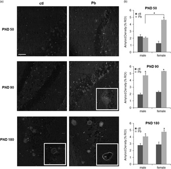
Amyloid-β load density, encompassing both immature aggregates of amyloid deposits as well as later plaque formation, was used to determine changes in AD pathological severity. Immunofluorescent evaluation of FFPE hippocampi revealed increased amyloid density in Pb-exposed females at PND 50 but not in Pb-exposed males, indicative of increased pathological severity due to an earlier appearance of amyloid deposits compared with 3×TgAD controls (Fig. 3b). At PND 90 Pb exposure increased amyloid density in both males and females, with the first appearance of diffuse plaque-like amyloid aggregates only seen in the Pb-exposed groups (Fig. 3a, insets). By PND 180 the 3×TgAD controls had detectable plaques and increased amyloid density. Likewise, Pb-exposed animals also exhibited mature, hollow plaques, but to a greater degree than controls at the latest time point. There were no differences between control males and females at any time point. PND 180 could be considered relatively early in the 3×TgAD pathological progression; there have been previous reports of virtually no detectable plaques in 4–6-month 3×TgAD hippocampi and no discernable sex difference in plaque number until 13–14 months.Reference Overk, Perez and Ma 28 However, this is contrasted by the Pb-exposed females that had significantly increased amyloid burden at PND 50, and a general trend toward greater pathology than males at all time points (Fig. 3b). Altogether, Pb exposure during a critical window of postnatal development increased amyloid burden and AD pathology in both males and females at later time points. Furthermore, Pb-exposed females were affected earlier (PND 50) than Pb-exposed males (PND 90), potentially contributing to the sex-related bias seen in AD.
Developmental lead (Pb) exposure increased amyloid density in the hippocampus of male and female triple transgenic murine model for AD (3×TgAD) mice at postnatal day (PND) 90 and PND 180, and only in females at PND 50. (a) Representative immunofluorescence images of amyloid-β immunoreactivity at PND 50, 90 and 180. Insets, representative plaques that progressed from dense clusters (PND 90) to more mature, hollow configurations (PND 180). Scale bar, 50 μm. (b) Summary graphs of amyloid density at each time point. Data are represented as mean density±s.e.m. n=3 mice/sex/age/exposure. P<0.05 was considered statistically significant compared with age-matched control values. *, Significant between exposure; #, significant between sex; ROI, region of interest; ctl, control.

Discussion
Previous studies have established a correlation between microglia and AD, many of which interpret the pathological role of microglia as an inflammatory propagator due to increased overall activation.Reference Banati, Gehrmann, Schubert and Kreutzberg 29 – Reference Solito and Sastre 32 The gaining momentum of DOAD hypotheses has propelled investigations into microglial priming and altered inflammatory sensitivity following a developmental immune insult as potential contributors to adult disease.Reference Wu, Basha and Brock 33 – Reference Perry and Teeling 36 However, there is a disjointed understanding of this early role for microglia, particularly in neurodegenerative diseases, due to a lack of standardized markers for the various states of activation, compounded by numerous disease models and conflicting data collection time points.Reference Olah, Biber, Vinet and WGM 11 , Reference Walker and Lue 12 In this study, we investigated the role of early toxicant exposure during a critical window of development for microglia in the exacerbation of AD pathologies. Our results support the utility and validity of this DOAD exposure paradigm, namely through demonstrating a persistent change in pathological severity months after exposure. Furthermore, our results demonstrate a sex-related vulnerability seen only in females at early stages of AD that are paralleled by the sex differential in early microglial responsiveness following Pb exposure. As microglial proliferation and activation were once thought to contribute to pathology, this correlation highlights the necessity of utilizing DOAD models to better define the putative role of microglia in AD.
During normal brain development activated microglia migrate, proliferate and mature to a ramified morphology by PND 28.Reference Nayak, Roth and McGavern 37 Developmental microglial activation is functionally distinct from activation in the adult CNS due to its critical role in neuronal synaptogenesis and pruning, the absence of which can result in the development of atypical brain circuitry.Reference Paolicelli, Bolasco and Pagani 38 This perinatal critical window of microglial development may be further temporally segmented, varying in susceptibility by sex. Both rodent and human brains develop in a sexually dimorphic manner, and microglia are critical to this process through various feed-forward mechanisms, such as those involving estrogens and prostaglandins.Reference Lenz, Nugent, Haliyur and McCarthy 13 , Reference Schwarz and Bilbo 39 Likewise, microglial-mediated processes, such as phagocytosis and proliferation, also proceed in surges that temporally vary in concert with steroid hormone signaling.Reference Lenz and McCarthy 40 At PND 4, male mice experience a testosterone surge that coincides with a peak in microglia numbers in various brain regions like the hippocampus, whereas female mice experience this microglial migration and proliferation later in development, around PND 21.Reference Schwarz, Sholar and Bilbo 27 , Reference Crain, Nikodemova and Watters 41 Although there are data to suggest females are less vulnerable to long-term changes compared with males following early postnatal infection,Reference Bilbo 4 sex-related microglial vulnerability may be more dependent on the concentration and type of toxicant rather than timing only. Our results demonstrate an increase in amyloid-β burden in Pb-exposed mice, with female mice exhibiting greater and earlier pathological severity associated with developmental toxicant exposure (Fig. 3). The Pb-related exacerbation of pathology was not seen in male mice at PND 50. An inverse sex bias was also present at PND 50 for markers of microglial activation, with greater microglial responsiveness in Pb-exposed males than females (Figs 1 and 2), implying that the early heightened microglia responsiveness in males was likely neuroprotective against signs of AD neuropathology. Conversely, the detectable sex differences at PND 50 supports the identification of a window of vulnerability for female microglia during a period of development previously mainly attributed to male vulnerability. The mechanisms by which females were more vulnerable than males may be consequent to the low concentration of Pb (100 ppm) and later exposure window (PND 5–15) used in our study.
A recent study by Sobin et al.Reference Sobin, Montoya and Parisi 42 reported atypical and differential microglial disruption depending on a high (330 ppm) or low (30 ppm) postnatal exposure to Pb, with no evidence of neuroinflammatory propagation at PND 28. Furthermore, dose-dependent reduction in microglia numbers correlated with decreased dentate gyrus volume, suggesting reduced later-life resiliency and cognitive function.Reference Sobin, Montoya and Parisi 42 This contrasts sharply with many investigations into the role of microglia in developmental Pb exposure paradigms that implicate microglia as propagators of bystander neuronal death and neuroinflammation.Reference Liu, Liu and Wang 17 , Reference Bunn, Parsons, Kao and Dietert 43 – Reference Kumawat, Kaushik, Goswami and Basu 45 Our results provide a similar early glimpse into microglial disruption following acute, low levels of Pb. By PND 90 Pb-exposed males still have higher CD11b expression compared with females, but by PND 180 there is no sex- or Pb-related effect detectable (Fig. 1). This could be due, in part, to latent and persistent neurotoxicity, resulting in male microglia eventually developing a similar dysfunctional phenotype to females over time; CD11b-associated phagocytosis decreases and amyloid deposition increases. As an alternative there could be fewer total number of microglia present to differentiate from controls. However, although Pb-exposed females had lower microglial density at PND 180 than males, there was no significant difference between control and Pb-exposed mice at that time in either sex (Fig. 2). Further studies are warranted to determine sex differences in microglial functionality, if any, at later stages of AD.
Increases in amyloid density following developmental Pb exposure persisted over time, although there was no direct age-related correlation with markers of microglial activation. This suggests that if there were a causative role for microglia in pathological exacerbation, as our results imply, it would occur very early in pathological progression. Many AD studies that evaluate microglia function typically do so at later time points, after pathology has been well established, producing conflicting data in terms of how the presence of microglia changes amyloid pathology. For example, one study showed prevention of neuronal loss and no change in amyloid-β plaques following selective microglial elimination, whereas another demonstrated that formation and maintenance of amyloid-β plaques were not influenced by the presence or absence of microglia.Reference Grathwohl, Kälin and Bolmont 46 , Reference Spangenberg, Lee and Najafi 47 Conversely, the colocalization of activated microglia with senile plaques has been extensively cited, with the pro-inflammatory phenotype frequently implicated as the major instigator of chronic inflammation and neurotoxicity in AD.Reference Banati, Gehrmann, Schubert and Kreutzberg 29 , Reference Cras, Kawai and Siedlak 48 – Reference Marlatt, Bauer and Aronica 53 Our data support the inverse association between microglial activation and early pathological exacerbation, inferring changes in classical markers of inflammation, such as interleukin 1 beta (IL-1β), tumor necrosis factor alpha (TNF-α) and inducible nitric oxide synthase (iNOS), evaluated at single point in time may not necessarily indicate a causative role for microglial inflammation in AD. Had we evaluated time points only at PND 90 or later, when the 3×TgAD model was anticipated to exhibit cognitive decline,Reference Oddo, Caccamo, Kitazawa, Tseng and LaFerla 19 any sex differences or correlations with microglial activation would not have been as significant or detectable, as developmental damage to microglia would have already occurred. This could potentially contribute to the mass of conflicting reports for the role of microglia in AD, or even account for the inefficiencies of non-steroidal anti-inflammatory drugs (NSAIDs) as AD therapeutics.
Along with demonstrating a link between very early microglia dysfunction and exacerbated amyloid burden, our results revealed a female bias in early susceptibility to AD following postnatal Pb exposure. For both amyloid density and microglial activation, the sex-related bias was most significant at PND 50, suggesting an early role for microglia. Our exposure window, at the overlap of previously established male/female microglial windows of vulnerability, as well as the relatively low concentration of Pb, likely contributed to the increased susceptibility in females via steroid hormones, imbalances in neuronal apoptosis and neurogenesis or even changes in transcriptional regulation of microglial activation. Additional studies to determine how steroid hormones skew microglia to varying responses throughout life and how early-life toxicants disrupt this process are warranted.
The role of microglia in the developmental etiopathology of AD is unclear and, as shown here, complicated by a variety of intrinsic factors, like sex biases and mechanistic roles, as well as extrinsic factors, such as appropriate models, exposure scenarios and timing of data collection. Here, we attempted to characterize a standardized double-hit model for exploring the role of microglia in the developmental origins of AD following early postnatal Pb exposure. The results indicated that microglia are activated by postnatal toxicant exposure in a sexually dimorphic manner, with activation in males at the earliest time point coinciding with greater neuroprotection against amyloid accumulation. Likewise, the critical window of Pb exposure used in this study defined a period of female microglial vulnerability and correlated with earlier pathological severity, suggesting that microglia likely are early contributors to AD via dysfunctional responsiveness to pathological stimuli and impaired neuroprotectiveness. Further investigations are warranted to determine the causative v. correlational role for microglia in AD, and the mechanisms of early sex bias to disease progression.
Acknowledgments
The authors acknowledge Dr Kym Gowdy and Dr Hu Huang for editing and proof reading the manuscript.
Financial Support
This research was supported by the Harriet and John Wooten Laboratory for Alzheimer’s and Neurodegenerative Diseases Research (internal grant).
Conflicts of Interest
None.
Ethical Standards
The authors assert that all procedures contributing to this work comply with the ethical standards of the relevant national guidelines on the care and use of laboratory animals, as delineated by the National Research Council’s Institute for Laboratory Animal Research, and has been approved by the institutional committee, the East Carolina University’s IACUC.