Introduction
Phyllobothriid tapeworms parasitizing carcharhiniform, lamniform and orectilobiform sharks have become the focus of some recent intrigue as mounting global collections have revealed a greater diversity of these taxa than previously predicted. For example, among the most conspicuous of these, are three genera that are unique in their possession of strobilar scutes, vitelline follicles interrupted by the ovary, relatively anterior genital pores, and serrate gladiate spinitriches on their proximal bothridial surfaces. The first described of these was Paraorygmatobothrium Ruhnke, Reference Ruhnke1994, a genus erected by Ruhnke (Reference Ruhnke1994) to house three species parasitizing carcharhiniform and certain lamniform sharks. Descriptions of 11 additional species from a diversity of other carcharhiniform sharks have since appeared (e.g. Ruhnke, Reference Ruhnke1996; Ruhnke & Thompson, Reference Ruhnke and Thompson2006; Ruhnke et al., Reference Ruhnke, Caira and Carpenter2006a, Reference Ruhnke, Healy and Shaperob; Ruhnke & Carpenter, Reference Ruhnke and Carpenter2008; Cutmore et al., Reference Cutmore, Bennett and Cribb2009; Malek et al., Reference Malek, Caira and Haseli2010). Orectolobicestus Ruhnke, Caira & Carpenter, 2006 was erected by Ruhnke et al. (Reference Ruhnke, Caira and Carpenter2006a) to house six species parasitizing orectolobiform sharks of the genus Chiloscyllium Müller & Henle. Unlike species of the former genus, these bear marginal bothridial loculi. The currently monotypic Ruhnkecestus Caira & Durkin, Reference Caira and Durkin2006 was erected by Caira & Durkin (Reference Caira and Durkin2006) for a species taken from the carcharhinid shark Scoliodon laticaudus Müller & Henle. This genus differs conspicuously from the former two in that the bothridia lack apical suckers and also in that each bothridium bears six facial loculi.
New collections from carcharhinid sharks from the Persian Gulf and northern Australia have revealed what we believe is yet another genus of tapeworms, bearing serrate gladiate spinitriches on their proximal bothridial surfaces, strobilar scutes, vitelline follicles interrupted by the ovary, and anterior genital pores. However, the members of this genus exhibit a particularly unusual scolex morphology in that their bothridia are essentially tubular in form; like Ruhnkecestus, their bothridia also lack apical suckers. A new genus is established below to house these shark-hosted species. While one of these species is new to science and is described below, the other is a known species, which is redescribed below and transferred to the new genus.
Materials and methods
A total of six specimens of Rhizoprionodon acutus (Rüppell), consisting of one female 84 cm in total length (TL) and five males ranging in TL from 50.5 to 79.5 cm, were collected using gill nets in conjunction with Darwin Fisheries near Darwin, Australia in August 1997. In addition, 35 specimens of Carcharhinus cf. dussumieri, ranging in TL from 48 to 95 cm, were collected from the Persian Gulf (26°15′–27°04′N, 53°02′–57°01′E) in November and December 2007 by the fishing trawler Ferdous I. The body cavity of each shark was opened with a midventral incision and the spiral intestine was removed, placed in a bag with 10% seawater-buffered formalin and shaken for approximately 30 s. After ~1 week, specimens were transferred to 70% ethanol for storage.
Whole mounts of a subset of tapeworm specimens from each shark species were prepared as follows. They were hydrated in a graded ethanol series, stained in Delafield's haematoxylin, dehydrated in a graded ethanol series, cleared in methyl salicylate, and mounted on glass slides in Canada balsam. The scolex of one specimen of each species was prepared for scanning electron microscopy (SEM) as follows. They were hydrated in a graded series of filtered ethanols, placed in 1.5% OsO4 overnight, dehydrated in a graded series of filtered ethanols, transferred to hexamethyldisilazane (Ted Pella, Inc., Redding, California, USA) and allowed to air dry in a fume hood. They were subsequently mounted on aluminium stubs using double-sided adhesive carbon tape, sputter coated with 35 nm of gold/palladium and examined with a LEO/Zeiss DSM982 Gemini field emission scanning electron microscope. Strobilae of both SEM specimens were prepared as whole mounts as described above. In order to explore the nature of the bothridia in more detail, frontal sections were prepared of the scolex of one specimen taken from R. acutus. The scolex was embedded in paraffin and frontal sections were made at 7–8 μm intervals using an Olympus CUT4060 rotary microtome. These were placed on glass slides, flooded with 2.5% sodium silicate, and allowed to air dry on a slide warmer. They were subsequently stained in Delafield's haematoxylin and eosin, cleared in xylene, and mounted on glass slides under coverslips in Canada balsam. The strobila of this specimen was prepared as a whole mount as described above.
All measurements were made using SPOT software (version 4.5) and a SPOT digital camera mounted on a Zeiss Axioskop 2 compound microscope. Measurements are in micrometres unless otherwise stated and are given in the descriptions as the range followed in parentheses by the mean, standard deviation, number of worms examined (n) and total number of measurements (n) when more than one measurement was made per worm. Proglottid measurements were taken from terminal proglottids only. Photographs were also taken using the SPOT digital camera system. Drawings were made using a camera lucida mounted on a Zeiss Axioskop 2.
Museum abbreviations are as follows: BMNH, The Natural History Museum, London, England; LRP, Lawrence R. Penner Parasitology Collection, Department of Ecology and Evolutionary Biology, University of Connecticut, Storrs, Connecticut, USA; MPM, Meguro Parasitology Museum, Meguro, Japan; QM, Queensland Museum, Brisbane, Queensland, Australia; USNPC, US National Parasite Collection, Beltsville, Maryland, USA; ZUTC, Collection of the Zoological Museum, University of Tehran, Tehran, Iran. Microthrix terminology follows Chervy (Reference Chervy2009). Statistics were calculated using SPSS version 13 (SPSS Inc., Chicago, Illinois, USA). Prevalence, mean abundance and mean intensity were calculated according to Bush et al. (Reference Bush, Lafferty, Lotz and Shostak1997).
Doliobothrium n. gen.
Diagnosis. Tetraphyllidea, sensu Euzet (Reference Euzet, Khalil, Jones and Bray1994); Phyllobothriidae Braun, 1900, sensu Euzet (Reference Euzet, Khalil, Jones and Bray1994); Phyllobothriinae de Beauchamp, 1905, sensu Euzet (Reference Euzet, Khalil, Jones and Bray1994) as emended by McKenzie & Caira (Reference McKenzie and Caira1998) and subsequently Healy et al. (Reference Healy, Caira, Jensen, Webster and Littlewood2009). Worms euapolytic. Neck present. Scolex with four bothridia. Bothridia lacking apical suckers and marginal loculi, tubular in form with relatively large distal opening and smaller proximal opening; distal bothridial opening with band of musculature running parallel to anterior and posterior margin, proximal bothridial opening with muscular rim. Proximal and distal bothridial surfaces with serrate gladiate spinitriches and capilliform filitriches. Neck and proglottids conspicuously scutellate; scutes consisting of densely arranged capilliform filitriches, pointed at their tips. Proglottids craspedote. Immature proglottids wider than long, mature proglottids longer than wide. Testes numerous in poral and aporal fields anterior to ovary, one row deep in cross-section. Genital pores anterior, uni-lateral or occasionally irregularly alternating. Genital atrium shallow. Cirrus sac elongate oval, containing armed cirrus. Vagina opening anterior to cirrus sac. Ovary posterior, H-shaped in frontal view, tetralobed in cross-section. Uterus ventral, reaching level of cirrus sac. Vitellarium follicular; follicles distributed in two lateral fields; each field consisting of multiple follicles, interrupted by cirrus sac and ovary. Parasites of spiral intestine of carcharhinid sharks.
Taxonomic summary
Type species. Doliobothrium haselii n. gen., n. sp.
Etymology. This genus is named for the jar, or barrel-like shape of the bothridia (i.e. dolio, L., jar).
The possession of pouch-like tubular bothridia and the lack of bothridial apical suckers or apical loculi, readily distinguish Doliobothrium n. gen. from the following genera considered by Euzet (Reference Euzet, Khalil, Jones and Bray1994) to belong to the Phyllobothriinae as modified by McKenzie & Caira (Reference McKenzie and Caira1998) and Healy et al. (Reference Healy, Caira, Jensen, Webster and Littlewood2009): Calyptrobothrium Monticelli, 1893, Ceratobothrium Monticelli, 1892, Crossobothrium Linton, 1889, Dinobothrium van Beneden, 1889, Gastrolecithus Yamaguti, Reference Yamaguti1952, Marsupiobothrium Yamaguti, Reference Yamaguti1952 as redescribed by Ivanov (Reference Ivanov2006), Monorygma Diesing, 1863, Orygmatobothrium Diesing, 1863, and Phyllobothrium van Beneden, 1850. In these respects the new genus also differs from the more recently described Bibursibothrium McKenzie & Caira, Reference McKenzie and Caira1998, Flexibothrium McKenzie & Caira, Reference McKenzie and Caira1998, Orectolobicestus Ruhnke, Caira & Carpenter, 2006 and Guidus Ivanov, Reference Ivanov2006. Doliobothrium n. gen. conspicuously differs from Ruhnkecestus and Cardiobothrium McKenzie & Caira, Reference McKenzie and Caira1998 in lacking facial loculi, and in the latter case also marginal loculi, and from Anthobothrium Van Beneden, 1850 in lacking elongate bothridial stalks and in that the bothridia bear gladiate spinitriches that are serrate, rather than smooth on their proximal surfaces. In the possession of pouch-like bothridia lacking apical suckers, it resembles Scyphophyllidium Woodland, 1927, and Carpobothrium Shipley & Hornell, 1906. However, Doliobothrium n. gen. differs from the former two genera in the possession of bothridia that are essentially tubular, bearing both proximal and distal apertures.
In the possession of tubular bothridia, Doliobothrium n. gen. most closely resembles Pithophorus Southwell, Reference Southwell1925, the type species of which ( = Pithophorus tetraglobum [Southwell, Reference Southwell1912] Southwell, Reference Southwell1925) was described by Southwell (Reference Southwell1912) from Rhynchobatus djiddensis (Forsskål) in Sri Lanka (as Ceylon Pearl Banks). However, Pithophorus is poorly known and there is some ambiguity surrounding some of the key features of its morphology. Perhaps most importantly, Southwell (Reference Southwell1912, fig. 11a) originally described and illustrated P. tetraglobum (as Orygmatobothrium tetraglobum Southwell, 1912) with bothridia each bearing an accessory sucker. However, in the generic diagnosis of Pithophorus, Southwell (Reference Southwell1925, p. 244) included the statement ‘accessory suckers absent’. It is curious that Southwell made no mention of the discrepancy between the specific description of the type species and his corresponding generic diagnosis with respect to bothridial apical suckers, suggesting that the inconsistency on his part was unintentional. We propose that the fact that Southwell (Reference Southwell1925) concluded his treatment of Pithophorus by suggesting that Linton's (Reference Linton1908) ‘undetermined cestode’, which, based on the figures of Linton to which Southwell specifically referred (i.e. figs 5 and 6), clearly bears bothridia with accessory suckers, is evidence that Southwell (Reference Southwell1925) was likely mistaken about the lack of bothridial accessory suckers in the generic diagnosis of Pithophorus, and we believe the genus will be found to bear apical suckers. Unfortunately, the existence of type material of P. tetraglobum is somewhat uncertain. Four of Southwell's slides, one bearing a scolex and three bearing portions of strobilae, labelled ‘TYPE’ were obtained from the collection of Louis Euzet and have been deposited in the Natural History Museum in London (BMNH 2010.3.3.1–4). However, since the specific identity given on these slides reads ‘Pithophourus tetraglobus n. g., n. sp., Southwell, 1924’, it is somewhat doubtful that the slides are of Southwell's original 1912 material unless the labels post-date the slides themselves. While there is some evidence of apical suckers and tubular bothridia on the scolex represented by the slides, the specimens are in very poor condition and do not allow definitive assessment of most morphological features. Nonetheless, if these slides are of P. tetraglobus, they suggest that the measurements presented in the original description of Southwell (Reference Southwell1912) may also have been in error. For example, the description suggests that P. tetraglobus is a veritable giant among tetraphyllideans, with a total length of 95 mm, and a scolex that is 4–6 mm in width. However, the mounted worms suggest that Southwell's measurements may have been off by an order of magnitude; this notion is supported by Southwell's illustrations (Southwell, Reference Southwell1912, fig. 11 and fig. 11a). Based on the uncertainty surrounding this genus, we propose that Pithophorus be considered a genus inquirendum until such time as worms conforming with Southwell's original (1912) description of this species can be recollected from the type host and locality. Nonetheless, we note that a number of additional features conspire to distinguish Pithophorus from Doliobothrium. The proglottid figured by Southwell (Reference Southwell1925), and also those seen on the slides mentioned above, exhibit vitelline follicles that are not interrupted by the ovary. Furthermore, there is no precedent among phyllobothriids for a genus to include species that parasitize both sharks and rays. Thus the fact that the type host of P. tetraglobus is a rajiform guitarfish, while both species of Doliobothrium parasitize sharks of the order Carcharhiniformes, lends further support to the notion that the genera are distinct.
With respect to other species of Pithophorus, several are poorly described and illustrated (e.g. P. pakistanensis Zaidi & Khan, Reference Zaidi and Khan1976, P. trygoni Jadhav, Shinde & Jadhav, Reference Jadhav, Shinde and Jadhav1992 and P. yamaguttii Shinde, Reference Shinde1978) and would be difficult to recognize based on the information provided in their descriptions; all three are hereby considered species inquirenda until such time as their type material can be located and studied in detail. Examination of specimens identified as Pithophorus vulpeculae Yamaguti, Reference Yamaguti1952 by Yamaguti (MPM No. 7333) reveals that the cup-like bothridia of this taxon each bear an apical sucker, as was reported by Yamaguti (Reference Yamaguti1952). While not necessarily confirming its placement in Pithophorus, this feature eliminates P. vulpeculae as a potential member of Doliobothrium n. gen. Subhapradha's (Reference Subhapradha1955) detailed description and illustrations of Pithophorus musculosus Subhapradha, Reference Subhapradha1955 provide excellent insight into the morphology of this species which, lacking the apical suckers of Pithophorus, appears to be fully consistent with Doliobothrium. Thus, despite our inability to locate type material, following examination of newly collected material from the type host, this species is transferred to the new genus below.
Doliobothrium haselii n. gen. n. sp.
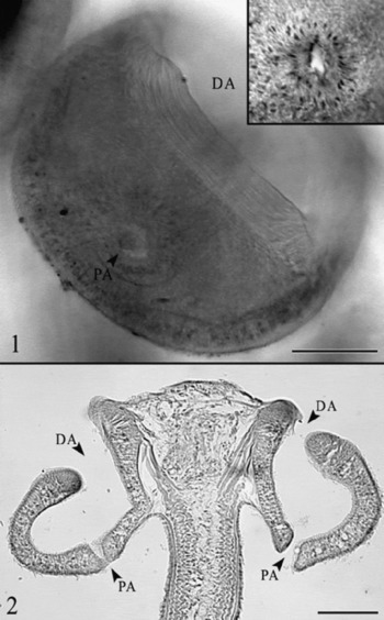
Doliobothrium haselii n. gen. n. sp. and Doliobothrium musculosum n. comb. 1, Posteroproximal view of bothridium of Doliobothrium haselii n. gen. n. sp. showing proximal and distal bothridial apertures (scale bar = 50 μm); inset shows detail of proximal aperture. DA, distal bothridial aperture; PA, proximal bothridial aperture. 2, Frontal section through scolex of Doliobothrium musculosum n. comb. (scale bar = 50 μm). DA, distal bothridial aperture; PA proximal bothridial aperture.

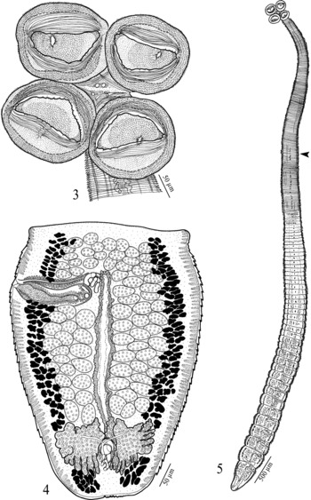
Doliobothrium haselii n. gen. n. sp. 3, Scolex; 4, terminal proglottid; 5, entire worm, arrow indicates boundary between neck and strobila.

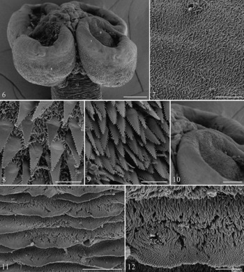
SEMs of Doliobothrium haselii n. gen. n. sp. 6, Scolex (scale bar = 50 μm); 7, margin of bothridium on proximal surface; note cilium in upper centre (scale bar = 2 μm); 8, posterior proximal surface of bothridium (scale bar = 2 μm); 9, distal surface of bothridium (scale bar = 2 μm); 10, apical region of bothridium showing lack of sucker (scale bar = 25 μm); 11, neck scutes (scale bar = 5 μm); 12, enlarged view of neck scutes showing comprised of capiliform filitriches (scale bar = 2 μm).

Based on whole mounts of three adults and one immature worm, and one scolex examined with SEM and the mature strobilar voucher.
Worms euapolytic, slightly craspedote, 9.9–12.7 (11.7 ± 1.3; n = 4) mm long; 40–54 (48 ± 6; n = 4) proglottids per worm; maximum width 425–446 (440 ± 10.2; n = 4) at level of proglottid. Scolex consisting of four bothridia. Bothridia with single loculus, 161 (n = 1) long by 123–390 (189 ± 91.1; n = 4, n = 7) wide, without apical sucker, tubular in form, with proximal and distal apertures; anterior and posterior margins of distal aperture with muscular bands. Distal bothridial surfaces with serrate gladiate spinitriches and capiliform filithriches (fig. 9). Margins of proximal bothridial surfaces with acicular filitriches (fig. 7); remainder of proximal bothridial surfaces with serrate gladiate spinitriches and capilliform filitriches (fig. 8). Cephalic peduncle not seen. Neck 2.3–3.8 (3 ± 0.8; n = 3) mm long, scutellate (fig. 11); scutes comprised of capilliform filitriches with pointed tips (fig. 12).
Strobila scutellate along its entire length. Immature proglottids wider than long, becoming longer than wide with maturity. Terminal proglottids 513–777 (637 ± 135.2; n = 4) long by 328–446 (393 ± 59.2; n = 4) wide; length:width ratio 1.4–1.8:1 (1.6 ± 0.1; n = 4). Testes 74–92 (85 ± 10; n = 3) in total number, 17–34 (24 ± 4.4; n = 5) in postporal field, 13–37 (24 ± 6; n = 3, n = 12) long by 26–51 (33 ± 8; n = 3, n = 12) wide, length:width ratio 0.5–1.2:1 (0.74 ± 0.22; n = 3, n = 12), arranged in 5–7 irregular columns anterior to cirrus sac, 2–3 columns each in postvaginal and aporal fields. Cirrus sac oblong, 132–195 (153 ± 35.9; n = 3) long by 27–43 (35 ± 11.3; n = 3) wide, containing coiled cirrus. Cirrus expanded at base, with spinitriches. Vas deferens minimal, coiled between anteriomedial margin of cirrus sac and vagina. Genital pores unilateral in all three measured specimens, 76–83% (81 ± 0.04; n = 2) of proglottid length from posterior end. Vagina weakly sinuous, extending from ootype along midline of proglottid to anterior margin of cirrus sac then laterally along anterior margin of cirrus sac to open into common genital atrium. Ovary near posterior of proglottid, H-shaped in frontal view, 113–240 (178 ± 63.8; n = 4) long by 130–220 (182 ± 46.4; n = 4) wide, tetralobed in cross-section. Ovicapt 22–27 (25 ± 2.5; n = 3) in diameter. Vitellarium follicular; follicles arranged in two lateral bands, 6–17 (11 ± 3; n = 3, n = 12) long by 9–28 (17 ± 6.6; n = 3, n = 12) wide; each band consisting of multiple columns of follicles, extending throughout length of proglottid, interrupted by cirrus sac and ovary. Gravid proglottids not observed. Excretory ducts lateral.
Taxonomic summary
Type host. Carcharhinus cf. dussumeri (Müller & Henle).
Type locality. Persian Gulf, Iran.
Prevalence. 14.3%
Mean abundance. 0.17 ( ± 0.08)
Mean intensity. 1.2 ( ± 0.2)
Site of infection. Spiral intestine.
Specimens deposited. Holotype (ZUTC platy.1070), 1 paratype (ZUTC 1071); 1 paratype (USNPC 102947); 1 paratype (LRP 7358), strobilar voucher of specimen examined with SEM (LRP 7359), scolex examined with SEM retained in the senior author's personal collection.
Etymology. This species is named in honor of Mohamad Haseli of the University of Tehran, in Tehran, who collected the type material of this species.
Doliobothrium musculosum (Subhapradha, Reference Subhapradha1955) n. comb. redescription
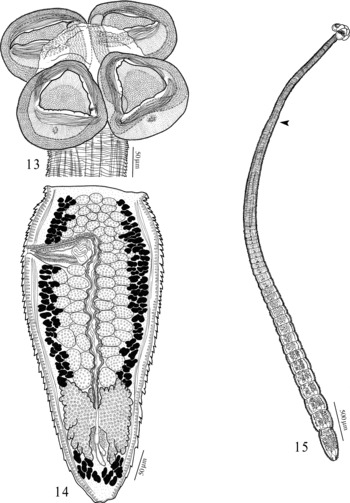
Doliobothrium musculosum n. comb. 13, Scolex; 14, terminal proglottid; 15, entire worm, arrow indicates boundary between neck and strobila.

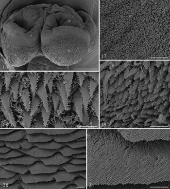
SEMs of Doliobothrium musculosum n. comb. 16, Scolex (scale bar = 50 μm); 17, margin of bothridium on proximal surface (scale bar = 2 μm); 18, posterior proximal surface of bothridium (scale bar = 2 μm); 19, distal surface of bothridium (scale bar = 2 μm); 20, neck scutes (scale bar = 5 μm); 21, enlarged view of neck scutes showing comprised of capiliform filitriches (scale bar = 2 μm).

Synonyms. Pithophorus musculosus Subhapradha, Reference Subhapradha1955.
Based on whole mounts of four adult worms, frontal sections of one scolex and its strobilar voucher, and one scolex examined with SEM and its mature strobilar voucher. Measurements of Subhapradha (Reference Subhapradha1955) are given in square brackets.
Worms euapolytic, slightly craspedote, 4.8–6.3 (5.6 ± 0.8; n = 3) mm long [up to 12 mm]; 18–29 (25 ± 5; n = 4) [18–25] proglottids per worm; maximum width 160–224 (198 ± 27.2; n = 4) at level of terminal proglottid. Scolex consisting of four bothridia. Bothridia with single loculus, 94–163 (129 ± 27.8; n = 2, n = 8) [133] long by 85–114 (105 ± 13.5; n = 3, n = 4) [max. 133] wide, without apical sucker, tubular in form, with proximal and distal apertures; anterior and posterior margins of distal aperture with muscular bands. Distal bothridial surfaces with serrate gladiate spinitriches and capiliform filitriches (fig. 19). Margins of proximal bothridial surfaces with acicular filitriches (fig. 17); remainder of proximal bothridial surfaces with serrate gladiate spinitriches and capilliform filitriches (fig. 18). Cephalic peduncle not seen. Neck 2.0–2.3 (2.1 ± 0.7; n = 4) mm long, scutellate (fig. 20); scutes comprised of capilliform filitriches with pointed tips (fig. 21).
Strobila scutellate along its entire length. Immature proglottids wider than long, becoming longer than wide with maturity. Terminal proglottids 336–552 (446 ± 95.7; n = 4) [800–950] long by 156–224 (198 ± 27.2; n = 4) [283] wide; length:width ratio 2–2.7:1 (2.2 ± 0.29; n = 4). Testes 58–70 (64 ± 6; n = 4) [69–74] in total number, 17–27 (22 ± 4.4; n = 5) in postporal field, 14–22 (17 ± 2.5; n = 3, n = 12) long by 13–22 (18 ± 2.5; n = 3, n = 12) wide, length:width ratio 0.8–1.3:1 (0.96 ± 0.2; n = 3, n = 12), arranged in 5–7 irregular columns anterior to cirrus sac, two irregular columns each in postvaginal and aporal fields. Cirrus sac slightly pyriform, 83–100 (93 ± 8.6; n = 3) long by 19–55 (35 ± 18.2; n = 3) wide, containing coiled cirrus. Cirrus expanded at base, with spinitriches. Vas deferens minimal, coiled between anteriomedial margin of cirrus sac and vagina. Genital pores unilateral in three of four specimens [unilateral in three], irregularly alternating in one specimen, 72–79% (76 ± 0.03; n = 4) of proglottid length from posterior end. Vagina weakly sinuous, extending from ootype along midline of proglottid to anterior margin of cirrus sac then laterally along anterior margin of cirrus sac to open into common genital atrium. Ovary near posterior of proglottid, H-shaped in frontal view, tetralobed in cross-section, 100–130 (115 ± 21.2; n = 3) long by 105–115 (110 ± 7.1; n = 3) wide. Ovicapt 13.6 (n = 1) in diameter. Vitellarium follicular; follicles arranged in two lateral bands, 6–12 (9 ± 2; n = 3, n = 12) long by 7–14 (10 ± 2.1; n = 3, n = 12) wide; each band consisting of multiple columns of follicles, extending throughout length of proglottid, interrupted by cirrus sac and ovary. Gravid proglottids not observed. Excretory ducts lateral.
Taxonomic summary
Type host. Rhizoprionodon acutus (Rüppell) [as Carcharias acutus].
Additional hosts. None.
Type locality. Madras Coast, India.
Additional locality. Fog Bay, Timor Sea, Northern Australia, 12°45′33″S, 130°21′7″E.
Site of Infection. Spiral intestine.
Specimens deposited. Two vouchers (QM G231738 and QM G231739); 1 voucher (USNPC 102946); 1 voucher (LRP 7360), cross-sections of scolex of 1 voucher (LRP 7363) and whole mount of its strobila (LRP 7362), strobila of voucher examined with SEM (LRP 7361); scolex examined with SEM retained in the senior author's collection.
Etymology. Not given by Subhapradha (Reference Subhapradha1955).
Unfortunately, the whereabouts of the type material of P. musculosus is unknown. Nonetheless, the specimens examined here, taken from Rhizoprionodon acutus off Australia, are generally consistent with Subhapradha's (Reference Subhapradha1955) detailed description and illustrations of D. musulosum (as P. musculosus) from the Madras Coast of India. The only difference is in the maximum length of the terminal proglottid, which we believe suggests that Subhapradha's material may have included specimens that were slightly more mature than ours. Thus, the present record extends the distribution of this tapeworm species to include northern Australia. We consider R. acutus to be the type host of this species. However, following R. acutus (as Carcharias acutus), Subhaprada (Reference Subhapradha1955) listed Carcharias limbatus [sic], Carcharias walbeehmi Bleeker [sic] and Rhynchobatus djeddensis [sic] as hosts for P. musculosus. While Carcharias walbeehmi is considered to be a synonym of R. acutus (see Compagno, Reference Compagno1984) and is thus unproblematic, we recommend that the latter two species be considered as provisional hosts until specimens verified to be conspecific with D. musculosum have been confirmed to infect these host species. Doliobothrium musculosum n. comb. differs conspicuously from D. haselii in that it is shorter in total length (4.8–6.3 vs. 9.9–12.7 mm), possesses fewer proglottids (18–29 vs. 40–54) and fewer testes (58–70 vs. 74–92).
Discussion
The existence of ‘tubular’ bothridia, which have also been described as ‘open both anteriorly and posteriorly’ (e.g. Yamaguti, Reference Yamaguti1952) and ‘open at both ends’ (e.g. Southwell, 1912, 1925; Subhapradha, Reference Subhapradha1955; Zaidi & Khan, Reference Zaidi and Khan1976; Shinde, Reference Shinde1978), has developed into somewhat of a mythical concept among cestodologists. This is, in large part, because the descriptions of such taxa are mostly brief and their accompanying illustrations schematic (e.g. Zaidi & Khan, Reference Zaidi and Khan1976; Shinde, Reference Shinde1978; Jadhav et al., Reference Jadhav, Shinde and Jadhav1992). Perhaps more importantly, however, this is the result of the fact that specimens of such taxa do not appear to be represented in collections globally. As a consequence, it has been impossible to determine if descriptions of this bothridial form are accurate, or are merely the result of misinterpretations as, for example, was found to be the case for the ‘cylindrical’ bothridia of Phoreiobothrium Linton, 1889 as Cylindrophorus Diesing, 1863 (see Caira, Reference Caira1985). While this morphology remains to be confirmed in most species of Pithophorus, it clearly exists in Doliobothrium species. The terminology employed here assumes that the bothridia are reflexed distally (i.e. away from the scolex proper) and thus, rather than referring to the bothridial apertures as ‘anterior’ and ‘posterior’, we have employed the terms ‘distal’ and ‘proximal’.
An interesting contrast exists between scolex morphology and proglottid morphology among species of Orectolobicestus, Paraorygmatobothrium, Ruhnkecestus, and now also Doliobothrium. In all four cases, proglottid resemblance is striking: all 22 of the included species exhibit a cirrus sac that is conspicuously anterior in position, a uterus that extends only to the level of the cirrus sac and vitelline follicles that are interrupted by (or at least reduced at the level of) the ovary. However, scolex variation among the four genera is remarkable. Collectively they differ in their possession of: apical suckers, facial loculation, marginal loculation, and proximal and distal apertures. Yet, all 22 known species bear serrate gladiate spinitriches on their proximal bothridial surfaces. Species in all four genera parasitize sharks; the latter three genera are restricted to carcharhiniform sharks. If formal phylogenetic analysis reveals these genera to comprise a phylogenetic unit, which seems likely given their morphology and host associations, the evolution of these scolex features will be interesting to explore in that phylogenetic context.
At this point, little is known of the cestodes of elasmobranchs of the Persian Gulf. In fact, this is only the second report of cestodes from elasmobranchs of this region; the first report (see Malek et al., Reference Malek, Caira and Haseli2010) was of two new Paraorygmatobothrium species also from C. cf. dussumieri. We have referred to this shark with a tentative specific identification, because it appears to differ somewhat in form from individuals of Carcharhinus dussumieri collected elsewhere around the globe (e.g. see MM-304 vs. BOD-41 in the Host Specimen Database, the link for which is available at https://web2.uconn.edu/tapeworm/hosts.php). In the absence of a formal treatment of the elasmobranchs of the Persian Gulf, the specific identity of this shark remains to be confirmed. These results, though very preliminary, suggest that the cestode fauna of elasmobranchs of the Persian Gulf has affinities with those of more eastern areas of the Indo-Pacific region.
Acknowledgements
We are grateful to Dr T. Valinasab from the Fisheries Organization of Iran for providing sampling facilities in Iran and also to the Office of Research Affairs of the University of Tehran and the Department of Environment of Iran for financial support. The collections of elasmobranchs from Australia would not have been possible without the able assistance of Richard Mounsey and Julie Lloyd, formerly of Darwin Fisheries. This work was supported in part with funds from NSF PEET award DEB 0118882 and NSF PBI awards DEB 0818696 and DEB 0818823.