Introduction
Helminth infection modulates the host's immune responses. Several studies have demonstrated that parasites can activate, suppress or polarize these responses (Barriga, Reference Barriga1984; Finkelman et al., Reference Finkelman, Shea-Donohue, Goldhill, Sullivan, Morris, Madden, Gause and Urban1997; Allen & MacDonald, Reference Allen and MacDonald1998; Maizels & Yazdanbakhsh, Reference Maizels and Yazdanbakhsh2003). Helminth infection is believed to be characterized by its persistence in the host and by a complex life cycle frequently associated with suppression of the host's immune responses. Helminth-produced compounds can down-regulate the immune system via several mechanisms; for example, secretion of protease inhibitors can block antigen processing, modulate antigen-presenting cells, interfere with cytokine production and mimic the effect of regulatory cytokines (Maizels & Yazdanbakhsh, Reference Maizels and Yazdanbakhsh2003).
Helminths can strongly enhance the T-helper type 2 (Th2) immune response by inducing high levels of immunoglobulin E (IgE) antibodies, the cytokines interleukin (IL)-4, -5, -10 and -13, eosinophilia and mastocytosis. Due to this strong Th2 response, chronic helminth infection may induce clinical manifestations of allergic diseases. In contrast, epidemiological studies have demonstrated an inverse correlation between helminth infection and atopy. In those studies, children with helminth infection presented less allergic manifestations than did uninfected children; further, anthelmintic drugs increased childrens' sensitization to environmental allergens. In addition, experimental Ascaris infection or injection of A. suum adult worm extract promoted intensive suppression of antibody production toward bystander antigen (Crandall, Reference Crandall1976; de Macedo & Mota, Reference de Macedo and Mota1980; Macedo & Barbuto, Reference Macedo and Barbuto1988; Paterson et al., Reference Paterson, Garside, Kennedy and Lawrence2002). These findings suggest that helminth infection provides immunomodulatory benefits (Lynch et al., Reference Lynch, Palenque, Hagel and DiPrisco1997; van den Biggelaar et al., Reference van den Biggelaar, Rodrigues, van Ree, van der Zee, Hoeksma-Kruize, Souverijn, Missinou, Borrmann, Kremsner and Yazdanbakhsh2004). Several studies have demonstrated that the reduction of allergic disease by helminth infection may reflect powerful suppression of the host immune response by induction of regulatory processes that affect regulatory T cells, B cells, macrophages and dendritic cells (Fallon & Mangan, Reference Fallon and Mangan2007; Smits et al., Reference Smits, Everts, Hartgers and Yazdanbakhsh2010; Ludwig-Portugall & Layland, Reference Ludwig-Portugall and Layland2012). Helminth infection can also interfere with the immune response to vaccines (Elias et al., Reference Elias, Akuffo and Britton2006; Urban et al., Reference Urban, Steenhard, Solano-Aguilar, Dawson, Iweala, Nagler, Noland, Kumar, Anthony, Shea-Donohue, Weinstock and Gause2007) and to co-infecting parasites (Hartgers & Yazdanbakhsh, Reference Hartgers and Yazdanbakhsh2006). Studies of products secreted by helminths have increased our understanding of the immunomodulatory mechanisms induced by chronic infection (Falcone et al., Reference Falcone, Loukas, Quinnell and Pritchard2004; Maizels et al., Reference Maizels, Balic, Gomez-Escobar, Nair, Taylor and Allen2004). These immunomodulatory factors can affect the host's inductive and effective immune response (de Macedo Soares & de Macedo, Reference de Macedo Soares and de Macedo2007; Soares & Araujo, Reference Soares and Araujo2008; Araujo & Macedo-Soares, Reference Araujo, Macedo-Soares and Pereira2012).
The suppressive effect of adult worm extract on the humoral and cellular immune response has been well characterized, especially in the heterologous response to ovalbumin (OVA). Immunization with OVA and A. suum extract promoted suppression of anti-OVA antibodies, suppressed delayed-type hypersensitivity in mice challenged with OVA, inhibited lymphocyte proliferation and suppressed allergic lung inflammation (de Macedo & Mota, Reference de Macedo and Mota1980; Macedo & Barbuto, Reference Macedo and Barbuto1988; Soares et al., Reference Soares, Mota and Macedo1992; Lima et al., Reference Lima, Perini, Garcia, Martins, Teixeira and Macedo2002). We have generated a monoclonal antibody, MAIP-1, that recognizes protein 1 from Ascaris suum (PAS-1) (Oshiro et al., Reference Oshiro, Rafael, Enobe, Fernandes and Macedo-Soares2004). We have found that PAS-1 is present in A. suum whole-body extract, that it suppresses the humoral (T-cell-dependent) and cellular (delayed-type hypersensitivity) immune responses and lung allergic inflammation, and that its immunosuppressive effect is likely to involve regulatory cells, which secrete IL-10 and transforming growth factor (TGF)-β1 in experimental models of inflammation (Itami et al., Reference Itami, Oshiro, Araujo, Perini, Martins, Macedo and Macedo-Soares2005; Araujo et al., Reference Araujo, Perini, Martins, Macedo and Macedo-Soares2008; de Araujo et al., Reference de Araujo, Perini, Martins, Macedo and Macedo-Soares2010).
Our group has characterized the immunomodulation induced by A. suum in experimentally infected mice (Enobe et al., Reference Enobe, Araujo, Perini, Martins, Macedo and Macedo-Soares2006). In mice, A. suum infection induces production of Th2 cytokines (IL-4 and IL-5), IgE and IgG1 antibodies, and IL-10. Larvae do not differentiate into adult worms in mice, as they do in their natural hosts (swine). Instead, A. suum infection is self-limiting, possibly due to larval swelling in the lung and elimination in the faeces. However, all larval stages are similar in mice and swine, and therefore mice provide a relevant experimental model of the immune response induced by A. suum. In this model we demonstrated that PAS-1, initially purified from adult worms, is also present in larvae and suppresses the humoral immune response to OVA. These findings provide evidence that PAS-1 produced during different life-cycle stages suppresses the host immune response. We investigated whether PAS-1 is an excretory/secretory (E/S) product of A. suum and assessed its immunosuppressive effect in an acute inflammation model in mice. We found that PAS-1 is an E/S protein, present in adult body fluid and larval culture supernatant, that suppresses lipopolysaccharide (LPS)-induced inflammation, suggesting that PAS-1 is secreted by A. suum at both larval and adult stages.
Materials and methods
Experimental infections and sample collection
Our experiments were performed in 6- to 8-week-old BALB/c mice with a weight range of 18–22 g. The mice were maintained in a temperature-controlled environment and received food and water ad libitum. Following the procedures of Williams & Soulsby (Reference Williams and Soulsby1970), A. suum eggs were collected from the lower uteri of adult worms in swine at a slaughterhouse (Frigorifico Itapecerica S.A., Itapecerica da Serra, SP, Brazil). Eggs were stored in 0.1 n H2SO4 for 4–5 weeks at room temperature, to allow development into infective eggs containing L3-stage larvae (Geenen et al., Reference Geenen, Bresciani, Boes, Pedersen, Eriksen, Fagerholm and Nansen1999; Fagerholm et al., Reference Fagerholm, Nansen, Roepstorff, Frandsen and Eriksen2000).
To obtain E/S products from L3 larvae, infective eggs were incubated overnight with 0.5% (w/v) sodium hydroxide and 5% sodium hypochlorite (pH 13–14) at room temperature. During incubation the eggs were agitated to induce de-shelling, using a modification of the protocol reported by Hinck & Ivey (Reference Hinck and Ivey1976). Treated eggs were bubbled with CO2 for 2 h at room temperature to promote egg hatching (Jaskoski, Reference Jaskoski1962), which was verified and quantified microscopically. Eggs found to hatch at a rate ≥ 90% were cultured for 23 days in six-well plates (104 treated eggs/well) using 3 ml of unsupplemented Dulbecco's modified Eagle medium (DMEM) per well, and incubated at 37°C, 5% CO2. Culture supernatants were collected every 2–3 days for 3 weeks. Infective and unhatched eggs were used as controls. Ascaris suum body fluid (BF) samples were obtained from freshly recovered adult worms. The worm cuticle was incised, and the worm was then centrifuged at 16,000 × g for 10 min; the supernatant was stored at − 70°C.
Induction of air pouches and quantification of cytokines
BALB/c mice were anaesthetized and their backs were shaved; air pouches were then induced by injecting 2.5 ml of sterile air into their dorsal subcutaneous tissues. Three days later, an additional 2.5 ml of sterile air was injected into the cavity. The air pouches of four groups of mice (n= 5 per group) were then injected simultaneously with 1 μg LPS (phenolic extract of Escherichia coli 026:B6, Sigma-Aldrich Inc., St. Louis, Missouri, USA) in 0.5 ml of sterile phosphate-buffered saline (PBS) and body fluid (BF) from adult worms; larval culture supernatant (LCS) or purified PAS-1 from body fluid (PAS-1[BF]) or from larval culture supernatant (PAS-1[LCS]) at a concentration of 300 μg total protein. Air pouches injected with 0.5 ml of PBS or 1 μg LPS alone were used as controls. Inflammatory exudate was collected 3 h after injection by washing the air pouches with 1 ml of PBS containing 2.5 UI/ml of heparin and centrifuging the exudates at 350 × g for 10 min. The pellet was used for cell counts. Cells were stained with Turk's solution (for total cell counts) or with haematoxylin–eosin (for differential counts of neutrophils, monocytes and eosinophils). Pro-inflammatory cytokines (tumour necrosis factor (TNF)-α, IL-1β and IL-6) and regulatory cytokine (IL-10) were measured in the supernatants by specific two-site sandwich enzyme-linked immunosorbent assay (ELISA) (DuoSet, R&D Systems Inc., Minneapolis, Minnesota, USA).
Purification and N-terminal sequencing of PAS-1
PAS-1 was isolated from A. suum adult BF and LCS by affinity chromatography, using the monoclonal antibody MAIP-1 coupled to Sepharose 4B, as described previously (Oshiro et al., Reference Oshiro, Rafael, Enobe, Fernandes and Macedo-Soares2004). Less than 0.06 EU/mg of isolated PAS-1 was detected by using the Limulus amoebocyte lysate test (BioWhittaker Inc., Walkersville, Maryland, USA).
The N-terminal sequence of PAS-1 isolated from BF was obtained at the Peptide and Protein Sequencing Unit of Butantan Institute (Sao Paulo, SP, Brazil) by using the Expert Protein Analysis System (http://www.expasy.org) and was aligned with other helminth proteins. PAS-1 was analysed by reverse-phase high-performance liquid chromatography (RP-HPLC) (LC Solution, Shimadzu, Kyoto, Japan) in a C4 column (Vydac C4; 5 μm particle; 4.6 mm × 250 mm). The proteins were eluted in a gradient of 0–90% acetonitrile in 0.1% trifluoroacetic acid (TFA) at a flow rate of 1 ml/min, and monitored at 214 nm. After RP-HPLC, PAS-1 (500 pmol) was analysed by Edman degradation (Edman, Reference Edman1949) using a PPSQ-21A protein sequencer (Shimadzu).
Data analysis
Values were expressed as mean ± SEM. Data sets were statistically compared by using two-tailed, unpaired t-tests in GraphPad Prism software v. 5.0 (GraphPad Software, La Jolla, California, USA). Differences were considered to be statistically significant at P < 0.05.
Results
PAS-1 secreted by larval and adult A. suum suppresses inflammation
We investigated whether PAS-1 was present in A. suum larval (L3 stage) culture. Infective eggs were treated chemically and examined microscopically to determine the hatching rate. Eggs that hatched at a rate ≥ 90% were cultured in unsupplemented DMEM for 23 days at 37 °C in 5% CO2. Infective and unhatched eggs were also cultured to investigate whether eggs in culture secrete PAS-1. Culture supernatants were collected every 2–3 days and PAS-1 concentration was measured by ELISA using MAIP-1, an anti-PAS-1 monoclonal antibody (Oshiro et al., Reference Oshiro, Rafael, Enobe, Fernandes and Macedo-Soares2004).
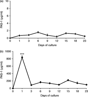
We found that chemical treatment of infective eggs in unsupplemented DMEM favoured larval release. These larvae secreted PAS-1 during the full time course of culture, but secretion was greatest on day 1 (fig. 1b), suggesting that in A. suum, secretion of PAS-1 is greatest during the early larval stage (L3). Larvae were motile for the first 2 weeks and less motile during the following weeks, but remained morphologically intact in culture. PAS-1 was not detected in cultures of unhatched eggs (fig. 1a), indicating that PAS-1 is secreted only after larvae are released.
Secretion of PAS-1 by A. suum third-stage larvae from (a) infective and unhatched eggs and (b) hatched eggs cultured in unsupplemented DMEM for 23 days at 37°C, 5% CO2. Data represent mean ± SEM of three independent experiments. ***P < 0.001 in comparison with days 3, 6, 9, 12, 15, 18, 23.

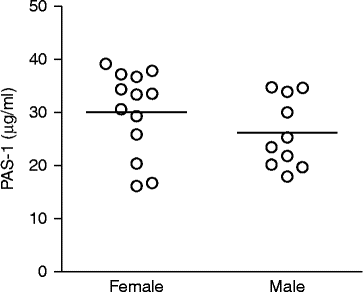
To evaluate whether A. suum adult worms secrete PAS-1, we measured PAS-1 in the BF of male and female worms by ELISA with the MAIP-1 monoclonal antibody. Similar quantities of PAS-1 were detected in BF from males and females (fig. 2). These results demonstrated that PAS-1 is secreted during adulthood as well as at early larval stages after release from eggs, suggesting that this protein may play a role in A. suum development.
Presence of PAS-1 in the body fluid of adult Ascaris suum. Data represent mean ± SEM of 13 adult female and 10 adult male worms.

To evaluate the biological effect of proteins secreted during different stages of the A. suum life cycle, we investigated the immunosuppressive effect of products present in LCS and BF and of PAS-1 purified from both sources (PAS-1[LCS] and PAS-1[BF]), by using LPS-induced inflammation in air pouches as a model. Inflammation was induced by injecting LPS into the air pouches. Three hours after injection of LPS and/or LCS, BF, PAS-1[LCS] or PAS-1[BF] into the air pouches, the inflammatory infiltrate was collected to determine the total and differential counts of inflammatory cells.
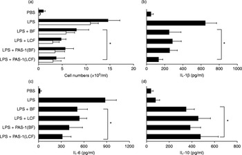
Inflammatory infiltrates, comprising predominantly neutrophils, were greater after injection of LPS than after injection of PBS. In contrast, air pouches injected with LPS + LCS, LPS + BF, LPS + PAS-1[LCS] or LPS + PAS-1[BF] had cellular infiltrates significantly lower than those in LPS-injected pouches (fig. 3a). These results indicated that PAS-1 suppresses leucocyte migration in this acute inflammation model.
Suppression of LPS-induced inflammation by PAS-1 isolated from third-stage larvae and adult worms of A. suum. Cells were recovered from air pouch exudates after injection of LPS alone, LPS +A. suum body fluid (BF), LPS + larval culture supernatant (LCS), LPS + PAS-1 isolated from adult worms, LPS + PAS-1 isolated from body fluid (PAS-1[BF]) or LPS + PAS-1 isolated from larval culture supernatant (PAS-1 [LCS]): (a) total cells (black bars) and neutrophils (white bars) were counted, and cytokines (b) IL-1β, (c) IL-6 and (d) IL-10 were measured in air pouch exudate supernatants. Data represent mean ± SEM of three independent experiments (n= 5 mice/group in each experiment). *P < 0.05 compared with LPS-injected group.

To assess the effect of A. suum modulatory factors on cytokine production, we measured pro-inflammatory cytokines (IL-1β and IL-6) and an anti-inflammatory cytokine (IL-10) in air pouches by ELISA. Supernatants from LPS-injected air pouches had higher levels of IL-1β and IL-6 than did PBS-injected pouches (fig. 3b, c). In air pouches injected with LPS + LCS, LPS + BF, LPS + PAS-1[LCS] or LPS + PAS-1[BF], production of these pro-inflammatory cytokines was significantly decreased. However, levels of IL-10 were greater than those in LPS-injected pouches (fig. 3d). These results suggested that PAS-1 is responsible for the suppression of LPS-induced inflammation by LCS and BF.
MAIP-1 abolishes PAS-1 suppression of LPS-induced inflammation in air pouches
To support our hypothesis that PAS-1 plays a crucial role in suppressing LPS-induced inflammation in air pouches, we injected MAIP-1, a monoclonal antibody to PAS-1, into the air pouches and compared the suppression of LPS-induced inflammation.
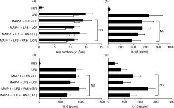
When MAIP-1 antibody was injected into air pouches together with LPS + LCS, LPS + BF, LPS + PAS-1[LCS] or LPS + PAS-1[BF], the inflammatory infiltrate was similar to that in LPS-injected pouches (fig. 4a), indicating that MAIP-1 had neutralized the PAS-1-mediated suppression of LPS-induced inflammation.
Neutralization of the PAS-1 suppressive effect on LPS-induced inflammation. Cells were recovered from air pouch exudates after injection of LPS alone; LPS + neutralizing MAIP-1 antibody; MAIP-1+ LPS + BF; LPS + larval culture supernatant (MAIP-1+ LPS + LCS); LPS + PAS-1 isolated from body fluid (MAIP-1+ LPS + PAS-1[BF]); or LPS + PAS-1 isolated from larval culture supernatant (MAIP-1+ LPS + PAS-1[LCS]): (a) total cells (black bars) and neutrophils (white bars) were counted, and cytokines (b) IL-1β, (c) IL-6 and (d) IL-10 were measured in air pouch exudate supernatants. Data represent mean ± SEM of three independent experiments (n= 5 mice/group in each experiment). NS = no significant difference compared with LPS-injected group.

We also found that injection of MAIP-1 into pouches containing LPS + LCS, LPS + BF, LPS + PAS-1[LCS] or LPS + PAS-1[BF] increased the production of IL-1β and IL-6 cytokines, as compared to that in LPS-injected pouches (fig. 4b, c). Conversely, MAIP-1 treatment decreased IL-10 production (fig. 4d). Abrogation of the PAS-1 anti-inflammatory effect by MAIP-1 indicates that PAS-1 is important in suppressing the production of pro-inflammatory cytokines and increasing IL-10 levels.
PAS-1 is homologous to other helminth-secreted proteins
A sequence of 11 amino acids of the N-terminal end of PAS-1 (His–His–Phe–Thr–Leu–Glu–Ser–Ser–Leu–Asp–Thr) was aligned with those of other helminth proteins. The PAS-1 primary sequence exhibited homology to ABA-1, a protein of Ascaris lumbricoides (sequence ID: gb|AAD13647.1|) and to TBA-1, a protein of Toxocara canis (sequence ID: dbj|BAA14015.1|) (fig. 5).
PAS-1 homology to ABA-1 (allergenic protein from A. lumbricoides) and TBA protein from Toxocara canis. The N-terminal structure of PAS-1 and its homology with the N-terminal sequences of ABA-1 and TBA are shown.

Discussion
Parasitic infection is known to down-modulate immune responses, often promoting the parasite's survival (Ogilvie & Wilson, Reference Ogilvie and Wilson1976). For example, levels of antibody to tetanus toxoid vaccine were found to be lower in Plasmodium-infected children than in healthy children (Barr et al., Reference Barr, Inselburg, Green, Kansopon, Hahm, Gibson, Lee-Ng, Bzik, Li and Bathurst1991). In the case of helminth infection, several studies have investigated the effect of secreted helminthic products on the immune response (Maizels et al., Reference Maizels, Balic, Gomez-Escobar, Nair, Taylor and Allen2004; Soares & Araujo, Reference Soares and Araujo2008; Araujo & Macedo-Soares, Reference Araujo, Macedo-Soares and Pereira2012). Our study demonstrated that PAS-1 is an E/S protein product of A. suum that has anti-inflammatory effects in an LPS-induced acute inflammation model.
Our studies have focused on the immunosuppressive effect of A. suum infection and the role of A. suum-derived products in inflammatory diseases. Previously, we isolated the protein PAS-1 from adult A. suum and demonstrated its role in suppressing the humoral and cellular immune response to bystander antigens, and in inhibiting lung allergic inflammation. In this study, we examined whether PAS-1 is an E/S protein and whether it is produced during larval life-cycle stages. We identified PAS-1 in the body fluid of both male and female adult worms and in the culture supernatants of hatched eggs, indicating that PAS-1 is secreted during both larval and adult stages.
Larval stages of A. suum differentiated as L1, L2, L3 and L4 from infective eggs and are morphologically identical in the natural hosts (Sylk et al., Reference Sylk, Stromberg and Soulsby1974; Stromberg et al., Reference Stromberg, Khoury and Soulsby1977; Urban & Douvres, Reference Urban and Douvres1981; Douvres & Urban, Reference Douvres and Urban1983; Slotved et al., Reference Slotved, Eriksen, Murrell and Nansen1998; Kasuga-Aoki et al., Reference Kasuga-Aoki, Tsuji, Suzuki, Isobe and Yoshihara2000). Levine & Silverman (Reference Levine and Silverman1969) demonstrated that A. suum larvae grow in Eagle's minimum essential medium. In our experiments, Dulbecco's modified Eagle's medium without supplementation was sufficient to allow the differentiation of larvae into the L2–L3 stages (day 15 of culture). We adapted a protocol reported by Hinck & Ivey (Reference Hinck and Ivey1976) to promote the hatching of A. suum eggs in vitro. Jaskoski (Reference Jaskoski1962) reported a hatching rate of almost 100% in medium saturated with CO2, while Hinck & Ivey (Reference Hinck and Ivey1976) found, in their study of proteinase activity in hatched eggs and E/S products of A. suum, that eggs must be deshelled with sodium hypochlorite before CO2 treatment to obtain efficient hatching. In our hands, hatching efficiency was approximately 90%, similar to the rate reported by Hinck & Ivey (Reference Hinck and Ivey1976).
Nematodes are known to produce TRPs (tandemly repetitive polyproteins), which are protein precursors that contain identical or similar repetitive amino acid sequences. They are cleaved after translation to yield several copies of functionally similar proteins. Nematode TRPs known as NPAs (nematode polyprotein allergens) bind to small lipid molecules such as fatty acids and retinol (vitamin A) and probably transport these insoluble compounds to different tissues. As nematodes do not produce these lipid molecules, it is possible that NPAs must be used to obtain them from the host (Spence et al., Reference Spence, Moore, Brass and Kennedy1993; Kennedy, Reference Kennedy2000; Xia et al., Reference Xia, Spence, Moore, Heaney, McDermott, Cooper, Watson, Mei, Komuniecki and Kennedy2000; Meenan et al., Reference Meenan, Ball, Bromek, Uhrin, Cooper, Kennedy and Smith2011). Examples of NPAs include ABA-1 and TBA-1, allergenic repetitive polyproteins found in the pseudocoelomic fluid of Ascaris spp. and T. canis, respectively (Christie et al., Reference Christie, Dunbar and Kennedy1993; Kennedy et al., Reference Kennedy, Brass, McCruden, Price, Kelly and Cooper1995). Here we demonstrated that the N-terminal sequence of PAS-1, a 200-kDa protein, is highly similar to the N-terminal end of the polyproteins ABA-1 and TBA-1. The similarity of the N-terminal sequences of ABA-1 and TBA-1 suggest that PAS-1 may be a repetitive polyprotein of A. suum. We found that the biological functions of PAS-1 differ from those of ABA-1 and TBA-1, as PAS-1 strongly suppresses LPS-induced inflammation. To our knowledge, this is the first study demonstrating nematode TRPs that have immunosuppressive properties in the context of an inflammatory onset.
We also evaluated the immunosuppressive effect of A. suum-derived products in a mouse model of acute inflammation induced by LPS in air pouches. We showed that LCS, BF, PAS-1[LCS] and PAS-1[BF] strongly suppressed leucocyte infiltration and inflammatory cytokines (IL-1β and IL-6) (fig. 3a–c). IL-1β is a potent pro-inflammatory cytokine that is crucial for the acute phase of inflammation. Administration of IL-1 elicits endotoxaemia-like responses (Dinarello, Reference Dinarello1991, Reference Dinarello2011). IL-6 is customarily considered a regulator of acute-phase inflammatory responses and is released in response to IL-1β and TNF-β (Heinrich et al., Reference Heinrich, Behrmann, Haan, Hermanns, Muller-Newen and Schaper2003; Rose-John, Reference Rose-John2012). In addition, PAS-1 stimulates the secretion of IL-10 in the air pouches, suppressing LPS-induced inflammation (fig. 3d). IL-10 plays an important role in the immune system due to its anti-inflammatory properties. It can be produced by macrophages and by several T-cell subsets, including CD4+CD25+FoxP3+ regulatory T (Treg) cells and Tr1, T-helper type 1 (Th1) cells, which exert a suppressive effect on the immune response (Moore et al., Reference Moore, de Waal Malefyt, Coffman and O'Garra2001; Groux, Reference Groux2003; Sabat et al., Reference Sabat, Grutz, Warszawska, Kirsch, Witte, Wolk and Geginat2010).
We previously reported the crucial role of IL-10 in suppressing inflammation induced by adult-worm-derived PAS-1 (Itami et al., Reference Itami, Oshiro, Araujo, Perini, Martins, Macedo and Macedo-Soares2005; Oshiro et al., Reference Oshiro, Macedo and Macedo-Soares2005; Araujo et al., Reference Araujo, Perini, Martins, Macedo and Macedo-Soares2008). Here we have demonstrated that PAS-1, either secreted at early life stages or adulthood, suppresses inflammatory responses via an IL-10-dependent mechanism. CD4+CD25+FoxP3+ Treg cells may be the main cellular source of IL-10 in PAS-1 suppression of inflammation, as suggested by our group's work (de Araujo et al., Reference de Araujo, Perini, Martins, Macedo and Macedo-Soares2010).
To ensure that PAS-1 was the protein in A. suum LCS and BF that suppressed the immune response in air pouches, we injected MAIP-1, an antibody to PAS-1, into the air pouches. MAIP-1 failed to inhibit the inflammatory infiltrate and the production of IL-1β and IL-6 in the air pouches (fig. 4), supporting the involvement of PAS-1 in the immunosuppressive effect on inflammation.
Collectively, our results demonstrated that PAS-1 is an E/S protein, produced by A. suum at larval and adult stages, which suppresses LPS-induced inflammatory responses by an IL-10-dependent immune mechanism. These findings suggest that PAS-1 may be useful as a therapeutic intervention for inflammatory diseases.
Financial support
This work was supported by CNPq, the Brazilian National Council of Technological and Scientific Development (M.F.M.S., grants 302060 and 305179).
Conflict of interest
None.
Ethical standards
The authors assert that all procedures contributing to this work comply with the ethical standards of the relevant national guides on the care and use of laboratory animals (CONCEA, Conselho Nacional de Controle de Experimentacao Animal) and have been approved by the institutional committee (CEUAIB, Comissao de Etica no Uso de Animais do Instituto Butantan).