Introduction
Ischaemic heart disease is a major cause of morbidity and mortality worldwide. Over recent decades, large epidemiological studies in a number of countries have consistently reported that low birth weight is associated with an increased risk of cardiac disease in adulthood.Reference Barker, Winter, Osmond, Margetts and Simmonds 1 – Reference Leon, Lithell and Vagero 6 In these studies, for each kilogram increase in birth weight there is a 10–20% lower risk of ischaemic heart disease.Reference Huxley, Owen and Whincup 4 These adverse associations are strongest when there is accelerated body growth after birth.Reference Eriksson, Forsen and Tuomilehto 2 , Reference Yajnik 3 Low birth weight and small size for gestational age (SGA, birth weight below the 10th centile) are often the result of intrauterine growth restriction (IUGR), which affects 6–12% of babies in developed countries.Reference Kramer 7 The majority (85–90%) of IUGR infants experience catch-up growth in childhood.Reference Albertsson-Wikland, Boguszewski and Karlberg 8 To date, the mechanisms leading to the increased vulnerability to cardiac disease in adult individuals that were born IUGR are not well understood. This increased vulnerability of the adult IUGR myocardium to disease may relate to a decreased complement of cardiomyocytes (CM) within the myocardium, which would likely adversely impact cardiac structure and function. Indeed, it has been shown in experimental animal models that IUGR reduces the complement of CM in the heart around the time of birth.Reference Black, Siebel, Gezmish, Moritz and Wlodek 9 – Reference Botting, McMillen, Forbes, Nyengaard and Morrison 12 Given that the proliferative capacity of CM throughout life is markedly reduced after birth,Reference Huttenbach, Ostrowski, Thaller and Kim 13 – Reference Kajstura, Urbanek and Perl 15 it is likely that this reduced complement of CM will persist into adulthood. This has not been examined to date, however. In rats, IUGR reduces the number of CM in the heart at birth.Reference Corstius, Zimanyi and Maka 10 This CM deficit persists at weaning when the growth restriction is maintained throughout lactation, but not when progeny are cross-fostered to control dams, removing nutritional constraint in early postnatal life.Reference Black, Siebel, Gezmish, Moritz and Wlodek 9 This early postnatal recovery of CM number in the rat is consistent with the later maturation of the heart muscle in rodents compared to humans. In the rat heart CM continue to proliferate in the first week after birth and then undergo a switch to maturation.Reference Clubb and Bishop 16 , Reference Li, Wang, Capasso and Gerdes 17 In the sheep, similar to humans, the majority of CM are mature and differentiated by birth,Reference Jonker, Zhang and Louey 18 , Reference Burrell, Boyn and Kumarasamy 19 but IUGR delays this process.Reference Louey, Jonker, Giraud and Thornburg 20 – Reference Morrison, Botting and Dyer 22 Despite this delayed maturation, the sheep heart does not appear to undergo catch up CM hyperplasia after birth. In lambs at 9 weeks of postnatal age, when CM maturation is complete, the number of CM in the left ventricle and adjoining interventricular septum was directly proportional to birth weight.Reference Stacy, De Matteo and Brew 11 Whether this persists to adult life is unknown, particularly given new evidence for a low level of CM proliferation (<1% per annum) persisting to adulthood in humans.Reference Bergmann, Zdunek and Felker 23 A reduced complement of CM in the adult heart would reduce functional reserve and therefore impair the adaptive and reparative capabilities of the myocardium. Overall, CM would be required to bear a higher than normal contractile force and as a result, CM hypertrophy would likely ensue. Studies in rats exposed to fetal hypoxia support this idea.Reference Li, Bae and Zhang 24 , Reference Li, Xiao and Estrella 25 In adult IUGR rats exposed to fetal hypoxia (a common cause of IUGR) there was hypertrophy of CM, but no difference in heart size compared to control rats. This implies a CM deficit in the myocardium of IUGR rats, although the number of CM in the hearts was not directly measured.Reference Li, Bae and Zhang 24 When the isolated hearts of these progeny were subjected to transient ischaemia (using a Langendorff apparatus), they were more vulnerable to myocardial infarction, post-ischaemic recovery was impaired and myocardial apoptosis was increased compared with hearts of control progeny.Reference Li, Xiao and Estrella 25
In this study, we therefore hypothesised that the reduced complement of CM in the IUGR heart at birth persists into adulthood and this is accompanied by CM hypertrophy. The specific aims were to examine in a sheep model, the effects of IUGR on the number, size and nuclearity of CM in the left ventricle and septum of the adult heart, which is the dominant ventricle in the postnatal heart.Reference Pinson, Morton and Thornburg 26 As there may be sexual dimorphism in the cardiac response to IUGRReference Rueda-Clausen, Morton and Davidge 27 , Reference van der Linde, Romano and Wadley 28 and as males are generally more vulnerable to cardiac disease in early adulthood,Reference Go, Mozaffarian and Roger 29 , Reference Maas and Appelman 30 we studied male offspring. To address these aims, we have investigated outcomes in progeny with variations in birth weight due to surgical restriction of placental size and nutrient delivery, induced by removal of the majority of the endometrial attachment sites before birthReference Alexander 31 , Reference Robinson, Kingston, Jones and Thorburn 32 and by twinning, which also reduces nutrient supply to the fetus and reduces fetal size from mid-gestation.Reference van der Linden, Sciascia, Sales and McCoard 33 This enabled us to study the long-term effects of the early environment on cardiac growth in a cohort of offspring that exhibited a wide range of birth weights but were raised in the same postnatal environment. Importantly, in the sheep, as in the majority of IUGR human infants,Reference Albertsson-Wikland, Boguszewski and Karlberg 8 restricted and low birth weight lambs experience catch up in body growth in the early postnatal period.Reference De Blasio, Gatford, McMillen, Robinson and Owens 34 , Reference De Blasio, Gatford, Robinson and Owens 35
Methods
Animal cohort
Placental size and function in primiparous Merino×Border Leicester ewes was restricted by surgical removal of all but four visible endometrial caruncles (placental attachment sites) from each uterine horn,Reference Alexander 31 , Reference Robinson, Kingston, Jones and Thorburn 32 at least 10 weeks before mating [placental restriction (PR)]. Control (CON) ewes remained unoperated. Ewes underwent timed mating, with pregnancy confirmed via ultrasound at ~60 days of gestational age (term ~150 days). Housing and nutrition of the ewes and progeny cohort has been described previously.Reference Wooldridge, Bischof and Meeusen 36 Lambs were weighed at 2-day intervals from birth to 16 days postnatal age and then weekly until weaning at 13 weeks of age, and then at ~5-week intervals until adulthood. Absolute neonatal growth rates and fractional neonatal growth rates between birth and 30 days of postnatal age, which is the period of catch-up growth in PR lambs, were calculated by linear regression.Reference De Blasio, Gatford, Robinson and Owens 35 Young adult sheep in the larger cohort, including those used for heart phenotype measures in the present paper, were surgically catheterized with femoral venous and arterial catheters and underwent a series of three metabolic tests to characterize insulin secretion (in response to glucose and arginine) and insulin sensitivity (by hyperinsulinaemic euglycaemic clamp), as described previously.Reference Liu, Schultz and De Blasio 37 Half of the animals in each group were then challenged with chronic mild hyperglycaemia for 16 days, maintained by clamping blood glucose at 150% of pre-challenge concentrations during a continuous glucose infusion, using the protocol we developed and evaluated in adolescent animals.Reference Gatford, De Blasio and How 38 Unchallenged animals were infused with matched volumes of saline, and metabolic tests were repeated in the final 6 days of the infusion. Only these saline-infused euglycaemic animals were included in the present paper due to potential effects of elevated glucose on the heart. At the end of the 16-day infusion period, young adult sheep were humanely killed by intravenous injection of an overdose of sodium pentobarbitone at 359±5 days of age. In the present study we studied cardiac structural outcomes at young adulthood in five intact male offspring from CON pregnancies and five intact male offspring from PR pregnancies. These animals were selected from saline-infused animals in the larger cohort on the basis that the heart had been collected and perfusion fixed at post-mortem at 1 year of age, and that no siblings from the same mother were included. We also balanced litter size, by including two singletons and three twin males in each treatment group.
Heart sampling
At necropsy hearts were rapidly excised, perfusion-fixed in 4% paraformaldehyde and stored in 70% ethanol. At the time of sampling, the fixed hearts were weighed after removal of excess fat and connective tissue. The atria were then excised, ventricles weighed and the ventricles cut into 5 mm transverse slices. A digital photograph was taken from the slice 5 mm below the plane of the valves and left ventricular (LV) free wall thickness and interventricular septum subsequently measured using image analysis Image-Pro Plus Version 6.0, Media Cybernetics.Reference Bensley, Stacy, De Matteo, Harding and Black 39 The LV plus septum (LV+S) was then sampled using a smooth fractionator approach;Reference Stacy, De Matteo and Brew 11 , Reference Gundersen, Bagger and Bendtsen 40 sampled tissue was embedded in glycolmethacrylate or paraffin.
Estimation of cardiomyocyte number
Glycolmethacrylate blocks were sectioned at 20 μm and every 10th section stained with haematoxylin. The total number of CM nuclei within the LV+S was then estimated using an optical disector/fractionator approach utilising Computer Aided Stereological Toolbox (CAST) stereological software (Olympus, Denmark).Reference Stacy, De Matteo and Brew 11 , Reference Bensley, Stacy, De Matteo, Harding and Black 39 Total CM number was then calculated by dividing total nuclei number by the average number of nuclei per CM (see below).
Measurement of cardiomyocyte size and nuclearity
Paraffin sections, 20-µm-thick, were stained with DAPI (Molecular Probes, Invitrogen, USA) to identify nuclei and wheat germ agglutinin-Alexa Fluor 488 conjugate (Molecular Probes) to stain cellular membranes.Reference Bensley, De Matteo, Harding and Black 41 CM were visualised using a Nikon C1 confocal microscope (Nikon, Japan). Utilizing NIS Elements Software (Nikon) the heart was systematically sampled and images of CM in cross-section and longitudinal section collected for analysis. Using FIJI imaging software (Max Planck Institute of Molecular Cell Biology and Genetics, Dresden, Germany), the cell boundaries of 200 CM with visible nuclei were traced for each heart and the number of nuclei in each CM counted.Reference Bensley, De Matteo, Harding and Black 41 , Reference Drenckhahn, Strasen and Heinecke 42
Statistical analysis
Data were analysed initially by two-way ANOVA to assess effects of treatment (CON cf. PR) and litter size (singleton births cf. twin births). Litter size did not affect any outcomes except for left ventricular free wall thickness, and final models therefore analysed outcomes for effects of treatment only. Effects of treatment on single measures on each animal were compared using a Student’s t-test for normally distributed continuous data and Kruskal–Wallis non-parametric test for proportions of each CM type (mono-, bi-, tri- or tetranucleated). Effects of treatment and CM nuclearity on cardiomyocyte size were assessed using repeated measures ANOVA, and relationships between variables were assessed by Pearson’s correlation, using one-tailed tests of the a priori hypotheses that the size and CM number of the adult heart would correlate positively with size at birth and with adult body weight, and that CM size would correlate negatively with CM number in the adult heart due to compensatory hypertrophy.
Results
Size at birth and postnatal growth
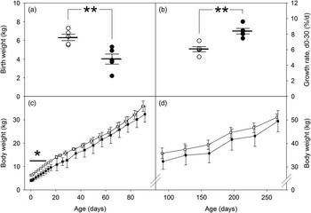
PR reduced size at birth (Fig. 1a, P=0.007), with a range of birth weights in each group (CON: 5.5–7.3 kg, PR: 2.2–5.3 kg). Fractional growth rates during the neonatal period were increased in PR lambs (Fig. 1b, P=0.001), such that these PR males achieved similar weights as control males by 14 days (Fig. 1c) and weights remained similar in both groups until adulthood (Fig. 1d). Adult body weights at postmortem were similar between groups (Table 1).
Birth weights (a), fractional growth rates in the first 30 days of postnatal life (b), and body weights before (c) and after weaning (d), in control (white bars and symbols) and placental restriction (black bars and symbols) male sheep included in the present study. Panels (a and b) show individual data for each animal with mean±s.e.m. shown in bars, and panels (c and d) show means±s.e.m. for each group. Differences between groups are indicated by *P<0.05; **P<0.01.

Characteristics of the adult heart

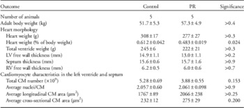
LV, left ventricle; RV, right ventricle; CM, cardiomyocyte.
Data are means±s.e.m.
Adult heart phenotype
Adult body and absolute heart and ventricle weights and wall thicknesses did not differ between PR and CON sheep (Table 1). Relative to adult body size, heart weight was higher in control than PR adults (Table 1). Left ventricle wall was thicker in males from singleton births than those from twin births (mean±s.e.m., singletons: 16.2±0.9 mm, twins: 12.4±0.8 mm, P=0.012), but no other measures of the adult heart differed between litter sizes. The total number of CM, average numbers of nuclei per CM, and longitudinal and cross-sectional areas of CM also did not differ between groups (Table 1).
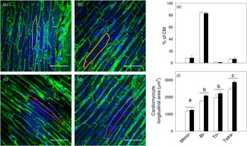
CM with between one and four nuclei (Fig. 2a–2d) were observed in the left ventricle and septum of adult hearts from both CON and PR sheep, with binucleated CM the most common type, and mononucleated CM comprising ~8% of total CM in both groups (Fig. 2e). Cardiomyocyte longitudinal surface area was lowest in mononucleated CM, intermediate in bi- and trinucleated CM, and greatest in tetranucleated CM (Fig. 2f). Consistent with this, average cardiomyocyte longitudinal surface area correlated positively with average nuclei number overall (R=0.609, slope B=1454 µm2/n, P=0.031). Average cardiomyocyte longitudinal (R=−0.398, P=0.127) and cross-sectional (R=−0.139, P=0.351) areas did not correlate with total cardiomyocyte number.
Confocal images of CM viewed in longitudinal cross-section. Cell membranes were stained with wheat germ agglutinin-Alexa Fluor 488 and nuclei with DAPI. Cardiomyocytes that were orientated longitudinally and were bound by a complete cell membrane were used for nuclearity analysis. All images of CM include a scale bar of 20 μm; mononucleated CM (a), binucleated CM (b), trinucleated CM (c), tetranucleated CM (d). Cardiomyocyte number of each nuclearity as a percentage of total CM number by treatment (e), and CM longitudinal surface area by treatment and nuclearity (f) are shown for control (white bars) and placental restriction (black bars) male sheep. Data are means±s.e.m., bars with different superscripts differ, P<0.05. CM, cardiomyocyte.

Relationships between size at birth, adult heart size and heart phenotype
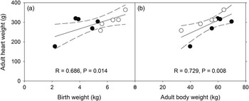
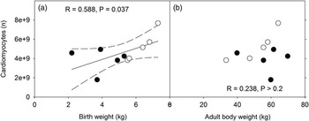
Adult body weight at necropsy did not correlate with birth weight (R=0.295, P>0.4). The weight of the fixed adult heart correlated positively with birth weight across the cohort (Fig. 3a) and in CON progeny (R=0.925, slope B=46.114 g/kg, P=0.024), but not in PR progeny analysed separately (R=0.664, P>0.2). The weight of the fixed adult heart correlated positively with adult weight across the cohort (Fig. 3b) and in both CON (R=0.946, slope B=3.068 g/kg, P=0.015) and PR (R=0.901, slope B=4.847 g/kg, P=0.037) progeny analysed separately. Numbers of CM in the left ventricle free wall and septum correlated positively with birth weight, overall (Fig. 4a), and in CON progeny (R=0.968, slope B=1.94×10⁹ n/kg, P=0.007), but not in PR progeny analysed separately (R=0.053, P>0.9). Numbers of CM in the left ventricle free wall and septum did not correlate with adult weight, overall (Fig. 4b), or in PR progeny (R=0.152, P>0.8), although these variables tended to correlate positively in CON progeny (R=0.872, slope B=1.14×10⁸ n/kg, P=0.054). Cardiomyocyte size did not correlate with birth weight or adult weight (data not shown).
Adult heart weight correlates positively with (a) birth weight and (b) adult body weight. Control males are shown in white circles and placental restriction males in black circles. Dotted lines indicate 95% confidence interval for the regression line.

Birth weight positively predicts numbers of CM in the left ventricle and septum of the adult heart (a), but CM number does not correlate with adult weight (b). Regression lines are shown for significant relationships; dotted lines indicate 95% confidence interval for the regression line. CM, cardiomyocyte.

Discussion
This study is the first to demonstrate that the complement of CM in the adult heart is dependent on birth weight. In comparing the postnatal growth response of CM in the LV+S (the dominant ventricle postnatally) in sheep that exhibited a wide range in birth weights, we clearly demonstrated that intrauterine growth directly influences the complement of CM in the adult heart, whereas body weight in adulthood had no effect. This relationship appears stronger within the normal birth weight range than within the more restricted animals, suggesting a possible threshold below which additional restriction does not further reduce CM numbers. This pattern is consistent with a study in control and PR fetuses, where cardiomyocyte numbers in the right ventricle decreased rapidly with decreasing birth weight across the normal size range but remained relatively constant within low birth weight ranges.Reference Botting, McMillen, Forbes, Nyengaard and Morrison 12 Contrary to our second hypothesis, however, there was no evidence of increased CM size in the adult IUGR heart, nor was CM size related to CM number; thus indicating that a reduced complement of CM does not directly lead to compensatory CM hypertrophy in the young adult.
There have been a number of experimental studies (in rodents and sheep) that have reported a reduced complement of CM in the fetal, newborn and early postnatal (pre-weaning) heart following restricted intrauterine growth,Reference Black, Siebel, Gezmish, Moritz and Wlodek 9 – Reference Botting, McMillen, Forbes, Nyengaard and Morrison 12 but until now it was unknown whether this CM deficit persisted into adulthood. Previous studies have reported CM hypertrophy, but no difference in heart size in adult IUGR rats thus implying that there is a reduced CM endowment;Reference Li, Bae and Zhang 24 however, such indirect findings are not conclusive, given that other cell types are present within the myocardium and the levels of fibrosis were not examined.
Although the proliferative capacity of CM is markedly reduced after birth there does remain a low level of CM proliferation postnatally.Reference Kajstura, Urbanek and Perl 15 In the human heart the number of CM approximately triples between birth and early adulthood, and recent studies demonstrate that childhood is the period when the postnatal levels of CM proliferation are highest in the human heart,Reference Mollova, Bersell and Walsh 43 while CM numbers appear to remain stable in adulthood.Reference Bergmann, Zdunek and Felker 23 A burst of CM proliferation has also been recently described in the murine heart during pre-adolescence.Reference Naqvi, Li and Calvert 44 Hence, at the commencement of this study, it was conceivable that there may be restoration of the CM deficit in low birth weight offspring during childhood and adolescence. Our findings demonstrate that this is not the case; although there was catch up in body growth in low birth weight offspring, the number of CM within the LV+S in adulthood was directly proportional to birth weight. Of course, given that the sheep were allowed to grow to adulthood we were unable to measure their CM numbers in the LV+S at birth. Hence, based on findings in fetusesReference Botting, McMillen, Forbes, Nyengaard and Morrison 12 and our own findings in younger animals,Reference Stacy, De Matteo and Brew 11 , Reference Bensley, Stacy, De Matteo, Harding and Black 39 we have made the assumption that CM number at the time of birth was directly related to birth weight and this has then persisted into adulthood.
Based on previous CM counts in lambs, it appears likely that in the present study there was modest postnatal proliferation of CM, leading to an overall increase in the number of CM as the heart grew postnatally. The total number of CM in the LV+S of adult sheep in the present study was approximately two-fold greater than reported in two previous studies of lambs at 9 weeks of age, where CM number was estimated using similar stereological methods.Reference Stacy, De Matteo and Brew 11 , Reference Bensley, Stacy, De Matteo, Harding and Black 39 This increase with postnatal age is similar to that observed in human hearts, where the number of CM triples during this postnatal period.Reference Mollova, Bersell and Walsh 43 The observation that ~10% of CM in the adult hearts of both CON and PR sheep were mononucleated also supports the concept that the postnatal heart retains a capacity for CM proliferation, given that mononucleated CM are generally considered as immature and capable of proliferation in the developing heart.Reference Thornburg, Jonker and O’Tierney 45 Postnatally, however, CM may only enable replacement during turnover of existing cells, with limited capacity to increase CM numbers, since the balance of evidence suggests that CM numbers remain stable in the adult human.Reference Bergmann, Zdunek and Felker 23 The fact that CM numbers were lower in low birth weight adult sheep in the present study, consistent with previous findings in lambs at 9 weeks of age,Reference Stacy, De Matteo and Brew 11 suggests that the presence of mononucleated CM has not enabled recovery of normal CM numbers in the adult heart after IUGR. It is interesting to note, that although our results suggests that some proliferation of CM occurred in the hearts as the animals grew postnatally, this appeared to be regulated independently from postnatal body growth, given that there was no correlation between adult body weight and the number of CM in the adult heart.
Examining twins and singletons in the PR and CON groups enabled us to study a wider range of birth weights within both experimental groups, with the birth weights of some lambs in the IUGR group similar to those of lambs in the control group. Using this approach, we clearly demonstrated that there was a direct positive correlation between birth weight and the number of CM in the adult LV+S, irrespective of whether the sheep were exposed to experimentally induced PR. Twinning, like surgical removal of placental attachment sites from the maternal endometrium, reduces the number of placentomes in the ovine placenta and limits placental transfer capacity.Reference van der Linden, Sciascia, Sales and McCoard 33 Fetal weights of twin sheep do not differ between twins and singleton sheep fetuses at 53–56 days of gestation,Reference MacLaughlin, Walker, Roberts, Kleemann and McMillen 46 but are 16–20% lower in twins in late gestation (day 140 of a 149–150-day gestation),Reference van der Linden, Sciascia, Sales and McCoard 33 , Reference Vonnahme, Arndt, Johnson, Borowicz and Reynolds 47 implying that fetal growth is reduced in twins during the period when cardiomyocyte maturation occurs in the fetal sheep heart.Reference Jonker, Zhang and Louey 18 , Reference Burrell, Boyn and Kumarasamy 19 Indeed, even when one fetus is killed in early pregnancy, after placental attachment, so that the remaining fetus does not experience substantial competition for maternal nutrients despite its smaller placenta, birth weight of the remaining twin is reduced compared to those of singleton lambs.Reference Hancock, Oliver, McLean, Jaquiery and Bloomfield 48 Due to the limited numbers of hearts available we needed to include both twins and singleton adult sheep in the present study and the spontaneous IUGR in twin fetuses and consequent variation in birth weight within each treatment group limited our ability to detect differences in outcomes between CON and PR groups. In cohorts of mixed CON and PR late gestation fetuses, fetal weight and fetal arterial oxygen concentrations each correlate positively and strongly with placental weight, providing additional evidence that low birth weight reflects the degree of constraint of nutrient supply via the placenta.Reference Poudel, McMillen, Dunn, Zhang and Morrison 49 Measures of fetal oxygenation or nutrient supply were not available in the present study as the progeny were not catheterised before birth, but it would be interesting to relate these to long-term cardiac outcomes. Our findings also suggest that placental restriction per se does not directly affect total CM number, size or nuclearity in adulthood, given that there were no significant differences between the PR and control groups in the cross-sectional area, longitudinal area and total number of CM in the LV+S, nor in the proportion of mononucleated, binucleated, trinucleated and tetranucleated CM. The latter finding requires confirmation in a larger cohort of animals, however, as other studies in the present cohort limited the numbers of hearts available within each treatment group and therefore our capacity to detect differences between CON and PR or between litter sizes in the present study.
The findings of this study are the first to directly show that the size of the infant at birth directly influences the complement of CM in the left ventricle of the adult heart. Hence, a lifelong CM deficit is likely to be a key contributor to the cardiac vulnerability in individuals that were born small.
Acknowledgements
The authors thank Laboratory Animal Services for their excellent care of animals throughout the project. They acknowledge Monash Micro Imaging and the Monash Histology Platform at Monash University, for the provision of instrumentation, training and technical support.
Financial Support
This work was supported by project grants (ID 627123 and 1011767) from the National Health and Medical Research Council of Australia. S.V. was the recipient of a Monash University Jubilee Honours scholarship and H.L. was supported by a University of Adelaide Faculty of Health Sciences Postgraduate Scholarship.
Conflicts of Interest
None.
Ethical Standards
All experimental and surgical procedures were approved by the University of Adelaide Animal Ethics Committee (M-2009-145 and M-2011-055) and SA Pathology/CHN Adelaide Health Sciences (M/135a/09) and conducted in accordance with the Australian Code for the Care and Use of Animals for Scientific Purposes. 50