Introduction
Attention deficit hyperactivity disorder (ADHD) is characterized by age-inappropriate symptoms of inattention, impulsivity and hyperactivity (APA, 2000). One of the key neuropsychological deficits is in working memory (WM) (Martinussen et al. Reference Martinussen, Hayden, Hogg-Johnson and Tannock2005), defined as the temporary storage and manipulation of information (Baddeley, Reference Baddeley1996).
Children with ADHD have consistently shown deficits in verbal–auditory and visuo-spatial WM tasks (Rommelse et al. Reference Rommelse, Altink, Oosterlaan, Buschgens, Buitelaar and Sergeant2008; Toplak et al. Reference Toplak, Jain and Rosemary2008; Gau & Shang, Reference Gau and Shang2010; Rhodes et al. Reference Rhodes, Park, Seth and Coghill2011; for meta-analyses, see Martinussen et al. Reference Martinussen, Hayden, Hogg-Johnson and Tannock2005 and Willcutt et al. Reference Willcutt, Doyle, Nigg, Faraone and Pennington2005) including visual–object (Pasini et al. Reference Pasini, Paloscia, Alessandrelli, Porfirio and Curatolo2007) and phonological n-back tasks (Klein et al. Reference Klein, Wendling, Huettner, Ruder and Peper2006; Pasini et al. Reference Pasini, Paloscia, Alessandrelli, Porfirio and Curatolo2007; Kobel et al. Reference Kobel, Bechtel, Weber, Specht, Klarhofer, Scheffler, Opwis and Penner2009). These deficits have been shown to be underpinned by functional magnetic resonance imaging (fMRI) evidence for reduced fronto-striatal and temporo-parietal activation during visuo-spatial (Silk et al. Reference Silk, Vance, Rinehart, Egan, O'Boyle, Bradshaw and Cunnington2005; Vance et al. Reference Vance, Silk, Casey, Rinehart, Bradshaw, Bellgrove and Cunnington2007) and verbal WM tasks (Kobel et al. Reference Kobel, Bechtel, Weber, Specht, Klarhofer, Scheffler, Opwis and Penner2009).
The stimulant methylphenidate (MPH) and the non-stimulant atomoxetine (ATX) are the most commonly prescribed pharmacological treatments for ADHD, showing comparable efficacy in 65–70% of cases (Hazell et al. Reference Hazell, Kohn, Dickson, Walton, Granger and van Wyk2010). However, the drug-specific mechanisms of action on brain function in ADHD patients are unknown.
MPH blocks dopamine (DA) transporters in the striatum (Volkow et al. Reference Volkow, Wang, Fowler, Gatley, Logan, Ding, Hitzemann and Pappas1998) and norepinephrine (NE) transporters (NET) in NET-rich regions including prefrontal regions, where it enhances both catecholamines (Hannestad et al. Reference Hannestad, Gallezot, Planeta-Wilson, Lin, Williams, van Dyck, Malison, Carson and Ding2010). ATX is a selective pre-synaptic NET blocker affecting NE and DA in the prefrontal cortex (Bymaster et al. Reference Bymaster, Katner, Nelson, Hemrick-Luecke, Threlkeld, Heiligenstein, Morin, Gehlert and Perry2002) and NE in the thalamus, locus coeruleus and cerebellum, with minimal striatal effects (Gallezot et al. Reference Gallezot, Weinzimmer, Nabulsi, Lin, Fowles, Sandiego, McCarthy, Maguire, Carson and Ding2011).
Placebo-controlled studies in medication-naive ADHD patients showed that a single dose of MPH upregulated and/or normalized the fronto-striatal, temporo-parietal and cerebellar underactivations during cognitive control tasks (Rubia et al. Reference Rubia, Halari, Christakou and Taylor2009a ,Reference Rubia, Halari, Cubillo, Mohammad and Taylor b , Reference Rubia, Halari, Cubillo, Mohammad, Brammer and Taylor2011a ,Reference Rubia, Halari, Mohammad, Taylor and Brammer b ), with similar effects being reported in previously medicated children compared with off-medication status (Vaidya et al. Reference Vaidya, Austin, Kirkorian, Ridlehuber, Desmond, Glover and Gabrieli1998; Shafritz et al. Reference Shafritz, Marchione, Gore, Shaywitz and Shaywitz2004; Epstein et al. Reference Epstein, Casey, Tonev, Davidson, Reiss, Garrett, Hinshaw, Greenhill, Glover, Shafritz, Vitolo, Kotler, Jarrett and Spicer2007).
Few fMRI studies, however, have tested for MPH effects on WM in ADHD children, with inconsistent results. Thus, in previously medicated ADHD children compared with controls, MPH normalized the inferior and medial frontal cortices (IFC/MFC) and striatal underactivation during a delayed matched to sample task (Prehn-Kristensen et al. Reference Prehn-Kristensen, Krauel, Hinrichs, Fischer, Malecki, Schuetze, Wolff, Jansen, Duezel and Baving2011) and upregulated activation and functional integration in fronto-parietal WM networks during a Sternberg WM task (Wong & Stevens, Reference Wong and Stevens2012). However, a single dose of MPH downregulated MFC and parietal activation during a delayed WM match-to-sample task in female ADHD subjects (Sheridan et al. Reference Sheridan, Hinshaw and D'Esposito2010) or had no effects in ADHD boys (Kobel et al. Reference Kobel, Bechtel, Weber, Specht, Klarhofer, Scheffler, Opwis and Penner2009). These studies, however, did not include a placebo condition, had small sample sizes and recruited patients with a previous history of stimulant medication, shown to affect both brain activation and structure (Nakao et al. Reference Nakao, Radua, Rubia and Mataix-Cols2011; Hart et al. Reference Hart, Radua, Mataix-Cols and Rubia2012, Reference Hart, Radua, Nakao, Mataix-Cols and Rubia2013).
In the only placebo-controlled cross-over study comparing the effects of single-dose challenges of MPH and ATX in medication-naive children with ADHD, MPH showed drug-specific upregulation and normalization effects during motor response inhibition in the right IFC and cerebellum, while both drugs normalized left IFC underactivation (Cubillo et al. Reference Cubillo, Smith, Barrett, Giampietro, Brammer, Simmons and Rubia2012b ).
However, no fMRI study has as yet investigated the effects of ATX on brain activation in ADHD patients during WM, or compared its effects with those of MPH. In healthy adults, acute ATX administration upregulates the IFC and superior temporal gyrus (STG) activation during inhibitory tasks (Chamberlain et al. Reference Chamberlain, Hampshire, Muller, Rubia, Del Campo, Craig, Regenthal, Suckling, Roiser, Grant, Bullmore, Robbins and Sahakian2009; Graf et al. Reference Graf, Abler, Freudenmann, Beschoner, Schaeffeler, Spitzer, Schwab and Gron2011), and using multivariate pattern recognition analyses ATX had relatively stronger deactivation effects on the default-mode network (DMN) while MPH has relatively stronger upregulating effects on WM networks (Marquand et al. Reference Marquand, De Simoni, O'Daly, Williams, Mourao-Miranda and Mehta2011).
We therefore conducted a randomized, double-blind, placebo-controlled, cross-over pharmacological fMRI study to test for drug-specific effects of a single clinical dose of either MPH or ATX on brain activation of medication-naive boys with ADHD during a WM task. We chose a parametric verbal WM task, given: (a) the impact of verbal WM deficits on academic performance; (b) prior evidence for impairments in this task in ADHD children (Martinussen et al. Reference Martinussen, Hayden, Hogg-Johnson and Tannock2005; Willcutt et al. Reference Willcutt, Doyle, Nigg, Faraone and Pennington2005; Pasini et al. Reference Pasini, Paloscia, Alessandrelli, Porfirio and Curatolo2007; Kobel et al. Reference Kobel, Bechtel, Weber, Specht, Klarhofer, Scheffler, Opwis and Penner2009); and (c) evidence for more pronounced impairments in ADHD with increasing WM load (Goldberg et al. Reference Goldberg, Mostofsky, Cutting, Mahone, Astor, Denckla and Landa2005; Kobel et al. Reference Kobel, Bechtel, Weber, Specht, Klarhofer, Scheffler, Opwis and Penner2009; Gau & Shang, Reference Gau and Shang2010). The focus on single rather than long-term drug effects on neurofunctional mechanisms avoids potential confounds including side effects, symptomatic improvement or chronic effects on brain activation. We furthermore compared brain activation during the WM task in patients under each drug condition with that of age-matched healthy controls to test for potential drug normalization effects. Based on previous evidence on the n-back task, we hypothesized that ADHD patients would be more impaired with increasing WM load (Kobel et al. Reference Kobel, Bechtel, Weber, Specht, Klarhofer, Scheffler, Opwis and Penner2009). Based on previous fMRI studies of WM in ADHD children (Prehn-Kristensen et al. Reference Prehn-Kristensen, Krauel, Hinrichs, Fischer, Malecki, Schuetze, Wolff, Jansen, Duezel and Baving2011), we hypothesized that ADHD boys would show underactivation relative to controls in dorsolateral prefrontal cortex (DLPFC) and parietal regions. Furthermore, based on previous findings of fronto-striatal upregulation by single doses of MPH of brain activation in ADHD (Vaidya et al. Reference Vaidya, Austin, Kirkorian, Ridlehuber, Desmond, Glover and Gabrieli1998; Shafritz et al. Reference Shafritz, Marchione, Gore, Shaywitz and Shaywitz2004; Rubia et al. Reference Rubia, Halari, Christakou and Taylor2009a , Reference Rubia, Halari, Cubillo, Mohammad, Brammer and Taylor2011a ,Reference Rubia, Halari, Mohammad, Taylor and Brammer b ; Prehn-Kristensen et al. Reference Prehn-Kristensen, Krauel, Hinrichs, Fischer, Malecki, Schuetze, Wolff, Jansen, Duezel and Baving2011) and of ATX on frontal brain activation in healthy adults (Chamberlain et al. Reference Chamberlain, Hampshire, Muller, Rubia, Del Campo, Craig, Regenthal, Suckling, Roiser, Grant, Bullmore, Robbins and Sahakian2009; Graf et al. Reference Graf, Abler, Freudenmann, Beschoner, Schaeffeler, Spitzer, Schwab and Gron2011), we hypothesized that both drugs would upregulate and normalize the reduced activation in the lateral PFC and that MPH would additionally enhance basal ganglia activation.
Method
Subjects
A total of 20 medication-naive right-handed boys (aged 10–17 years) were recruited from clinics who had a clinical diagnosis of ADHD, inattentive/hyperactive-impulsive combined subtype, as assessed by an experienced child psychiatrist using the standardized Maudsley diagnostic interview that assesses ADHD according to Diagnostic and Statistical Manual of Mental Disorders, 4th edition, Text Revision (DSM-IV-TR) criteria and is of standard use in the UK (Goldberg & Murray, Reference Goldberg and Murray2002). ADHD boys scored above the clinical threshold for hyperactive-impulsive/inattentive symptoms on the Strengths and Difficulties Questionnaire for parents (SDQ; Goodman & Scott, Reference Goodman and Scott1999), the Conners' Parent Rating Scale (CPRS-R; Conners et al. Reference Conners, Sitarenios, Parker and Epstein1998), and below the clinical threshold on the Social Communication Questionnaire (SCQ; Rutter et al. Reference Rutter, Bailey, Berument, LeCouteur, Lord and Pickles2003) (Supplementary Table 1). Patients were scanned in a double-blind, placebo-controlled, cross-over design. On each scanning session, they received a single dose of either placebo (vitamin C: 50 mg), MPH (Equasym: 0.3 mg/kg, range 5−20 mg) or ATX (Strattera: 1 mg/kg, range 16–66 mg), in a pseudo-randomized order, and remained medication-free between scans. A Latin square design was used for the randomization to achieve counterbalanced effects. Dosages were determined following NICE guidelines of clinical efficacious dosages with minimal side effects at the time of the study (National Institute for Heath and Clinical Excellence, 2008). Based on pharmacokinetic evidence, both medications were administered 1.5 h before the scan to allow for maximum absorption (Chan et al. Reference Chan, Swanson, Soldin, Thiessen, Macleod and Logan1983; Witcher et al. Reference Witcher, Long, Smith, Sauer, Heilgenstein, Wilens, Spencer and Biederman2003). The same or similar dosages and time lapses between drug administration and scan have shown to be sufficient to observe changes in brain activation and performance in ADHD patients (MPH) (Lijffijt et al. Reference Lijffijt, Kenemans, ter Wal, Quik, Kemner, Westenberg, Verbaten and van Engeland2006; Rubia et al. Reference Rubia, Halari, Cubillo, Mohammad, Brammer and Taylor2011a ,Reference Rubia, Halari, Mohammad, Taylor and Brammer b ) and healthy controls (ATX) (Chamberlain et al. Reference Chamberlain, Del Campo, Dowson, Muller, Clark, Robbins and Sahakian2007, Reference Chamberlain, Hampshire, Muller, Rubia, Del Campo, Craig, Regenthal, Suckling, Roiser, Grant, Bullmore, Robbins and Sahakian2009). All three medications were over-encapsulated using the same capsules by the pharmacist.
Performance data for 20 healthy control boys and 20 boys with ADHD under each medication condition

ADHD, Attention deficit hyperactivity disorder; MPH, methylphenidate; ATX, atomoxetine.
A total of 20 right-handed healthy boys (aged 10–17 years) were recruited through advertisement in the same geographical South London area of the UK. They scored below clinical cut-offs for the SDQ, SCQ and CPRS-R. They were scanned once, unmedicated.
Exclusion criteria for all participants were an intelligence quotient (IQ) below 70 on the Wechsler Intelligence Scale for Children, 4th version (WISC-IV; Wechsler, Reference Wechsler2004) (administered in the clinics: n = 7) or in the Wechsler Abbreviated Scale of Intelligence (Wechsler, Reference Wechsler1999) (administered in our laboratory: n = 13), history of substance abuse or neurological deficits, presence of psychiatric disorders (except for ADHD and conduct disorder/oppositional defiant disorder in the ADHD group: n = 2), learning disability, and reading, speech or language disorder.
One-way analyses of variance (ANOVAs) showed no between-group differences for age (F 1,38 = 0.88, p < 0.35) but did so for IQ (F 1,38 = 42, p < 0.001) (Supplementary Table 1). Low IQ is associated with ADHD (Bridgett & Walker, Reference Bridgett and Walker2006). Although commonly conducted in case-control studies, analysis of covariance is statistically illegitimate when the covariate is an attribute of the disorder and when groups were not randomly selected (Evans & Anastasio, Reference Evans and Anastasio1968; Miller & Chapman, Reference Miller and Chapman2001; Dennis et al. Reference Dennis, Francis, Cirino, Schachar, Barnes and Fletcher2009). Furthermore, WM has been shown to be closely associated with IQ and is part of the WISC-IV (Wechsler, Reference Wechsler2004). Therefore, co-varying for IQ during this task would in fact mean co-varying for any potential group differences in WM. Consequently, all analyses were conducted without IQ as a covariate. However, in order to test for potential effects of outliers, we repeated the fMRI analyses excluding subjects with an IQ more than 2 s.d. above or below average (i.e. four subjects in each group).
Participants were paid £50 for each scanning session. Parental/child informed consent/assent and approval from the local ethical committee were obtained.
WM task (n-back)
Subjects practised the task once before scanning. The 6-min block design WM task (Ginestet & Simmons, Reference Ginestet and Simmons2011) consists of four conditions. During ‘1-back’, ‘2-back’ and ‘3-back’ conditions, subjects are presented with series of letters (A–Z) (1 s duration, inter-trial interval = 2 s) and must respond with their right thumb using a button box whenever the letter presented is the same as one, two or three before it, respectively (e.g. 2-back: B/J/A/J). This requires both storage and continuous updating of stimuli being held in WM. In the baseline vigilance ‘0-back’ condition, subjects must respond to each X that appears on the screen. The task consists of 12 randomized blocks. Before each block, written instructions of 3 s duration are shown as to which condition is next (i.e. ‘0-back’, 1-back’, etc.). In each of the WM blocks of 30 s duration only one WM condition is presented (i.e. 2-back), and contains 15 stimuli: three targets and 12 non-targets. Each condition is presented three times. Performance data were recorded during scanning. The dependent variable is accuracy (percentage of correctly identified targets).
fMRI acquisition and analyses
Gradient-echo echoplanar MR imaging (EPI) data were acquired on a GE Signa 3 T Horizon HDx system (General Electric, USA) at the Centre for Neuroimaging Sciences, Institute of Psychiatry, King's College London, UK. A semi-automated quality-control procedure ensured consistent image quality (Simmons et al. Reference Simmons, Moore and Williams1999). A quadrature birdcage head coil was used for radio frequency transmission and reception. In each of 39 non-contiguous planes parallel to the anterior–posterior commissure line, 186 T2*-weighted MR images depicting blood oxygen level-dependent (BOLD) contrasts covering the whole brain were acquired (echo time = 30 ms, repetition time = 2 s, flip angle = 75°, in-plane resolution = 3 mm, slice thickness = 3.5 mm, slice-skip = 0.5 mm). This EPI dataset provided complete brain coverage.
Blocked fMRI data were acquired in randomized block presentation, and analysed using the non-parametric XBAM software (Brammer et al. Reference Brammer, Bullmore, Simmons, Williams, Grasby, Howard, Woodruff and Rabe-Hesketh1997; Bullmore et al. Reference Bullmore, Brammer, Rabe-Hesketh, Curtis, Morris, Williams, Sharma and McGuire1999, Reference Bullmore, Long, Suckling, Fadili, Calvert, Zelaya, Carpenter and Brammer2001; Brain Image Analysis Unit, 2011). Methods for individual fMRI analyses and group mapping are described in the Supplementary text. For between-group comparisons, a 2 × 3 split plot-design ANOVA (groups: controls, patients; WM load: 1-back, 2-back, 3-back, each separately contrasted with 0-back) was conducted. For within-group comparisons, a 3 × 3 factorial-design repeated-measures ANOVA (drug condition: placebo, MPH, ATX; WM load: 1-back, 2-back, 3-back, separately contrasted with 0-back) was conducted. Statistical measures of BOLD response were extracted for each participant in each of the clusters of activation differences for each of the three contrasts, and post hoc analyses were conducted to clarify the direction of the differences. Within patients, repeated-measures ANOVAs on the extracted BOLD response measures were conducted to test for potential order effects.
Performance analysis
For the main performance measure of accuracy within patients, repeated-measures ANOVAs were conducted with drug condition (placebo, MPH, ATX) and WM load (0-back, 1-back, 2-back, 3-back) as within-subject factors. For case-control comparisons, three repeated-measures ANOVAs (controls versus ADHD under placebo; controls versus ADHD under MPH and controls versus ADHD under ATX) were conducted with WM load as the within-subjects factor and group as the between-subjects factor. Repeated-measures ANOVAs were used to test for potential scan order effects.
Results
Performance data
Across all participants, there was a significant WM load effect in accuracy (F 3,114 = 49, p < 0.001), showing that accuracy decreased with increasing WM load (Table 1).
In case-control comparisons, no group effects were found. However, as hypothesized, there was a significant interaction effect between WM load and group in accuracy when healthy controls were compared with patients under placebo (F 3,114 = 3, p < 0.035) due to patients being more accurate during 0-back and 1-back but less accurate during 2- and 3-back than healthy controls (Table 1).
Within patients, no effects of drug condition or WM load × drug condition were detected on performance variables.
No other group, group × WM load interaction or drug order effects were observed.
Brain activation
Motion
Participants were excluded if they had >3 mm in the X or Y dimensions. Multivariate ANOVA showed no significant group differences between controls and ADHD patients under each drug condition in the extent of mean rotation and translation movement parameters in the three-dimensional Euclidean space (F 6,152 = 1, p = 0.43).
Within-group brain activations
Within-group brain activations are shown in Supplementary Fig. 1 and described in the Supplementary text and Supplementary Table 2.
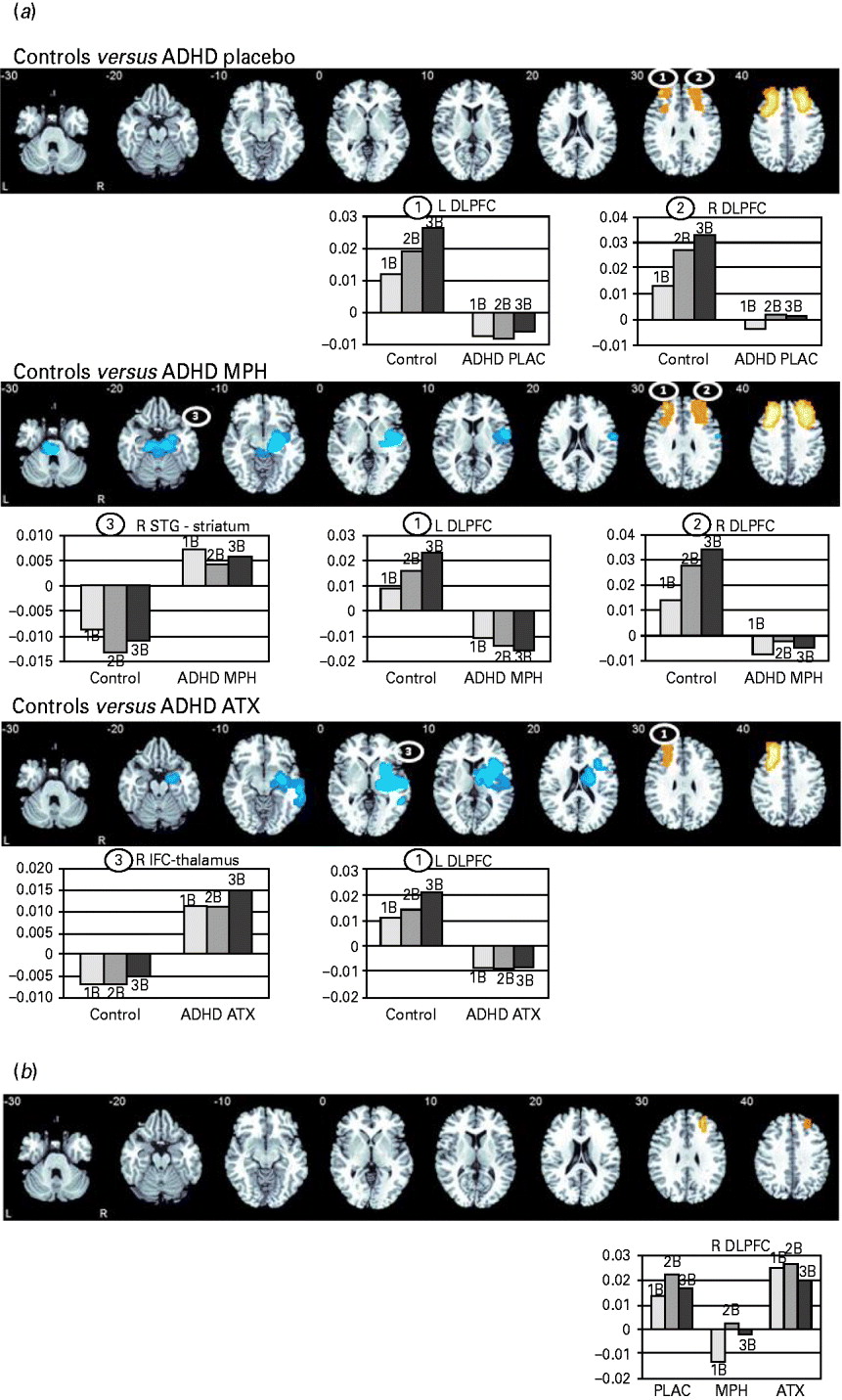
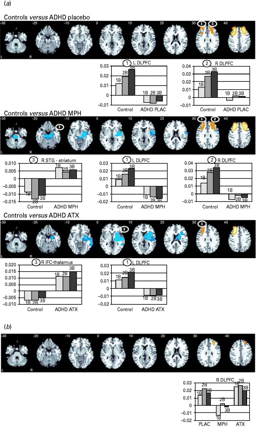
Results from the main group comparisons and the within-patient drug condition analysis. (a) Between-group analysis of variance (ANOVA) comparisons between healthy control boys and boys with attention deficit hyperactivity disorder (ADHD) under either placebo (PLAC), methylphenidate (MPH) or atomoxetine (ATX) (main effect of group differences): axial sections showing the ANOVA between-group differences in brain activation between healthy control boys and boys with ADHD under each drug condition (placebo, MPH, ATX). For ease of view, clusters and their correspondent bar chart are numbered. Clusters in orange denote areas where control boys showed enhanced activation compared with ADHD boys, clusters in blue denote areas where ADHD boys showed enhanced activation compared with control boys. The graphs show the statistical measure of blood oxygen level-dependent (BOLD) (brain activation) response in each area for each group and working memory (WM) condition. The x-axis of the graphs corresponds to the statistical measure of the BOLD response in this region. In the bar charts, the labels 1B, 2B and 3B denote the BOLD response of each group in the contrasts 1-back v. 0-back, 2-back v. 0-back and 3-back v. 0-back, respectively. (b) Within-patients ANOVA comparison (main effect of drug condition). Axial sections showing the repeated-measures ANOVA results of drug condition within ADHD boys. The graphs show the statistical measure of BOLD response in the cluster for drug condition and WM load condition. The x-axis of the graphs corresponds to the statistical measure of the BOLD response in this region. In the bar charts, the labels 1B, 2B and 3B denote the BOLD response of each group in the contrasts 1-back v. 0-back, 2-back v. 0-back and 3-back v. 0-back, respectively. Talairach z-coordinates are indicated for slice distance (in mm) from the intercommissural line. The right side of the image corresponds to the right side of the brain. The maps are thresholded to give less than one type I error three-dimensional cluster per map (p < 0.01). L, Left; DLPFC, dorsolateral prefrontal cortex; R, right; STG, superior temporal gyrus; IFC, inferior frontal cortex.
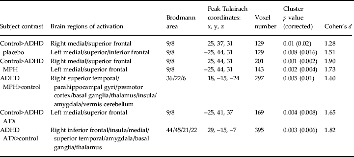
Group differences in brain activation for between-group ANOVA comparisons between controls and boys with ADHD under either the placebo, MPH or ATX condition a

ANOVA, Analysis of variance; ADHD, attention deficit hyperactivity disorder; MPH, methylphenidate; ATX, atomoxetine.
a The maps are thresholded to give less than one type I error three-dimensional cluster per map.
ANOVA between-group comparisons between healthy controls and ADHD boys under placebo, MPH or ATX
Placebo
Controls compared with patients under placebo showed enhanced activation in the bilateral DLPFC (Table 2, Fig. 1 a). There was no enhanced activation in patients compared with healthy controls.
Given that patients were less accurate than controls in the 2- and 3-back conditions, we hypothesized that accuracy would be positively correlated with DLPFC activation in these conditions. Hence, one-tailed Pearson correlations were conducted in both groups separately between statistical measures of the BOLD response in the left and right DLPFC and a composite score of their accuracy during 2- and 3-back. At a trend level, activation in the right DLPFC was correlated positively with accuracy within controls (r = 0.35, p < 0.06). No correlations were observed for left DLPFC activation.
MPH
After the single dose of MPH, patients compared with controls showed underactivation in the same left and right DLPFC clusters as under placebo. However, ADHD patients relative to controls showed additional, enhanced activation in a cluster comprising the right STG/premotor cortex, striatum/thalamus, insula, reaching into the cerebellar vermis (Table 2, Fig. 1 a). To test whether the increased right medial fronto-STG-striatal activation in patients under MPH was compensatory for the reduced bilateral DLPFC activation, statistical measures of BOLD response were extracted for each patient in these three clusters and correlated with the DLPFC activation clusters as well as with the accuracy composite score. Pearson correlations showed that the medial fronto-STG-striatal activation was negatively correlated with the left DLPFC activation (r =− 0.5, p < 0.012) and positively with accuracy (r = 0.37, p < 0.05).
ATX
Under the single dose of ATX, patients compared with controls still showed reduced activation in the left DLPFC. However, they no longer showed underactivation of the right DLPFC. In addition, abnormally enhanced activation was observed in ADHD relative to controls in a right-lateralized cluster of IFC and STG, insula, thalamus and striatum (Table 2, Fig. 1 a), which within patients was positively correlated with accuracy (r = 0.54, p < 0.007).
Repeated-measures ANOVA within patients, for all three contrasts, showed no significant scan order effects on the extracted BOLD response measures.
When those subjects whose IQ scores were more than 2 s.d. above or below average were excluded, all findings remained, although at a more lenient p value (p < 0.03).
Effect size comparisons of case-control conditions to test for significant ‘normalization’ effects
To establish whether the group differences between controls and patients under each drug condition were significantly different, we directly compared the effect sizes (ES) of the group differences from the three case-control comparisons (Matthews & Altman, Reference Matthews and Altman1996) (see details in the Supplementary text).
In the right DLPFC, the ES of the case-control contrast under ATX was significantly different from the ES of the case-control comparison under MPH (p < 0.03), suggesting that ATX significantly enhanced activation in this region relative to MPH. For the cluster of enhanced right fronto-STG-striatum activation under MPH relative to controls the ES of the case-control contrast under MPH was significantly higher than the ES of the case-control comparisons under placebo (p < 0.004) and ATX (p < 0.05). Finally, for the cluster of enhanced right IFC-STG-striato-thalamic activation under ATX relative to controls, the ES was significantly larger for the case-control contrast under ATX relative to those of the case-control comparisons under placebo (p < 0.007) and under MPH (p < 0.03) (Supplementary Table 3).
ANOVA within-patient comparison between placebo, MPH and ATX
Given the reduced activation in the left and right DLPFC, we tested for significant upregulation effects of both drugs in the frontal cortex. The Talairach Client (Lancaster et al. Reference Lancaster, Rainey, Summerlin, Freitas, Fox, Evans, Toga and Mazziotta1997, Reference Lancaster, Woldorff, Parsons, Liotti, Freitas, Rainey, Kochunov, Nickerson, Mikiten and Fox2000) was used to define a mask of the frontal lobe, restricting the analysis to those voxels present in the mask. A significant effect of drug condition was observed in a cluster in the right DLPFC [13 voxels, peak Talairach coordinates (x, y, z): 25, 44, 26; Brodmann area (BA) 10/9, p < 0.044], which was due to significantly enhanced activation when under ATX relative to MPH (p < 0.002), as well as when under placebo relative to MPH (p < 0.01) (Fig. 1 b). There were no significant correlations between brain activation changes and performance.
Interaction effects
Between-groups comparisons: WM load × group
No significant interaction effects were observed in the comparison between healthy controls and patients under placebo.
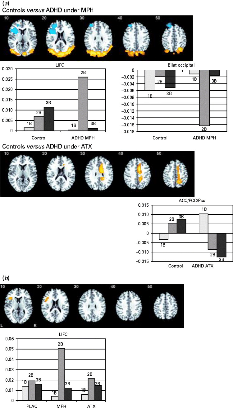
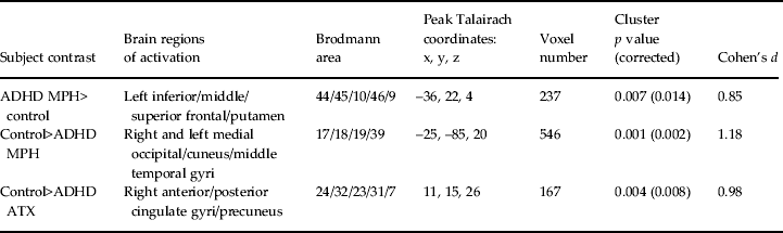
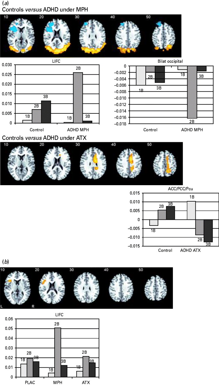
In the comparison between healthy and ADHD boys under MPH, a significant interaction of group × WM load was observed in the left IFC reaching into the DLPFC, putamen and anterior insula, due to enhanced activation in ADHD under MPH relative to controls during 2-back (p < 0.01). Additional interaction effects were observed in bilateral occipital regions extending to the cuneus, due to the enhanced deactivation of this cluster in patients under MPH compared with controls during 2-back (p < 0.001) (Fig. 2 a, Table 3).
Interaction effects with working memory (WM) load. (a) Axial sections showing the interaction effects between WM load and group for the between-group comparisons between healthy controls and boys with attention deficit hyperactivity disorder (ADHD) under methylphenidate (MPH), and between healthy controls and ADHD boys under atomoxetine (ATX). The graphs show the blood oxygen level-dependent (BOLD) response in each area for each group and WM load condition. In the bar charts, the labels 1B, 2B and 3B denote the BOLD response of each group in the contrasts 1-back v. 0-back, 2-back v. 0-back and 3-back v. 0-back, respectively. Clusters in orange denote areas where control boys showed enhanced activation compared with ADHD boys, clusters in blue denote areas where ADHD boys showed enhanced activation compared with control boys. (b) Axial section showing the interaction effects of WM load and drug condition for the within-ADHD ANOVA. The graph shows the BOLD response in each area for each drug condition during each WM load condition. In the bar charts, the labels 1B, 2B and 3B denote the BOLD response of each group in the contrasts 1-back v. 0-back, 2-back v. 0-back and 3-back v. 0-back, respectively. Talairach z-coordinates are indicated for slice distance (in mm) from the intercommissural line. The right side of the image corresponds to the right side of the brain. The maps are thresholded to give less than one type I error three-dimensional cluster per map (p < 0.01). L, Left; IFC, inferior frontal cortex; Bilat, bilateral; ACC/PCC/Pcu, anterior cingulate/posterior cingulate cortex/precuneus; PLAC, placebo.
Group × WM load interaction effects on brain activation for between-group ANOVA comparisons between controls and boys with ADHD under either the placebo, methylphenidate or atomoxetine condition a

WM, Working memory; ANOVA, analysis of variance; ADHD, attention deficit hyperactivity disorder; MPH, methylphenidate; ATX, atomoxetine.
a The maps are thresholded to give less than one type I error three-dimensional cluster per map.
In the comparison between controls and patients under ATX, significant interaction effects were observed in the anterior and posterior cingulate extending to the precuneus, due to significantly enhanced activation within patients relative to controls during 1-back (p < 0.004) and enhanced deactivation during 3-back (p < 0.005) (Fig 2 a, Table 3).
Within-subjects comparison: WM load × drug condition
Significant interaction effects were observed in the left IFC [14 voxels, peak Talairach coordinates (x, y, z): −36, 22, 9; BA 44/45, p < 0.019 (p < 0.04 Bonferroni corrected)]) (Fig. 2 b), due to significantly enhanced activation in this cluster during 2-back when under MPH compared with placebo (p < 0.001) and ATX (p < 0.015).
Repeated-measures ANOVAs showed no scan order effect on the extracted BOLD response measures.
Discussion
The present fMRI comparison between single-dose challenges of MPH and ATX on neural correlates of WM in children with ADHD shows both shared as well as drug-specific laterality effects on frontal activations. In line with previous studies, medication-naive ADHD boys under placebo relative to controls showed impaired performance under high but not low WM load. This was accompanied by placebo-associated underactivation in the left and right DLPFC relative to controls. Both drugs had no effect on within-subject performance, but normalized the case-control performance deficits in the more difficult 2- and 3-back conditions in ADHD patients relative to controls, which were no longer observed under either drug. ATX had drug-specific modulation effects on the right DLPFC, which was upregulated in ADHD patients relative to MPH and significantly normalized in its underactivation relative to controls. By contrast, MPH showed a drug-specific WM load-dependent effect on the left-lateralized IFC, which was upregulated during 2-back, both relative to controls (case-control comparison), and relative to ATX and placebo (within-subject contrast). Both drugs, in addition, elicited abnormally enhanced activation in patients relative to controls in different fronto-STG-striatal networks, which were associated with improved performance, suggesting compensatory effects. The findings therefore show both drug-specific laterality effects in the upregulation of prefrontal WM regions as well as shared effects of recruiting compensatory brain areas.
ADHD patients under placebo relative to controls showed no deficits during the first easy WM task condition. However, they were less accurate than controls during the 2-back and 3-back conditions, in line with prior evidence for more pronounced deficits in ADHD patients with increased task difficulty during verbal n-back (Kobel et al. Reference Kobel, Bechtel, Weber, Specht, Klarhofer, Scheffler, Opwis and Penner2009) and visuo-spatial WM tasks (Goldberg et al. Reference Goldberg, Mostofsky, Cutting, Mahone, Astor, Denckla and Landa2005; Gau & Shang, Reference Gau and Shang2010). Although neither drug improved performance within patients, the case-control analyses showed that both drugs normalized the between-group accuracy deficits in the 2- and 3-back condition, as they were no longer observed. The findings are in line with evidence that MPH normalizes the performance deficits shown by ADHD patients relative to controls in a verbal n-back (Kobel et al. Reference Kobel, Bechtel, Weber, Specht, Klarhofer, Scheffler, Opwis and Penner2009). The normalization effects suggest relatively small accuracy improvements that were probably underpowered to emerge in the within-subject analyses.
Compared with controls, patients showed underactivation in the bilateral DLPFC, a key region for WM, involved in the storage and coding of the temporal sequence of stimuli (Owen et al. Reference Owen, McMillan, Laird and Bullmore2005). The findings extend previous evidence of underfunctioning of the DLPFC in adult ADHD during WM (Valera et al. Reference Valera, Brown, Biederman, Faraone, Makris, Monuteaux, Whitfield-Gabrieli, Vitulano, Schiller and Seidman2010) and in ADHD children during other attention and executive function tasks (Rubia, Reference Rubia2011; Cubillo et al. Reference Cubillo, Halari, Smith, Taylor and Rubia2012a , Hart et al. Reference Hart, Radua, Mataix-Cols and Rubia2012; Christakou et al. Reference Christakou, Murphy, Chantiluke, Cubillo, Smith, Giampietro, Daly, Ecker, Robertson, Murphy and Rubia2013).
The most interesting findings are those of drug-specific lateralization effects on frontal activations. While ATX showed a drug-specific right-hemispheric DLPFC upregulation and normalization effect relative to MPH, MPH showed a drug-specific left-lateralized WM load-dependent upregulation effect on left IFC/DLPFC activation during the 2-back condition relative to ATX within patients and relative to controls. The findings suggest potential differences in their effects on underlying catecholaminergic systems. WM is mediated in the DLPFC by noradrenergic α 2 receptors that increase neural ‘signalling’ (increased firing to relevant stimuli) and by dopaminergic D1 receptors that decrease ‘noise’ (suppressing firing to irrelevant stimuli) (Gamo et al. Reference Gamo, Wang and Arnsten2010). Although ATX affects both DA and NE in the PFC (Bymaster et al. Reference Bymaster, Katner, Nelson, Hemrick-Luecke, Threlkeld, Heiligenstein, Morin, Gehlert and Perry2002), in non-human primates it increases ‘signalling’ more frequently than it decreases ‘noise’ (Gamo et al. Reference Gamo, Wang and Arnsten2010). This study thus shows for the first time that upregulation of right frontolateral activation is not only observed with a single dose of ATX in healthy adults (Chamberlain et al. Reference Chamberlain, Hampshire, Muller, Rubia, Del Campo, Craig, Regenthal, Suckling, Roiser, Grant, Bullmore, Robbins and Sahakian2009; Graf et al. Reference Graf, Abler, Freudenmann, Beschoner, Schaeffeler, Spitzer, Schwab and Gron2011), but also in children with ADHD, implying similar mechanisms of action in both healthy subjects and ADHD patients. Most importantly, however, we show that, during WM, the right frontal normalization and upregulation effects with ATX are drug-specific relative to MPH.
Interestingly, the finding of left IFC upregulation under the 2-back with MPH reflects the association between dopaminergic striatal function and left IFC activation during WM maintenance (Landau et al. Reference Landau, Lal, O'Neil, Baker and Jagust2009), in line with previous findings of predominantly left-hemispheric upregulation and normalization effects (Epstein et al. Reference Epstein, Casey, Tonev, Davidson, Reiss, Garrett, Hinshaw, Greenhill, Glover, Shafritz, Vitolo, Kotler, Jarrett and Spicer2007; Rubia et al. Reference Rubia, Halari, Christakou and Taylor2009a , Reference Rubia, Halari, Mohammad, Taylor and Brammer2011b ). Therefore, the findings could possibly suggest stronger effects of ATX on predominantly right-lateralized NE-mediated WM and attention networks (Tucker & Williamson, Reference Tucker and Williamson1984), while MPH showed stronger effects on predominantly left-lateralized fronto-striatal DA systems (Glick et al. Reference Glick, Ross and Hough1982; Flor-Henry, Reference Flor-Henry1986).
The enhanced deactivation with both drugs in regions of the DMN in ADHD relative to controls during the high WM load conditions suggest that catecholamine reuptake inhibitors not only increase task-positive but also switch off task-negative DMN activation. The findings extend prior evidence of enhanced DMN deactivation with MPH during motor inhibition in previously medicated patients relative to an off-medication condition (Liddle et al. Reference Liddle, Hollis, Batty, Groom, Totman, Liotti, Scerif and Liddle2010) to medication-naive patients in a placebo-controlled design for another task and furthermore show for the first time that this effect is shared by ATX.
Both drugs showed abnormally enhanced fronto-STG-striatal activation in ADHD boys relative to controls, although in somewhat different locations, affecting the premotor cortex with MPH and the right IFC with ATX. The findings echo previous findings of fronto-temporo-striatal upregulation/normalization in ADHD children with MPH during WM (Prehn-Kristensen et al. Reference Prehn-Kristensen, Krauel, Hinrichs, Fischer, Malecki, Schuetze, Wolff, Jansen, Duezel and Baving2011) and other cognitive control tasks (Vaidya et al. Reference Vaidya, Austin, Kirkorian, Ridlehuber, Desmond, Glover and Gabrieli1998; Epstein et al. Reference Epstein, Casey, Tonev, Davidson, Reiss, Garrett, Hinshaw, Greenhill, Glover, Shafritz, Vitolo, Kotler, Jarrett and Spicer2007; Rubia et al. Reference Rubia, Halari, Cubillo, Mohammad, Brammer and Taylor2011a ,Reference Rubia, Halari, Mohammad, Taylor and Brammer b ) and extend for the first time previous evidence of enhanced IFC/STG activation after a single-dose ATX challenge in healthy adults (Chamberlain et al. Reference Chamberlain, Hampshire, Muller, Rubia, Del Campo, Craig, Regenthal, Suckling, Roiser, Grant, Bullmore, Robbins and Sahakian2009) to a paediatric ADHD group.
The strength of the study is the double-blind, placebo-controlled design and the recruitment of medication-naive children with ADHD, which avoids the confound of a previous history of stimulant medication (Nakao et al. Reference Nakao, Radua, Rubia and Mataix-Cols2011; Hart et al. Reference Hart, Radua, Mataix-Cols and Rubia2012).
A limitation is the fact that groups differed in IQ. However, the findings survived when outliers were excluded from the analyses.
An important caveat is that while MPH has an immediate effect on ADHD symptoms (Greenhill et al. Reference Greenhill, Swanson, Vitiello, Davies, Clevenger, Wu, Arnold, Abikoff, Bukstein, Conners, Elliott, Hechtman, Hinshaw, Hoza, Jensen, Kraemer, March, Newcorn, Severe, Wells and Wigal2001), ATX reaches its maximum clinical efficacy after 12 weeks of treatment (Montoya et al. Reference Montoya, Hervas, Cardo, Artigas, Mardomingo, Alda, Gastaminza, Garcia-Polavieja, Gilaberte and Escobar2009). Given the differences of the two drugs in temporal courses to clinical efficacy, however, future studies should also compare their long-term effects on brain activation when they have reached maximum clinical efficacy. Also, while a within-group design was crucial to assess differential drug effects, it has the disadvantage that only one group was medication-naive in the second drug and none in the third drug condition. However, the 7 days' break between scans should have minimized carry-over effects. MPH has a 3.9 h half-life (Modi et al. Reference Modi, Lindemulder and Gupta2000). For ATX, the plasma half-life is 3 h for fast metabolizers and 16–25 h for poor metabolizers (single dose and steady state) (Sauer et al. Reference Sauer, Ponsler, Mattiuz, Long, Witcher, Thomasson and Desante2003; Witcher et al. Reference Witcher, Long, Smith, Sauer, Heilgenstein, Wilens, Spencer and Biederman2003) and hence effects should have been washed out after 1 week. While we used the smallest clinical dosages with minimal side effects at the time of the study as recommended by the NICE guidelines (National Institute for Heath and Clinical Excellence, 2008), some children with ADHD receive higher dosages in clinical practice, which could lead to more pronounced effects and long-term changes in catecholaminergic systems. A further limitation is that ADHD boys performed the task three times, while, for financial and ethical reasons, controls were scanned only once, unmedicated. However, the counterbalanced randomized design for the ADHD group adequately controlled for practice effects in patients, and we found no order effects for the within or the case-control studies, which suggests that these were unlikely to have confounded the between-groups analyses. Also, we included a relatively large age range. While this does not affect the within-subject analyses and controls were age-matched, a smaller age range may have resulted in less variance and more refined changes particular to specific ages, given developmental changes between late childhood and adolescence during WM and other executive functions (Crone et al. Reference Crone, Wendelken, Donohue, van Leijenhorst and Bunge2006; Bunge & Wright, Reference Bunge and Wright2007; Rubia, Reference Rubia2012). Furthermore, due to the higher prevalence of ADHD in boys (Merikangas et al. Reference Merikangas, He, Brody, Fisher, Bourdon and Koretz2010), we only tested male adolescents, which limits the generalizability of the findings.
In conclusion, the findings show both drug-specific as well as shared effects on task-positive and task-negative WM networks. ATX appears to have a drug-specific effect of upregulating and normalizing WM-related right DLPFC dysfunction in ADHD, while MPH appears to upregulate compensatory activation of left IFC activation, but only during the 2-back condition.
Supplementary material
For supplementary material accompanying this paper visit http://dx.doi.org/10.1017/S0033291713000676
Acknowledgements
The study was funded by the Department of Health via the National Institute for Health Research Biomedical Research Centre (NIHR BRC) for Mental Health at the South London and Maudsley NHS Foundation Trust and Institute of Psychiatry, King's College London and Lilly Pharmaceuticals. The NIHR BRC also supported A.C. and A.B.S. via a Ph.D. studentship and post-doctoral fellowship, respectively.
Declaration of Interest
K.R. has received speaker's honoraria from Shire, Novartis, Medice and Lilly. Lilly had no input into the design of the study, its analysis or interpretation.