Introduction
Spirocerca lupi Railliet & Henry, 1911 (Order Spirurida, Family Spirocercidae) is an endoparasite that causes spirocercosis in canids (Bailey et al., Reference Bailey, Cabrera and Diamond1963; Lobetti, Reference Lobetti2000; Mazaki-Tovi et al., Reference Mazaki-Tovi, Baneth, Aroch, Harrus, Kass, Ben-Ari, Zur, Aizenberg, Bark and Lavy2002). This nematode is prevalent in areas with a warm climate and has been reported throughout the world (van der Merwe et al., Reference van der Merwe, Kirberger, Clift, Williams, Keller and Naidoo2008). Infected canids pass eggs in faeces and these are consumed by coprophagous arthropods, the most documented being beetles from the family Scarabaeidae (Bailey, Reference Bailey1972; Chhabra & Singh, Reference Chhabra and Singh1973; Lobetti, Reference Lobetti2000; Dvir et al., Reference Dvir, Kirberger, Mukorera, van der Merwe and Clift2008). Scarabaeus sacer, S. variolosus and Gymnopleurus sturmi (Bailey et al., Reference Bailey, Cabrera and Diamond1963), Copris hispana (Seurat, Reference Seurat1916), Onthophagus pugionatus, O. sugillatus, O. ebenus, O. obtusicornis and Gymnopleurus virens (du Toit et al., Reference du Toit, Scholtz and Nyman2008) have been documented as intermediate hosts of S. lupi in the USA, Algiers and South Africa, respectively. A variety of domestic and wild animals, ranging from birds, hedgehogs, mice and rabbits, have been incriminated as paratenic hosts (Bailey, Reference Bailey1972).
The prevalence of S. lupi in dogs from a resource-limited urban community in Gauteng province of South Africa was 14.5% (Minnaar & Krecek, Reference Minnaar and Krecek2001) and 13% in Free State province (Minnaar et al., Reference Minnaar, Krecek and Fourie2002). The distribution of the disease in dogs is dependent on various factors (van der Merwe et al., Reference van der Merwe, Kirberger, Clift, Williams, Keller and Naidoo2008) with some reports of high prevalence in an urban area in Israel (Mazaki-Tovi et al., Reference Mazaki-Tovi, Baneth, Aroch, Harrus, Kass, Ben-Ari, Zur, Aizenberg, Bark and Lavy2002) and in rural areas in the USA (Dixon & McCue, Reference Dixon and McCue1967).
The genus Gongylonema Molin, 1857 (Order Spirurida, Family Gongylonematidae) has species that affect wild and domestic mammals and birds (Mowlavi et al., Reference Mowlavi, Mikaeili, Mobedi, Kia, Masoomi and Vatandoost2009). The life cycle is indirect with coprophagous arthropods as intermediate hosts (Anderson, Reference Anderson1988). Gongylonema ingluvicola Molin, 1857, affects both domestic and wild birds, and eggs are passed out with faeces of infected birds and contain the first-stage larvae (Anderson, Reference Anderson1988). Coprophagous beetles Copris minutus and Phanaeus vindex, and the cockroach Blatella germanica, have been reported as intermediate hosts (Soulsby, Reference Soulsby1965). In many communities in developing countries, chickens are allowed to scavenge freely and are exposed to arthropod intermediate hosts of several parasites, including G. ingluvicola, through ingestion of infected arthropods (Permin et al., Reference Permin, Esmann, Hoj, Hove and Mukaratirwa2002). A study conducted in Zimbabwe in free-range chickens reported the prevalence of G. ingluvicola to be 28% in young chickens and 56% in adult chickens (Permin et al., Reference Permin, Esmann, Hoj, Hove and Mukaratirwa2002), whereas in Ghana it was 62% (Poulsen et al., Reference Poulsen, Permin, Hindsbo, Yelifari, Nansen and Bloch2000).
Other arthropods also exhibit a coprophagous tendency, such as millipedes (Mwabvu, Reference Mwabvu1998) and omnivores and scavengers such as crickets and cockcroaches (Musgrave, Reference Musgrave and Carruthers2000), all of which could act as potential intermediate hosts for S. lupi and G. ingluvicola. Crickets burrow into the ground and feed on organic matter which may include infected faeces (Gorochov & Mostovski, Reference Gorochov and Mostovski2008). With several species of these arthropods existing in southern Africa (Musgrave, Reference Musgrave and Carruthers2000), no information is available on whether these species are involved in the transmission of the above-mentioned parasites.
The objective of this study was to evaluate experimentally the susceptibility of selected arthropods as potential intermediate hosts of S. lupi and G. ingluvicola.
Materials and methods
Dung beetles
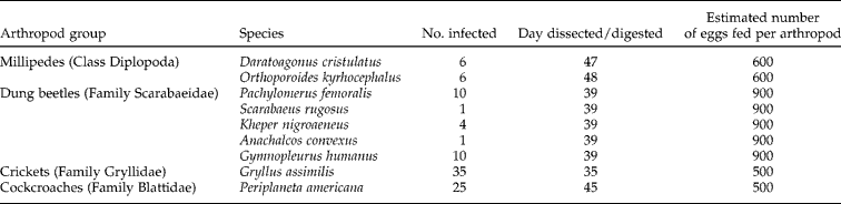
Dung beetles used in this study were collected manually into ventilated plastic contatiners from faecal deposits of wild ungulates in Hluhluwe game reserve of the KwaZulu-Natal province of South Africa and transferred to our laboratory. They were identified to species level following the descriptions by Musgrave (Reference Musgrave and Carruthers2000) and ten beetles representing species for each collection were selected randomly for dissection to rule out any infections prior to the study. A small proportion (3 out of 10) of the beetles was infected with larvae of rhabditiform soil nematodes which were easily identifiable from those of known parasite species. Beetles were sorted by species and were maintained in separate glass tanks filled with sterilized cow dung as substrate according to the parasite species they were exposed to. The substrate was moistened with tap water daily. The prevailing temperature in which they were housed ranged between 25 and 26°C. The range of dung beetle species and the number infected with S. lupi and G. ingluvicola are shown in tables 1 and 2.
Species of arthropods exposed to different doses of Spirocerca lupi eggs and day of dissection/digestion post-infection.
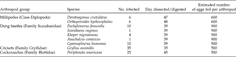
Species of arthropods exposed to different doses of Gongylonema ingluvicola eggs and day of dissection/digestion post-infection.

Estimated number of eggs fed per arthropod = 5000.
Millipedes
Millipedes used in this study were laboratory bred at the School of Biological and Conservation Sciences, University of KwaZulu-Natal, Durban, South Africa. Species and the number infected with S. lupi and G. ingluvicola are shown in tables 1 and 2. Millipedes were maintained in separate glass tanks according to the species of parasite eggs they were exposed to. The temperature in which they were maintained was between 25 and 26°C and they were fed mashed commercial rodent pellets mixed with water and ripe bananas until they were dissected.
Crickets
The cricket species used in this study, Gryllus assimilis, was commercially bred from a local pet shop and the crickets were housed in a glass tank with empty egg carton castles as shelter and covered with a mesh screen lid. The temperature in which they were maintained ranged between 25 and 26°C and they were fed as described by McCluney & Date (Reference McCluney and Date2008). The numbers of crickets infected with S. lupi and G. ingluvicola are shown in tables 1 and 2.
Cockroaches
Periplaneta americana used in this study was bred in the laboratory and housed in ventilated carton boxes. The temperature was maintained in the same range as for crickets and beetles. The number of cockroaches infected with S. lupi is shown in table 1.
Experimental infection with S. lupi
The method followed to infect dung beetles with S. lupi eggs was as described by Bailey et al. (Reference Bailey, Cabrera and Diamond1963). Faecal samples were collected from 20 stray dogs kept by the Society for the Prevention of Cruelty to Animals (Durban North branch, South Africa) and screened for S. lupi eggs. The formol–ether technique described by Knight et al. (Reference Knight, Hiatt, Cline and Ritchie1976) was used to concentrate the eggs for easy detection and enumeration. A positive dog was identified, shedding 1500 S. lupi eggs per gram of faeces, and this dog was used as the source of eggs.
The number of S. lupi eggs fed to each group of dung beetles, millipedes, crickets and cockroaches was estimated from a stock of infected dog faeces and is shown in table 1; calculation was based on the number of eggs per gram of faeces from the positive faecal sample. Dung beetles were fed infected dog faeces, which they readily consumed, as a substitute for the cow dung for two consecutive days, and were dissected/digested 39 days post-exposure.
Infection of millipedes and cockroaches was carried out by exposing them to a mixture of infected dog faeces with an equal portion of wet commercial rodent feed, which they readily consumed. Daratoagonus cristulatus millipedes were dissected to detect S. lupi larvae 47 days post-exposure, whereas Orthoporoides kyrhocephalus were dissected 48 days post-exposure.
Experimental infection with G. ingluvicola
Fifteen free-range chickens were collected from Maphumulo rural area in Durban, South Africa, and humanely slaughtered. Adult female G. ingluvicola located under the serosal surface of the crop were removed and collected in saline solution. Parasites were macerated using a mortar and pestle to release the eggs. The eggs were observed under a dissecting microscope and the proportion of mature (larvated) eggs was quantified. Mature egg concentration was estimated to be 5000 eggs/ml.
The estimated number of G. ingluvicola eggs fed to each group of dung beetles is shown in table 2. Exposure was carried out by thoroughly mixing a solution of infective eggs of G. ingluvicola with rodent commercial feed and beetles readily consumed all the infective mixture within 2 days.
The estimated number of G. ingluvicola eggs fed to the millipedes is shown in table 2. The infective solution was mixed with commercial rodent feed and exposed to the millipedes for 2 days, substituting their normal feed. Once the infective material was completely consumed, their regular feed was resumed.
Crickets were infected by spiking their normal feed of moistened rodent commercial feed with infective material. After infection, crickets were maintained by feeding them rodent commercial feed.
Dissection/digestion of arthropods exposed to infection
Infected beetles, millipedes, cockroaches and crickets were dissected in lukewarm saline and examined under a stereomicroscope for the presence of larvae. Warm saline facilitated the movement of larvae, which allowed easy visualization. Individual specimens that were negative on dissection were further processed by the HCl–pepsin digestion method described by Kaufmann (Reference Kaufmann1996). After digestion, pieces of exoskeleton were removed, the samples sieved through a 20 μm sieve and the contents that remained on the sieve were collected and preserved in 70% alcohol. Contents were observed under a stereomicroscope and the number of free larvae/encysted larvae of S. lupi or G. ingluvicola were enumerated for each arthropod. The infective larvae of S. lupi were recognizable by the presence of a pair of cephalic prominences, a narrow tubular buccal capsule, and a terminal caudal rosette of five closely set papillae (Anantaraman & Sent, Reference Anantaraman and Sent1966), and those of G. ingluvicola were as described by Yamaguti (Reference Yamaguti1971) and Chitwood & Chitwood (Reference Chitwood and Chitwood1974).
Results and discussion
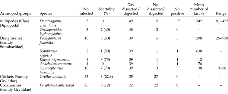
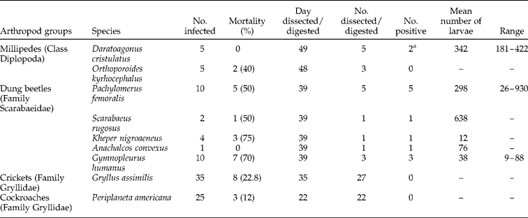
Larvae conforming to the descriptions of pre-infective and infective L3 of S. lupi were recovered from five species of beetles belonging to five genera, as shown in table 3. Mortality rate in the different species ranged from 0 to 75% with the highest rate being recorded for Kheper nigroaeneus. All surviving Pachylomerus femoralis beetles at day 39 post-infection were infected with S. lupi with varying intensities ranging from 26 to 930 larvae/beetle (table 3).
Compatibility parameters of exposed arthropod species to Spirocerca lupi eggs under experimental conditions.
a Depicts encysted larvae.
Five millipedes of the species D. cristulatus were dissected at day 49 post-infection and two had encysted larvae of S. lupi. The millipede species O. kyrhocephalus, the cricket Gryllus assimilis and the cockroach Periplaneta americana were negative for the presence of free or encysted larvae of S. lupi.
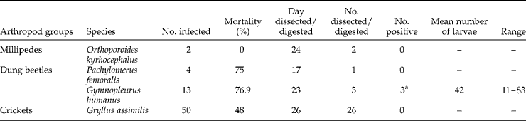
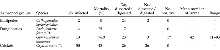
Two species of beetles infected with G. ingluvicola had a high mortality ranging between 75 and 76.9% (table 4). Gy. humanus beetles dissected at day 23 post-infection were positive for encysted larvae (ranging from 11 to 83 larvae) and P. femoralis was negative for any form of G. ingluvicola larvae. No encysted or free larvae of G. ingluvicola were observed in exposed millipede and cricket species.
Compatibility parameters of selected arthropod species to Gongylonema ingluvicola eggs under experimental conditions.
a Depicts encysted larvae.
Results from this study show the ability of P. femoralis, S. rugosus, A. convexus, and Gy. humanus to act as intermediate hosts for S. lupi under experimental conditions. The difference in the number of larvae of S. lupi found among the dung beetle species could be due to the difference in the size of the species, where P. femoralis beetles were relatively large (approximately 5 cm in length with an average weight of 3.65 g) followed by S. rugosus and A. convexus (approximately 4.5 cm with an average weight of 3.40 g), while Gy. humanus was relatively small (approximately 1 cm with an average weight of 0.17 g). Although K. nigroaeneus was of intermediate size, only 12 larvae were recovered and this could be due to the beetle not consuming much of the infective material compared to the other beetles that died during the course of the experiment. Based on these results, it is possible that the intensity of the infection (based on the number of larvae recovered) in the dung beetle intermediate host is proportional to the size of the beetle, with larger beetles generally having higher infections.
Although S. lupi showed little host specificity among the different species of dung beetles in this study, it was not clear which species were more suitable than others. Bailey et al. (Reference Bailey, Cabrera and Diamond1963) found that the different species of dung beetles vary in their susceptibility to S. lupi infection and this was attributed partly to the grinding efficiency of the mandibles of the different species (Miller, Reference Miller1961). This might also be explained by findings on the feeding biology of beetles by Holter et al. (Reference Holter, Scholtz and Wardhaugh2002) and Holter & Scholtz (Reference Holter and Scholtz2005). Holter et al. (Reference Holter, Scholtz and Wardhaugh2002) have shown that food particles in beetles are filtered through channels that connect to the molar surface through fissures before ingestion, and suggested that the size of these fissures, which can allow passage of eggs depending on the size, determine whether a beetle can be an intermediate host. Bailey (Reference Bailey1972) also attributed the difference in susceptibility to be related to the attractiveness of the faeces of different animals to the different species of dung beetles and Fincher et al. (Reference Fincher, Stewart and Davis1970) observed that the species Geotrupes sp. were not attracted to dog faeces and hence may not be a suitable intermediate host compared to other species of dung beetles under natural conditions.
The presence of encysted larvae of S. lupi in the millipede species Daratoagonus cristulatus is interesting and indicates that this species may act as an intermediate host of S. lupi under natural conditions. The presence of larvae in cyst form could be a strategy by the parasite to wait for a suitable paratenic or definitive host. Orthoporoides kyrhocephalus was negative for both the presence of encysted and free larvae of S. lupi.
Attempts to infect experimentally the cockroach species Blatta orientalis, Periplaneta americana and P. australasiae with S. lupi have been unsuccessful (Faust, Reference Faust1928). Our results are in agreement with the above author, as no free or encysted larvae were observed from infected P. americana.
Contrary to S. lupi developing in five species of beetles in our study, G. ingluvicola only successfully developed in Gy. humanus. This is in agreement with results reported by Mowlavi et al. (Reference Mowlavi, Mikaeili, Mobedi, Kia, Masoomi and Vatandoost2009) who found similar Gongylonema sp. larvae in naturally infected Gy. humanus. Beetles of the species P. femoralis were negative for the presence of free or encysted larvae of G. ingluvicola even though they consumed the infective material and were allowed the same time to feed as Gy. humanus. This could be due to P. femoralis being an unsuitable intermediate host for G. ingluvicola and might be an indication of intermediate host specificity of the parasite.
Only millipedes of the species Orthoporoides kyrhocephalus were exposed to G. ingluvicola and the results were negative. Mouthparts of a millipede are designed not only to bite but also to crush food particles effectively (Hopkin & Read, Reference Hopkin and Read1992). This crushing action may have affected the viability of the parasite eggs and hence the absence of G. ingluvicola larvae.
The cricket species Gryllus assimilis was refractory to infection with G. ingluvicola. This could also be due to the small but powerful mouthparts of the cricket (Gorochov & Mostovski, Reference Gorochov and Mostovski2008) that are able to destroy the parasite eggs.
The results of this study provide valuable information concerning which arthropod species to target on a local scale when screening for natural infections of S. lupi and G. ingluvicola.
Acknowledgements
The authors thank the National Research Foundation of South Africa for financial support and the School of Biological and Conservation Sciences for the field collection of dung beetles.