Introduction
The use of cartilage in middle-ear surgery is not a new concept. However, the last decade has seen renewed interest in this material as an alternative to more traditional grafting materials for tympanic membrane reconstruction.
Perichondrium and cartilage share with fascia the quality of being mesenchymal tissue, but they are thicker and stiffer. The greatest advantage of the cartilage graft has been thought to be its very low metabolic rate. However, in addition, it can receive its nutrients by diffusion, it is easy to work with because it is pliable, and it can resist deformation from pressure variations. The perceived disadvantage of the cartilage graft is that it creates an opaque tympanic membrane repair site, which could potentially hide a residual cholesteatoma.Reference Ghanem, Monroy, Alizadeh, Nicolau and Eavey1
Jansen first reported the use of auricular and nasal septal cartilage to reconstruct the ossicular chain in ears without a stapes suprastructure.Reference Jansen2 In 1963, SalenReference Salen3 and JansenReference Jansen4 reported the use of cartilage–perichondrial composite graft for tympanic membrane reconstruction. However, the main advocate of cartilage tympanoplasty was Heermann.Reference Heermann5 He claimed to have used the cartilage palisade technique for middle-ear and mastoid cavity reconstruction since 1960, in over 13 000 cases,Reference Heermann6 and published extensively on his experience. Most of his early publications were in the German literature.Reference Heermann7, Reference Heermann8
Rather than providing a chronological account of the development of cartilage tympanoplasty, the author would like to present a summarised review under the following, clinically important headings: (1) the fate of cartilage implants in the middle ear; (2) the use of cartilage struts for ossiculoplasty; (3) the use of cartilage for tympanic membrane reconstruction; and (4) the use of cartilage for bony canal wall reconstruction.
For topics one to three above, a Medline search of the literature from 1 January 1950 to 31 May 2007 was performed, using a combination of the key words ‘ossiculoplasty’, ‘tympanoplasty’, ‘myringoplasty’, ‘cartilage’ and ‘cartilage-perichondrium’. The content of each abstract was reviewed in order to identify studies relevant to each topic. All chosen articles were read in full, and their references were cross-checked for reports not identified by the Medline search. Topic four (use of cartilage for attic or bony wall reconstruction) is well established and represented the least controversial review subject; therefore, the author selected only those papers which best illustrated the different surgical techniques involved.
Fate of cartilage implants in the middle ear
One of the fundamental questions about cartilage tympanoplasty is whether cartilage grafts placed within the middle ear are eventually resorbed.
Guildford et al. inserted autogenous costal and auricular cartilage implants into the bullae of eight mongrel dogs. The implants were examined three to eight months later. Microscopic studies revealed that the structural patterns of the cartilage implants were essentially similar to their appearance before implantation. However, peripheral absorption of the auricular cartilage was observed in one instance.Reference Guildford, Shortreed and Halpert9
Symth and Kerr inserted struts of homologous septal cartilage and autologous auricular cartilage into the middle ears of six cats. The animals were sacrificed 1.5 to 18 months later and the cartilage grafts examined. The autologous cartilage struts remained viable, whilst the dead homologous struts remained as non-vital grafts. In two out of six homologous cartilage struts, there was some bending and scalloping of the cartilage graft.Reference Symth and Kerr10
Several authors studied the histology of cartilage explants retrieved at revision surgery, in order to detect signs of resorption. Symth et al. examined 11 explants of homologous septal cartilage struts which had been in the middle ear for more than two years. Four of the specimens showed fibrous tissue replacement or erosion of the cartilage, especially of the areas in contact with Silastic® sheets in the middle ear.Reference Symth, Kerr, Pahor and Law11
Don and Linthicum retrieved six autologous tragal cartilage grafts and two preserved homologous septal cartilage grafts six to 12 months after these had been used as columellar grafts. The autologous cartilage remained viable; chondrocytes were present and the amount of mucopolysaccharide was preserved. The homologous septal cartilages were non-viable; chondrocytes were absent and mucopolysaccharides were depleted.Reference Don and Linthicum12 Schuknecht and Shi also examined the histology of four cartilage graft explants (three conchal cartilage autografts and one septal cartilage homograft). There was no inflammatory reaction to the graft, but a loss of rigidity was observed in the cartilage grafts. Two of three cartilage autografts showed a high rate of chondrocyte survival.Reference Schuknecht and Shi13 The authors' opinion was that although cartilage implants lose rigidity they maintain mass and would thus serve well in repairing surgical defects. On the other hand, the loss of rigidity renders them unfit as sound conductors.
Using histochemical methods, Elwany studied the lactic dehydrogenase enzyme activity of cartilage autografts in 32 cats and also of cartilage strut explants in 59 patients. Lactic dehydrogenase enzyme activity was thought to be a good measure of the metabolic activity and viability of chondrocytes and chondroblasts. Elwany's study showed that the presence of perichondrium on both sides of the cartilage strut increased the chance of survival of chondrocytes. On the other hand, middle-ear infection had a very bad effect on chondrocyte viability.Reference Elwany14
One of the most negative reports on the fate of cartilage grafts within the middle ear came from Steinbach and Pulsalkar.Reference Steinbach and Pusalkar15 They studied 52 cartilage graft explants and correlated the histological findings with the clinical progress of the ears. The explants were either columellas or ‘L’-shaped struts; 39 were autologous tragal cartilage grafts and 13 were homologous septal cartilage grafts. In 31 out of the 52 cases, hearing deterioration had occurred three to seven years after surgery. Forty-six of the 52 grafts had gross macroscopic defects; three had completely disappeared. For the L-shaped struts, the vertical limbs were found to have an irregular appearance in some specimens, whereas the horizontal limb in contact with the tympanic membrane had maintained its shape. Microscopically, ‘empty’ chondrocytes were observed in many grafts. The authors concluded that cartilage grafts remained viable for only a certain length of time, and would eventually show changes in their character and function. The difference found between the homologous and autologous cartilages was that the Cialit-preserved homologous cartilage had completely lost its architecture and consistency. Steinbach and Pulsalkar also studied six cartilage–perichondrial grafts which had previously been used to reconstruct the lateral attic wall. The cartilage appeared normal even after 13 years; the authors attributed this fact to blood supply from the tympano-meatal flap.
From these studies, it seems that autologous cartilage struts are more suitable than homologous cartilage struts for use as ossicular prostheses. However, cartilage struts may lose their rigidity over time, especially in a hostile environment. On the other hand, cartilage–perichondrial composite autografts used for meatal wall repair tend to survive well and to maintain their form. Should one wish to use cartilage struts for ossicular reconstruction, it is advisable to preserve the perichondrium on at least one side.
The studies listed above should be interpreted with reservations. Animal studies only give information on the short-term outcome of the grafts, and examination of explants removed at revision or from failed tympanoplasties does not accurately represent the fate of all implanted cartilage grafts.
Use of cartilage struts in ossiculoplasty
Considering the long history of cartilage tympanoplasty, there are relatively few reports on the use of cartilage struts in ossicular reconstruction.
In 1963, Jansen reported his experience of using autologous and homologous cartilage columella for ossicular reconstruction. If the malleus was present, he interposed the cartilage columella between the malleus and the stapes head or footplate. If the malleus was absent, he interposed the cartilage strut between the reconstructed tympanic membrane and the stapes or footplate. The tympanic membrane graft was made from a thin cartilage disc with a small hole to accommodate the top end of the cartilage strut. Jansen claimed to have performed 100 operations over a three-year period. However, he did not provide any audiological results in his report.Reference Jansen4
Around the same time, Heermann started to publish his technique of cartilage palisade for tympanic membrane and ossicular reconstruction. He too did not report the audiological results of his cartilage ossiculoplasties.Reference Heermann7
In 1965, Brockman described his technique of ‘composite T columella’ for repairing the tympanic membrane and ossicles, using a single graft. Perichondrium was elevated from both sides of the tragal cartilage graft (in the manner of lifting the covers off a book), but remained attached at the edge (like the spine of a book). The exposed cartilage was then trimmed into a ‘T’ shape, with the horizontal part of the T still attached to the perichondrium. The wing-like perichondrial flaps were then used to repair the tympanum, whilst the vertical limb of the attached, T-shaped cartilage was placed on the footplate.Reference Brockman16 This technique was later adopted by Goodhill.Reference Goodhill17 Neither Brockman nor Goodhill provided audiological results in their original publications.
Eviatar modified Brockman's technique by changing the shape of the composite graft to an ‘inverted L’ (placed on the stapes footplate) or a ‘W’ (placed on the stapes head). Notably, Eviatar also used autologous and homologous ossicles for his ossicular reconstruction.Reference Eviatar18 He too provided no results for his cartilage ossiculoplasty.
Altenau and Sheehy reviewed the outcome of 564 tympanoplasties in which autologous tragal cartilage grafts had been used. The perichondrium was preserved on one side of the cartilage struts for the eardrum–footplate reconstruction and preserved on both sides of the cartilage block for the eardrum–stapes reconstruction. At six months, 75 per cent of the eardrum–stapes reconstructions and 67 per cent of the eardrum–footplate reconstructions had a residual conductive deficit of 20 dB or less. These authors emphasised the importance of placing the prosthesis under tension. Interestingly, Altenau and Sheehy also claimed that they were changing over to Plastipore prostheses because of their perceived superior results.Reference Altenau and Sheehy19
Accepting that a thin strut of auricular or septal cartilage may not be stiff enough as a sound conductor, Symth et al. inserted stainless steel wire into the cartilage to strengthen their cartilage struts.Reference Symth, Kerr, Pahor and Law11
In a more recent publication, Quaranta et al. defended the use of homologous costal cartilage in staged tympanoplasty. They claimed that the costal cartilage was thicker and more resistant than the conchal or tragal cartilage, and their chondroprostheses were T-shaped. They published long-term audiological results for 40 ears (18 partial and 22 total ossicular reconstructions). At 10-year follow up, 72.2 per cent of the partial and 72.7 per cent of the total ossicular reconstructions had an air–bone gap of 20 dB or better. However, these 40 cases only represented 27 per cent of the original cohort.Reference Quaranta, Feijoo, Piazza and Zini20
Chole and Kim also reported their experience of using presculpted homologous costal cartilage for partial and total ossicular reconstruction. They analysed 187 patients and did not observe any significant difference in hearing, comparing partial and total reconstructions. Post-operatively, 65.2 per cent of the ears had closure of the air–bone gap to within 20 dB. However, the authors did not indicate the follow-up period in their report. Also, they confessed that the inability of the cartilage to withstand extreme thinning was a major limitation in total ossicular reconstructions.Reference Chole and Kim21
Most of the above reports on cartilage ossiculoplasty are now more than 25 years old. Two more recent publications both reported results for homologous costochondrial cartilage, but long-term outcome data were limited. This reflects the fact that many otologists are not yet convinced that cartilage is a good ossiculoplasty material. Even amongst cartilage tympanoplasty enthusiasts (see the next section), many use alternative materials for ossicular reconstruction.
Use of cartilage for tympanic membrane reconstruction
The use of cartilage to reconstruct the tympanic membrane is gaining popularity amongst otologists. Cartilage can be used in the form of several parallel, full thickness strips (palisade technique) or in plates of different sizes and shapes; in the latter case, grafting can be modified by using composite cartilage–perichondrial grafts. Heermann was the first to introduce the cartilage palisade technique, in 1962.Reference Heermann5 With the perichondrium preserved on the outer surface, cartilage strips were placed parallel to the malleus until the middle-ear cavity was covered. Heermann's early publications comprised only a description of his technique, without reporting specific outcomes. Reference Heermann8, Reference Heermann22, Reference Heermann, Heermann and Kopstein23 Salen was one of the first surgeons to use septal cartilage plates for subtotal tympanic membrane perforation,Reference Salen3 and Overbosch was the first to describe a microslice technique in order to improve the acoustic properties of the reconstructed tympanic membrane.Reference Overbosch24
A number of surgeons have adopted the cartilage palisade technique. Amedee et al. reported on 52 cases of cartilage palisade tympanoplasty used to treat recurrent perforations or atelectasis of the tympanic membrane.Reference Amedee, Mann and Eiechelmann25 Milewski used the cartilage palisade technique for ‘difficult’ cases (i.e. large perforations, missing malleus, perforations above the tubal orifice, blunting, lateralisation or atelectasis of the tympanic membrane, and second revisions).Reference Milewski26 Dornhoffer modified Heermann's technique by using several cartilage plates pieced together, like the pieces of a jigsaw puzzle, to reconstruct the posterior part of the tympanic membrane. However, Dornhoffer grafted the anterior half of the tympanic membrane with conventional materials in order to allow post-operative surveillance and tube insertion, if necessary.Reference Dornhoffer27 Murbe et al. also described a modified cartilage plate technique, with several thin cartilage slices overlapping at their edges, like the petals of a tulip blossom.Reference Murbe, Zahnert, Bornitz and Huttenbrink28 These techniques of reconstruction are illustrated in Figure 1.
Various methods of tympanic membrane grafting using cartilage. (a) Full thickness cartilage plates for posterior half tympanic membrane, used by Dornhoffer;27 (b) full thickness cartilage palisades for whole tympanic membrane, used by Heermann et al.;23 (c) thin cartilage plates for whole tympanic membrane, used by Murbe et al. 28

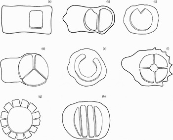
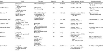
The techniques of using composite cartilage–perichondrial grafts or island grafts for tympanic membrane reconstruction are even more variable, and a number of designs have been reported in the literature. Goodhill described using perichondrial grafts with a circumferential cartilage batten in cases with a shallow middle-ear cavity, in order to avoid sagging of perichondrium onto the promontory.Reference Goodhill17 Other surgeons have trimmed the cartilage part of the composite cartilage–perichondrial graft into the shape of a ‘shield’,Reference Duckert, Muller, Makielski and Helms29 ‘double islands’,Reference Dornhoffer30 ‘Mercedes Benz’ sign,Reference Spielmann and Mills31 ‘wheel’,Reference Shin, Lee, Kim and Lee32 ‘coin with butterfly edges’,Reference Eavey33 ‘crowncork’Reference Hartwein, Leuwer and Kehrl35 or ‘lamellae’,Reference Neumann and Jahnke34 among others. Some of these designs are illustrated in Figure 2, and the outcomes of some of these techniques are summarised in Table I. Unfortunately, most authors reported on the overall results of tympanoplasties; relatively few analysed only type I tympanoplasty. In general, cartilage tympanic membrane grafts have high ‘take’ rates and the audiological results seem good. However, even cartilage grafts cannot prevent retraction pockets in some ears. Also, some authors reported rare cases of thinningReference Duckert, Muller, Makielski and Helms29 and even dissolution of the cartilage over time.Reference Dornhoffer27
Various designs of composite cartilage–perichondrial graft used for tympanic membrane reconstruction. (a) Single island graft, used by Linde;36 (b) double island graft, used by Dornhoffer;30 (c) ‘shield’-shaped graft, used by Duckert et al.;29 (d) ‘Mercedes Benz sign’-shaped graft, used by Spielmann and Mills;31 (e) graft with circumferential cartilage batten, used by Goodhill;17 (f) ‘wheel’-shaped graft, used by Shin et al.;32 (g) ‘crowncork’-shaped graft, used by Hartwein et al.;35 (h) ‘lamella’-shaped graft, used by Neumann and Jahnke.34

Use of cartilage grafts for tympanic membrane reconstruction: summary of retrospective studies

FU = follow-up period; cartlg = cartilage; perichond = perichondrium; perf = perforation; mth = months; ABG = air–bone gap; ET = eustachian tube; post-op = post-operative; post = posterior; sup = superior; av = average; OME = otitis media with effusion, ant = anterior; y = years
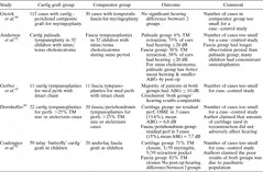
Some surgeons split the edge of the cartilage graft to create a groove at its circumference which the edge of the perforation can be slotted into, thus creating an inlay graft. Eavey was the first to describe the butterfly cartilage inlay grafting technique. The edges of the cartilage–perichondrial composite graft curve out like butterfly wings when the edge is split.Reference Eavey33 Fernandes also described a permeatal triple ‘C’ technique. The perichondrium is elevated only at the outer edge of the cartilage composite graft circumferentially. The edge of the perforation is then wedged into the groove created between the perichondrial and cartilaginous layers, in order to keep the composite graft in place.Reference Fernandes39 These inlay grafts are illustrated in Figure 3. The advantage of all these techniques is that the procedure is quick and can be performed permeatally. Both Eavey and Fernandes used such inlay grafts only for small to medium-sized central perforations in which the surgeon could see the whole of the perforation through the ear speculum. However, Ghanem et al. extended the indication for Eavey's butterfly inlay graft to include medium-sized and large perforations, including even marginal ones.Reference Ghanem, Monroy, Alizadeh, Nicolau and Eavey1 The overall results of these grafts appear good (Table II). The author could not identify any published report comparing such cartilage inlay grafts with more conventional fat inlay grafts.
Different methods of preparing the cartilage inlay graft. (a) Circumferential splitting of the cartilage at its edge, as advocated by Eavey;33 (b) separating perichondrium from cartilage at the edge of the composite graft, as advocated by Fernandes;39 (c) insertion of the inlay composite graft.

Use of inlay cartilage composite grafts for tympanic membrane reconstruction: summary of retrospective studies

FU = follow-up period; mth = months; cartlg = cartilage; perf = perforation; med = medium; incl = including; ABG = air–bone gap
The traditional material for myringoplasty is temporalis fascia. The rigidity of the cartilage graft has obvious benefits in reducing retraction of the tympanic membrane; however, it is unclear if the increase in rigidity and mass compromises the sound conduction properties of the graft. Concerns that the stiffness and mass of cartilage grafts may adversely affect hearing have not been substantiated in clinical outcome reports.
To date, there has been no randomised or prospective case–control study comparing the outcome of fascia and cartilage tympanoplasties. All the comparative studies currently available have been retrospective (Table III). The number of cases included in these comparative studies was small and not based on power analysis. Accepting the limitations of these studies, there was no obvious adverse effect of cartilage tympanoplasty compared with fascia tympanoplasty. Further research is necessary to determine if cartilage grafting is truly superior to temporalis fascia grafting in tympanoplasty, especially in cases with atelectasis.
Cartilage grafts vs conventional materials for tympanic membrane reconstruction: summary of retrospective studies

Cartlg = cartilage; perichond = perichondrium; TM = tympanic membrane; ABG = air–bone gap; y = year; post-op = post-operative; med = medium-sized; perf = perforation; OME = otitis media with effusion
Using laboratory models, some researchers investigated the acoustic transfer characteristics of cartilage plates and their resistance to pressure changes. Zahnert et al. examined the frequency response function of tragal and conchal cartilage plates, using a laser Doppler interferometer. They found no statistical difference in the acoustic transfer characteristics of conchal and tragal cartilage. There were transmission losses at lower frequencies when large tympanic membrane defects were reconstructed with thick pieces of cartilage. Reducing cartilage thickness led to an improvement of the acoustic transfer qualities. Using normal tympanic membrane as a reference, Zahnert et al. noted that cartilage plate with a thickness of less than 0.5 mm gave least acoustic transfer loss.Reference Zahnert, Huttenbrink, Murbe and Bornitz44 From the same research group, Murbe et al. also investigated the sound-induced vibrational amplitudes of four different tympanic membrane reconstruction techniques – cartilage plates of varying thickness, cartilage palisade, and large and small cartilage island transplants. Using an ear canal–eardrum model, the vibrations of the cartilage grafts were measured by scanning laser Doppler vibrometry. Slicing thick cartilage into thin plates or palisades decreased the first resonance frequency and increased its amplitude, reflecting improved sound transmission properties of the transplant. The 0.5 mm cartilage plate seemed preferable compared with the palisade technique. Cartilage island techniques showed vibration characteristics superior to those for plate or palisade techniques.Reference Murbe, Zahnert, Bornitz and Huttenbrink28 The results from these laboratory experiments are interesting. However, it is not clear how such information can be translated into real clinical settings. In real life, cartilage grafts placed within the middle ear become less rigid with time, and this could influence their vibrational properties.
Use of cartilage for bony meatal reconstruction
Of all the various indications for using cartilage grafts in the middle ear, reconstruction of defects in the bony meatus or outer attic wall is the most established.
Even in his early publications on cartilage palisade tympanoplasty, Heermann was already advocating the use of cartilage palisades for ‘antrum-plasty’ and ‘mastoid-plasty’. He used thick pieces of cartilage to reconstruct the bony meatal wall and/or to obliterate the mastoid cavity.Reference Heermann8
McCleve was the first to use cartilage–perichondrial composite autografts to reconstruct the superior or posterosuperior canal wall, in 20 cases, with one failure.Reference McCleve45 Beneficial results were subsequently reported in more than 100 patients.Reference McCleve46
Other otologists soon adopted the technique of using cartilage–perichondrial composite grafts to reconstruct bony defects of the meatal wall. Linde reported the use of conchal cartilage with perichondrium for repairing a dehiscent posterosuperior canal wall in 10 cases. He observed no retraction in the grafted area.Reference Linde36 Adkins and Osguthorpe also used cartilage–perichondrial composite grafting to correct defects in the attic wall.Reference Adkins and Osguthorpe47 Adkins then reported 102 cases of composite cartilage–perichondrial autografts to repair the attic wall in atticotomy (40 cases) and combined-approach tympanoplasty (62 cases), with one to six year follow up. Four failures were noted, with one further failure observed later. Adkins claimed that the failures (retraction of the cartilage graft) were mainly due to technical problems. He advised using adequate cartilage, with the perichondrium of the composite graft overlapping the bony margins so that the cartilage fitted into a bony shelf at the margin of the bony defect.Reference Adkins38
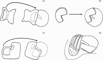
Some surgeons further modified the design of the composite cartilage–perichondrial graft by changing the shape of the cartilage island. Levinson used a ‘Pac Man’-shaped cartilage–perichondrial graft to reinforce the posterior tympanic membrane and attic in posterior marginal and/or attic retraction pockets.Reference Levinson48 He reviewed the outcome of 85 ears in 79 patients with a one-year follow up. There was no graft failure or recurrent retraction pocket at the grafted site. However, 13 per cent of the ears subsequently developed fresh retraction pockets in another area of the drum. In addition, six ears subsequently developed recurrent middle-ear effusions. Poe and Gadre reported results for another design of the composite cartilage–perichondrial graft.Reference Poe and Gadre49 Their graft had two cartilage islands attached to the same piece of perichondrium – a rectangular cartilage island for the posterosuperior quadrant of the tympanic membrane, and an adjacent, sausage-shaped cartilage island for the attic. They reported the outcomes of 37 ears with retraction pockets in the pars flaccida or the posterosuperior quadrant of the tympanic membrane. They did not specify the number of cholesteatoma cases in their report. The follow-up time ranged from eight to 42 months. In no patient did a retraction pocket form at the site of an intact cartilage graft; however, Poe and Gadre observed that nine ears subsequently developed new retraction pockets in different areas of the drum. Levinson and Poe and Gadre all noted that retraction pockets occurred less frequently when both the pars flaccida and the posterosuperior quadrant were supported by cartilage. Some of the techniques described above are illustrated in Figure 4.
Various designs of composite cartilage–perichondrial graft for reconstruction of attic and/or posterior retraction pockets. (a) Single island graft, used by Adkins;38 (b) ‘Pac Man’-shaped graft, used by Levinson;48 (c) two-island graft to reinforce attic and posterior tympanic membrane, used by Poe and Gadre;49 (d) cartilage palisades technique, used by Heermann et al. 23

It is clear that cartilage grafts are effective in preventing local retraction. However, they do not address the causal factors involved in initiation of the retraction process.
Conclusion
It can be concluded from the present review that cartilage tympanoplasty is already an established method for the reconstruction of the middle ear and mastoid. This method is particularly useful for repairing defects of the bony meatus and the tympanic membrane. The commonest techniques are the cartilage palisade and the cartilage island methods. Different designs of cartilage–perichondrial composite grafts have been reported. Most authors used cartilage tympanoplasty for atelectasis and large perforations or in revision cases. However, it is still unclear whether cartilage is indeed a better material than fascia for type I tympanoplasty, as the published reports to date were all retrospective studies. Because of the risk that cartilage may become softer with time, and may even be resorbed, few surgeons have used cartilage for ossicular reconstruction.