Introduction
A diagnostic technique based on the direct demonstration of parasite products such as circulating filarial antigens is one of the definitive ways to detect active filarial infection in humans (Theodore & Kaliraj, Reference Theodore and Kaliraj1996). Microscopic detection is a conventional method to diagnose microfilariae in samples taken during the night. Under circumstances such as a low level of filarial parasites this method is not suitable for diagnosis. This seems to be a major drawback (Nuchprayoon et al., Reference Nuchprayoon, Porksakorn, Junpee, Sanprasert and Poovorawan2003). As a part of the filarial genome project, our laboratory has been involved in screening a large number of molecular targets for diagnosis. Wuchereria bancrofti SXP-1 (Wb-SXP-1) was identified and established as a potential candidate for immunodiagnosis of human lymphatic filariasis. Wb-SXP-1 protein was identified as a parasite surface protein and was highly immunogenic, present in all the stages of the filarial parasite life cycle and sharing 82% homology with Brugia malayi SXP-1 (Bm-SXP-1) (Roa et al., Reference Rao, Eswaran, Ravi, Gnanasekhar, Narayana, Kaliraj, Jayaraman, Marson, Raghavan and Scott2000). An enzyme-linked immunosorbent assay (ELISA) developed with antibodies against the recombinant filarial proteins Bm-SXP-1 and Wb-SXP-1 has been applied successfully to quantify circulating filarial antigens in sera from bancroftian and brugian infections (Lalitha et al., Reference Lalitha, Eswaran, Gnanasekhar, Rao, Narayanan, Scott, Nutman and Kaliraj2002). Hence monoclonal antibodies against Wb-SXP-1 were developed using hybridoma technology (Janardhan et al., Reference Janardhan, Pandiaraja, Pandey, Karande and Kaliraj2011; Pandey et al., Reference Pandey, Madhumathi, Karande and Kaliraj2011). Although the clinical and commercial success of monoclonal antibodies have led to the large-scale production of hybridomas, the maintenance and production of monoclonal antibodies are expensive and time consuming. Hence a viable alternative to the production of complete antibodies is to generate recombinant antibody fragments in large amounts in Escherichia coli (Holliger & Hudson, Reference Holliger and Hudson2005).
Recombinant antibody fragments were simpler to construct and easy to structurally manipulate for improved specificity. Also, the purification and recovery of antibody fragments are effortless when fused with a tag. Thus a single-chain fragment variable (ScFv) module consisting of a single polypeptide chain with aligned antibody variable heavy (VH) and variable light (kappa) (Vκ) chain domains were linked together with a polypeptide linker to mimic Fv fragments (Hudson, Reference Hudson1998). The variable regions contain segments showing a statistically high degree of variability (hypervariable regions) (Capra & Kehoe, Reference Capra and Kehoe1974) and are predicted to be the complementarity-determining (CD) segments which make contact with the antigenic determinants. Variable sequences in the proximity of residues 30–35, 50–65 and 95–100 were known to be the CDRs (complementarity-determining regions). The rest of the residues are termed as framework regions. By taking advantage of the fact that the immunoglobulin variable region genes are flanked by conserved sequences at the 3′ end and by semi-conserved sequences at the 5′ end, it was possible to design consensus primers to selectively amplify variable region genes by polymerase chain reaction (PCR) (Orlandi et al., Reference Orlandi, Gussow, Jones and Winter1989). The conserved regions at each end of the nucleotide sequence encoding variable domains of mouse immunoglobulin heavy chain (VH) and light chain (kappa) (Vκ) were identified by aligning the entries of the nucleotide sequences of the variable region genes extracted from the Kabat database (Kabat et al., Reference Kabat, Wu, Reid-Miller, Perry and Gottesmann1987). The frequency of the most common nucleotide was scored for each site, and the information was used to design amplification primers. Recent developments in antibody engineering and recombinant DNA technology have made it possible to generate recombinant Fv antibody fragments with a high degree of specificity and affinity.
We report the cloning and sequencing of the antibody variable region of hybridoma cell line 2E12E3 as well as subsequent ScFv construction and expression. Single-chain antibodies were constructed by tethering the Vκ and the VH genes to a DNA sequence encoding a 15-residue polypeptide (Gly4Ser)3 as a linker. The recombinant ScFv was expressed and purified by an on-column refolding method without the presence of reducing agents. The affinity of recombinant ScFv towards the filarial antigen was equivalent to that of the monoclonal antibody which is determined by ELISA.
Materials and methods
Strains, plasmids, antigens and antibodies
The E. coli strain Rosetta 2 (DE3) (F–ompT hsdSB (rB –mB –) gal dcm lacY1 (DE3) pRARE6 (CmR)) was used as expression host. The plasmid pRSET A (Invitrogen, Carlsbad, California, USA), carrying an N-terminal His tag under the control of the T7 promoter, was used for the construction of the expression vector. Escherichia coli GJ1158 host was used as the expression system for production of recombinant Wb-SXP-1 antigen. A hybridoma cell line (clone 2E12E3) producing IgM type monoclonal antibody against recombinant Wb-SXP-1 antigen was provided by Professor Anjali A. Karande (Indian Institute of Science, Banglore). The hybridoma clone 2E12E3 was used for ScFv construction. Another hybridoma cell line (clone 2E4F1), producing monoclonal anti-Wb-SXP-1 antibody, was used for immunoblot assay. The rabbit polyclonal antibody used in this study was already developed in our laboratory (Pandey et al., Reference Pandey, Madhumathi, Karande and Kaliraj2011). The serum samples for capture assay were collected from Vellore, Thiruvannamalai and Nagarkoil, three towns in the southern Indian state of Tamil Nadu. The field samples were obtained from 30 microfilaraemic (Mf-positive) patients, 30 patients with chronic lymphatic disease (CP), 30 microfilariae-negative healthy individuals residing in endemic areas (EN) and 10 from healthy individuals residing in non-endemic areas (NEN).
Amplification of variable region genes
Total RNA was isolated from about 1 × 10⁸ freshly subcloned hybridoma 2E12E3 cells using RNeasy RNA isolation kit from Qiagen (Hilden, Germany). mRNA was purified from total RNA using Oligotex mRNA purification Kit from Qiagen. cDNA was constructed using CloneMiner cDNA library construction kit (Invitrogen, San Diego, California, USA) First-strand cDNA was synthesized separately for variable heavy chain (VH) and variable light chain (Vκ) from mRNA using primers VH1FOR and VK1FOR, respectively. The second strand of cDNA encoding the antibody variable domains (VH and Vκ) was amplified by a separate polymerase chain reaction using Taq DNA polymerase (Bangalore Genei, India). Amplification primers VH1FOR and VK1FOR were designed to be complementary to the mRNA of the J regions, and primers VH1REV and VK1REV to be complementary to the first-strand cDNA encoding the conserved N-terminal region (Orlandi et al., Reference Orlandi, Gussow, Jones and Winter1989). The amplified variable regions were cloned into the pKRX-T vector (Beijing SBS Genetech, China). The constructs were sequenced and a mouse immunoglobulin BLAST was performed for individual VH and Vκ sequences at the National Center for Biotechnology Information (NCBI) for sequence homology with known mouse variable region genes. The CDRs were identified by using the Kabat database numbering at NCBI.
Construction of the ScFv gene
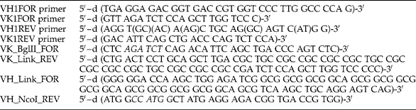
The genes encoding the variable regions in pKRX-T vector were independently modified by PCR reaction for the addition of linkers and restriction sites. VK_Link_REV and VH_Link_FOR carry overlapping sequences encoding the linker peptide sequence (Gly4Ser)3. Both the fragments were joined by overlap extension PCR to construct the ScFv gene in Vκ–Linker–VH orientation (Lee et al., Reference Lee, Park, Park and Kwak2002). The entire ScFv gene was directionally cloned between BglII and NcoI sites into pRSET A vector. The list of primers used is given in table 1.
List of primers used for amplification and construction of the ScFv gene.

Expression and purification of the ScFv gene
Bacterial cells harbouring recombinant ScFv plasmid were grown in Luria–Bertani (LB) medium containing 34 μg/ml chloramphenicol and 100 μg/ml ampicillin at 37°C with shaking. At 0.6 OD600nm, the culture was induced with 1 mm isopropyl β-d-thiogalactopyranoside (IPTG) and the culture was incubated at 37°C for 3 h. Finally, the culture was centrifuged and the pellet was processed for extraction of inclusion bodies by sonication. The clarified supernatant of inclusion bodies containing solubilized ScFv was loaded on to a Hi Prep IMAC Fast Flow 16/10 column (GE Healthcare, Little Chalfont, Bucks, UK). In vitro refolding and purification of ScFv was done using AKTA prime (Amersham Pharmacia Biotech, Amersham, Bucks, UK). The column was equilibrated with buffer containing 20 mm Tris–HCl, 0.5 m NaCl, 10 mm imidazole, 8 m urea, pH 8.0. Refolding was then performed by decreasing the concentration gradient from 8 m urea to 0 m urea. Refolded protein was eluted in 500 mm imidazole. The purified ScFv was analysed by 12% sodium dodecyl sulphate-polyacrylamide gel electrophoresis (SDS-PAGE) under reducing and non-reducing conditions. In non-reducing SDS-PAGE the sample buffer was devoid of β-mercaptoethanol (Sambrook & Russell, Reference Sambrook and Russell2001).
Immunoblot and ELISA analysis
To study the binding capacity, purified and refolded ScFv was electrophoresed on a 12% SDS-PAGE gel and the protein was transferred on to nitrocellulose membrane. Individual lanes were then challenged with recombinant Wb-SXP-1 antigen, crude Mf antigen and Mf-positive patient sera containing circulating Wb-SXP-1 antigen, respectively. Monoclonal anti-Wb-SXP-1 antibody from clone 2E4F1 was used as capture antibody and was developed with alkaline phosphatase (ALP)-conjugated antimouse IgM secondary antibodies (Sigma-Aldrich, St. Louis, Missouri, USA). Immunoblotting was done with suitable vector control (cell lysate of vector control) and blots challenged without antigens, to eliminate the question of cross-reactivity. ELISA was carried out to compare ScFv and monoclonal antibody (2E12E3 clone) with different antigenic concentrations (0.25–1 μg) (Pandey et al., Reference Pandey, Madhumathi, Karande and Kaliraj2011).
Capture assay for detection of filarial antigen in field samples
Microtitre ELISA plates were coated with purified ScFv (1 μg/well in phosphate buffered saline (PBS)) overnight at room temperature, followed by blocking with 0.5% gelatin in PBS. Serum samples diluted 1:50 in PBS were added to the wells (in duplicate) and incubated for 2 h, followed by washing and addition of rabbit polyclonal Wb-SXP-1 in PBS for 1 h. The plates were washed three times with PBS and incubated with horseradish peroxidase (HRP)-conjugated secondary antibodies (Dako, Glostrup, Denmark) for 1 h. The plates were washed extensively and the bound peroxidase activity was detected using tetramethyl benzidine (TMB) at a concentration of 60 μg/ml in citrate phosphate buffer, pH 5.5, containing 0.03% hydrogen peroxide. The reaction was stopped with 1 m sulphuric acid and the absorbance was read at 450 nm using a multiscan ELISA reader (Molecular Devices, Sunnyvale, California, USA).
Data analysis
All statistical analyses were done using GraphPad Prism software version 5. Comparisons between two individual data points were made using the Mann–Whitney test. For multiple comparisons, Bonferroni's post-hoc test was used. P≤ 0.05 was considered to be statistically significant.
Results
Construction of the ScFv gene
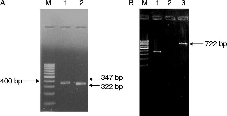
A set of oligonucleotide primers designed by Orlandi et al. (Reference Orlandi, Gussow, Jones and Winter1989) was used for amplification of VH and Vκ genes from the mRNA of the hybridoma clone 2E12E3. The amplified variable heavy-chain and light-chain fragments were 347 bp and 322 bp in length (fig. 1A). The fragments were sequenced and a mouse immunoglobulin BLAST was performed for individual VH and Vκ sequences at NCBI for sequence homology with known mouse variable region genes. The CDR was identified by using the Kabat database numbering at NCBI (KP053273, KP053274). ScFv was assembled as Vκ–Linker–VH, where the linker bridges the gap between the carboxy-terminal and the amino-terminal ends of the respective domains without interfering with the structure of Fv. The linker was designed in such a way that it spans a distance of 3.5 nm between its points of fusion to the variable domains without distortion of the native Fv, and it was also hydrophilic to preclude intercalation within or between variable domains during protein folding. Based on these requirements, a 15-residue linker comprising the pentameric units of –(Gly)4–Ser was designed (Huston et al., Reference Huston, Levinson, Mudgett-Hunter, Tai, Novotny, Margolies, Ridge, Bruccoleri, Haber and Crea1988). Individual VH and Vκ segments were amplified using primers that introduce overlapping segments of the linker region. These fragments were then purified and annealed by overlap PCR. The product obtained after overlap PCR was then amplified using 5′-end and 3′-end primers of ScFv to generate the full-length sequence (fig. 1B).
(A) PCR amplification of variable region genes from the cDNA of hybridoma clone 2E12E3. Lanes: 1, variable heavy-chain gene (VH); 2, variable light-chain gene (Vκ); M, 100 bp DNA ladder. (B) Analysis of full-length ScFv amplified by overlap extension PCR. Lanes: 1, positive control; 2, negative control; 3, Vκ–Linker–VH; M, 100 bp DNA ladder.
Cloning, expression and purification of the ScFv gene
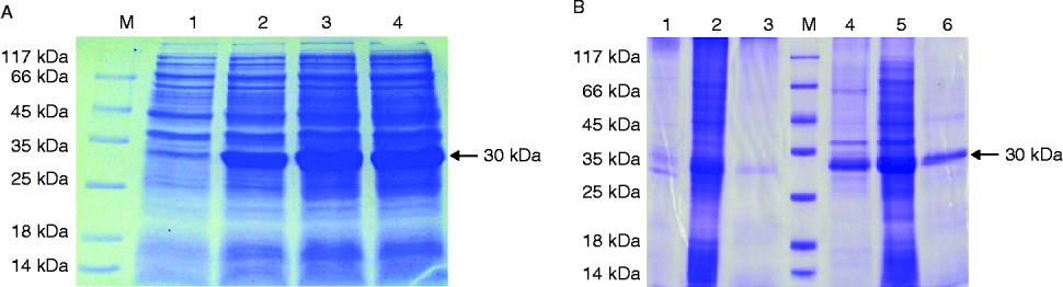
The ScFv gene was expressed as a fusion protein containing an N-terminal His tag in the cytoplasm of the Rosetta 2 (DE3) host. Maximum protein expression was obtained when the culture was induced with 1 mm IPTG at 0.6 OD600nm. Expressed ScFv formed inclusion bodies in the cytoplasm and was analysed by SDS-PAGE (fig. 2A). ScFv was expressed as a 30 kDa protein. The expression was confirmed by immunoblotting using anti-His antibodies. Insoluble ScFv was then solubilized with the chaotropic reagent 8 m urea, which disrupts the non-covalent interactions. The solubilized inclusion bodies of ScFv were purified using a Hi prep IMAC column. The denatured and bound ScFv was then subjected to refolding on IMAC column by decreasing the urea concentration to 0 m. A single peak was obtained in the A280 profile of the chromatogram. A single band was observed when purified and refolded ScFv samples were run on an SDS-PAGE gel under reducing and non-reducing conditions (fig. 2B). The purified ScFv moved faster under non-reducing conditions than reducing conditions.
SDS-PAGE analysis of (A) ScFv expression in E. coli Rosetta 2 (DE3) host. Lanes: 1, 10 μl of un-induced sample; 2, 10 μl of induced first-hour sample; 3, 10 μl of induced second-hour sample; 4, 10 μl of induced third-hour sample; M, protein molecular weight marker. (B) Purified ScFv under reducing and non-reducing conditions. Lanes: 1, crude cell lysate (non-reduced); 2, urea-solubilized ScFv (non-reduced); 3, refolded and purified ScFv (non-reduced); 4, crude cell lysate (reduced); 5, urea-solubilized ScFv (reduced); 6, refolded and purified ScFv (reduced); M, protein molecular weight marker.
Antigen binding capacity of ScFv
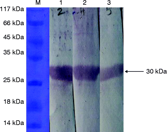
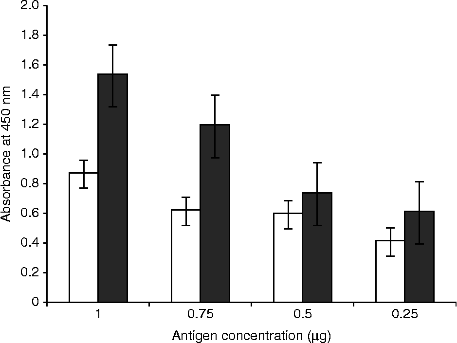
The antigen binding capacity of ScFv was confirmed with sandwich immunoblotting. Bound ScFv was able to recognize and bind recombinant Wb-SXP-1 antigen and crude Mf antigen. Monoclonal antibody against Wb-SXP-1 from hybridoma clone 2E4F1 was used as a capture antibody. A single band corresponding to 30 kDa was developed, confirming the affinity of ScFv towards Wb-SXP-1 antigen. Sera from Mf-positive patients, containing circulating Wb-SXP-1 antigen, was also used to confirm the antigen binding capacity of ScFv. Purified and refolded ScFv was able to recognize and bind circulating filarial Wb-SXP-1 antigen in sera from Mf-positive patients (fig. 3). Using sandwich ELISA, the detection specificity of ScFv was compared to that of monoclonal antibody. The colour intensity developed for both recombinant ScFv and monoclonal antibody is shown in fig. 4. From the absorbance readings of ELISA, it is evident that ScFv was able to detect antigen at lower concentration and it was similar to that of monoclonal antibody.
Immunoblotting analysis to determine the antigen binding capacity of ScFv. Lanes: 1, ScFv challenged with recombinant Wb-SXP-1 antigen; 2, ScFv challenged with crude Mf antigen; 3, ScFv challenged with serum from an Mf-positive patient, containing circulating Wb-SXP-1 antigen.
Comparison of the binding capacity of ScFv (white bars) with that of the monoclonal antibody (black bars).
Capture assay for detection of native Wb- SXP-1 antigen in field samples
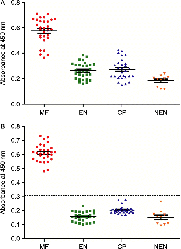
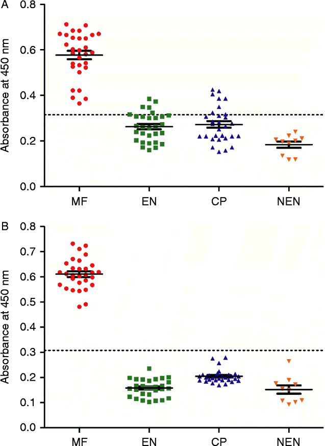
Sandwich ELISA was performed with ScFv and parental monoclonal antibody to compare the detection of native Wb-SXP-1 antigen in the field samples. Also, to verify the field samples, a standard Og4C3 kit was used for comparison. The mean optical density (OD) ± SD values are given in table 2. Mean OD +3SD of the negative control (non-endemic normal) was taken as cut-off, indicated by a line drawn parallel to the x-axis in fig. 5. The detection of circulating filarial antigen in microfilaraemic patients by ScFv was comparable to that by monoclonal antibody (fig. 5A, B).
Optical density values (mean ± SD) from ELISA assays of patient serum samples for the detection of Wb-SXP-1 antigen.

Capture assay for ScFv-based (A) and monoclonal antibody-based (B) detection of native Wb-SXP-1 antigen in patient serum samples: MF, microfilaraemic; EN, endemic normal; CP, chronic patients; NEN, non-endemic normal.
Discussion
Circulating filarial antigen in the sera from patients with lymphatic filariasis was detected by developing monoclonal antibodies against recombinant filarial antigen Wb-SXP-1 used in the Og4C3 ELISA kit (Lalitha et al., Reference Lalitha, Ravichandran, Suba, Kaliraj, Narayanan and Jayaraman1998). Polyclonal antibodies developed against recombinant Wb-SXP-1 were found to show some degree of cross-reactivity. Hence monoclonal antibodies against Wb-SXP-1 antigen were developed using hybridoma technology (Janardhan et al., Reference Janardhan, Pandiaraja, Pandey, Karande and Kaliraj2011, Pandey et al., Reference Pandey, Madhumathi, Karande and Kaliraj2011). Production of monoclonal antibodies using hybridomas is not only time consuming but also expensive. Hence ScFv molecules, which contain the entire antibody binding domain produced in E. coli, were developed as an alternative. This recombinant ScFv can be used as a diagnostic tool for detection of filarial infection.
The major challenge in cloning antibody variable-region genes was their specific amplification. This was due to the fact that mouse immunoglobulin genes are highly diverse in sequence. Therefore, the primers used for gene amplification should be universal (Wang et al., Reference Wang, Raifu, Howard, Smith, Hansen, Goldsby and Ratner2000). There have been numerous descriptions of primer design for PCR amplification of mouse Ig genes (Dattamajumdar et al., Reference Dattamajumdar, Jacobson, Hood and Osman1996). A set of oligonucleotide primers designed by Orlandi et al. (Reference Orlandi, Gussow, Jones and Winter1989) was used to amplify mouse immunoglobulin heavy- and light-chain variable regions from hybridomas by polymerase chain reaction. Primers were designed by making a systematic comparison of the aligned sequences of VH and Vκ genes from the Kabat database (Kabat et al., Reference Kabat, Wu, Reid-Miller, Perry and Gottesmann1987) and the nucleotide sequences at the 5′ end of both VH and Vκ genes that were relatively conserved were identified. Since JH and JL regions at the 3′ ends of the genes were also conserved, this enabled them to design primers based on these sequences. These primers were reported to be able to amplify most immunoglobulin mRNA of the mouse repertoire. The Vκ and the VH genes were joined together with a flexible linker. Several linkers with different lengths were tried by many groups and in most cases (Gly4Ser)3 has been proven to yield ScFv species that reproduce the binding affinity of the parent antibody (Glockshuber et al., Reference Glockshuber, Malia, Pfitzinger and Plueckthun1990). The serine residue confers extra hydrophilicity on the peptide backbone and the glycyl residues reduce steric hindrance so as to have a flexible conformation. For protein expression, the recombinant plasmid was transformed into E. coli Rosetta 2 (DE3), which codes for rare tRNAs of E. coli. Since variable region genes were amplified from mouse hybridoma, they contained many codons rarely coded by E. coli. Hence this host was chosen for expression.
The on-column refolding procedure was established for efficient recovery of active ScFv molecules. Formation of disulphide bonds plays a critical role in stabilization of the immunoglobulin fold (Swietnicki, Reference Swietnicki2006). The intermolecular interactions leading to aggregation of the protein were minimized when the refolding molecules were bound to the column via a His tag. Furthermore, the linear urea gradient provides a gentle environment for protein renaturation (Yang et al., Reference Yang, Chen, Wei, Zhang, Zhang, Jia, Yan, Luo, Cao, Yao, Sun, Xu and Yang2010). The absence of reducing agents enhanced the refolding efficiency. The mobility shift in the movement of ScFv under reducing and non-reducing conditions might be due to a conformational difference caused by disulphide bonds.
Immunoblotting analysis and ELISA reveal the antigen binding capability of expressed ScFv. The refolded and purified ScFv was also able to recognize and bind circulating filarial antigen in the sera from Mf-positive patients. Sandwich ELISA for detection of native Wb-SXP-1 antigen in field samples with ScFv was comparable to that with parental monoclonal antibody and the Og4C3 kit. It is apparent from the above experiments that the recombinant ScFv can act similarly to monoclonal antibody in the detection and diagnosis of filarial antigen.
In endemic countries like India, where there are both brugian and bancroftian infections, a filariasis detection kit identifying both the infections is very important for global programmes to eliminate lymphatic filariasis (GPELF). It is important to detect filarial antigens in the circulating bloodstream to identify active infection in the endemic population area. For detecting filarial antigen, monoclonal antibodies were developed and assays based on these monoclonal antibodies were reported (Janardhan et al., Reference Janardhan, Pandiaraja, Pandey, Karande and Kaliraj2011; Pandey et al., Reference Pandey, Madhumathi, Karande and Kaliraj2011; Weil et al., Reference Weil, Curtis, Fakoli, Fisher, Gankpala, Lammie, Majewski, Pelletreau, Won, Bolay and Fisher2013). Although these test kits are valuable tools for the GPELF programme, their short shelf life and cost are barriers to their use. The present investigation was an attempt to reduce the cost of these kits, one of the road blocks to usage.
In the present study, we report a simple approach that allows the rapid generation of a large amount of active ScFv molecule expressed in E. coli, which is able to bind Wb-SXP-1 antigen. Refolded and affinity purified ScFv could be used effectively as a diagnostic reagent for the detection of circulating filarial infection. This recombinant ScFv can be an alternative to monoclonal antibody in the detection of filarial infections. The application of the recombinant ScFv in filarial antigen detection kits was validated by field samples.
Acknowledgements
We sincerely thank Dr V. Gopalrathinam, Senior Entomologist, Zonal Entomological Research Laboratory, Vellore, Tamilnadu, for his cooperation and support in sample collection. We also thank Dr Murray Selkirk, Professor of Biochemical Parasitology, Division of Cell and Molecular Biology, Imperial College, London, UK, for kindly providing the non-endemic sera samples for the study.
Financial support
The work was supported by a grant from the University Grant Commission (UGC), Government of India. The fellowships granted to R.R. by the Council of Scientific and Industrial Research (CSIR), Government of India and to R.K. by the Department of Biotechnology (DBT) are gratefully acknowledged.
Conflict of interest
None.
Ethical standards
Blood samples were collected from volunteers after obtaining written consent and this was documented by the Department of Public Health and Preventive Medicine, Chennai, Tamil Nadu, India (Ref. No. 26433/VCII/SI//2012). The procedures followed were in accordance with the guidelines issued by the same department. The Institutional Review Board at the Centre for Biotechnology, Anna University, India has also approved the protocols.