Introduction
In humans, female overweight and obesity at fertile age are becoming a worldwide burden, which in turn associate increased risk of altered fetal growth (i.e., restriction or macrosomia). Compelling data show that fetal macrosomia (>4000 g) or large for gestational age (LGA) newborns from overweight/obese mothers have increased risk of neonatal morbidity,Reference Higgins, Greenwood, Wareing, Sibley and Mills 1 and the development of cardiovascular diseases at adulthood.Reference Reynolds, Allan and Raja 2 – Reference Ornellas, Souza-Mello, Mandarim-de-Lacerda and Aguila 5 Recent reports show that chorionic plate arteries from placentae of obese women have a reduced relaxation in response to exogenous nitric oxide (NO)Reference Hayward, Higgins and Cowley 6 and endothelial dysfunctionReference Schneider, Hernandez, Farias, Uauy, Krause and Casanello 7 compared with placentae from women with normal body mass index (BMI). However, there is no data addressing whether these changes at the placental bed from overweight women would represent an altered systemic vascular reactivity in the fetus.
A comprehensive analysis reveals that in humans pre-pregnancy BMI is one of the main factors associated with fetal macrosomia and LGA newborns with cardiometabolic consequences at long term.Reference Yu, Han and Zhu 8 Many rodent models have been proposed to address the detrimental effects of an increased maternal weight previous and during gestation. In those models, maternal obesity is generated by altering the maternal metabolic status with long-term exposure to pro-obese diets (i.e., high fat, sucrose or fructose), genetic manipulation or induction of diabetes.Reference Li, Reynolds, Sloboda, Gray and Vickers 9 – Reference Toop, Muhlhausler, O’Dea and Gentili 11 Interestingly, despite the strong metabolic intervention, maternal obesity in these models associates mainly with intrauterine growth restriction (IUGR) rather than macrosomia, in both mice and rats.Reference Sloboda, Howie, Pleasants, Gluckman and Vickers 12 – Reference Busso, Mascareno and Salas 15 Notably, guinea pigs have a spontaneous capacity of becoming obese, which ultimately impacts on their reproductive outcomes.Reference Michel and Bonnet 16 In fact a pre-pregnancy weight over 700 g in guinea pigs sows associates with increased adiposity, reduced fertility and litter size, but apparently increased offspring birth weight.Reference Michel and Bonnet 16 In this study, we propose that increased pre-pregnancy weight in guinea pig sows resembles the human maternal obesity gestational outcomes with a higher fetal weight at term and associates with altered vascular reactivity in umbilical and systemic arteries.
Materials and methods
Groups
Pirbright White female guinea pigs (Cavia porcellus) of 3–4 month of age were feed with guinea pig diet (3.53 kcal/g; 26.4% protein, 13.3% fat and 60.3% carbohydrates) (LabDiet, cat. no. 5025, US) in order to maintain the body weight between 500–700 g (25 gfood/day) or induce overweight (>800 g; 40 gfood/day). Sows were classified as control (CS, n=4) or high weight (HWS, n=5) based on pre-mating weight according to a previous report.Reference Michel and Bonnet 16 After confirmation of pregnancy by ultrasound at day 18 post-mating, diet was adjusted to 40 gfood/day until near to term for all pregnant sows. Sows from both groups have a similar amount of fetuses, with 3–4 fetuses per pregnancy. In utero fetal growth (biparietal diameter and abdominal circumference)Reference Turner and Trudinger 17 and umbilical resistance [pulsatility and resistance index in arbitrary units (a.u.)] were recorded during gestation by sonography and Doppler velocimetry, respectively (Sonoace R3 ultrasound system, Samsung Medison, Korea). At 0.9 gestation (60–63 days, term=68) sows were anaesthetized (sodium thiopenthone 100 mg/kg), and fetuses were extracted by c-section, weight and dissected for further analysis.
Wire myography
Carotid, femoral and umbilical arteries were dissected from control fetuses (CSF) from control sows and fetuses from HWS (HWSF). Vessel segments of 2 mm were mounted in a wire myograph (model 620 M; Danish Myo Technology A/S, Aarhus, Denmark), maintained at 37ºC in Krebs buffer (in mmol/l: 118.5 NaCl, 25 NaHCO3, 4.7 KCl, 2.5 CaCl2, 1.2 KH2PO4, 1.2 MgSO4, 5.5 D-glucose) with constant bubbling (5% CO2 in air). Isometric force was recorded using PowerLab data acquisition hardware (ADInstruments, Castle Hill, Australia) and LabChart (version 6; ADInstruments) software. After 30 min of equilibration, vessel internal circumferences were determined by measuring the maximal active force in response to KCl (65 mmol/l) as described.Reference Krause, Prieto and Munoz-Urrutia 18 This method allows the comparison between different vessels normalizing the vessel tone with similar in vivo levels.Reference Mulvany and Aalkjaer 19 In addition, the maximal wall tension was determined measuring the tension achieved to increasing concentration of KCl (5–125 mmol/l) and the vessel length as described by Delaey et al.Reference Delaey, Boussery and Van de Voorde 20 In order to determine the nitric oxide synthase (NOS)-dependent vasodilation, ring vessels were pre-constricted with half maximal KCl concentration (40.8 mmol/l) and the isometric force in response to cumulative concentrations of acetylcholine for carotid and femoral arteries (10−10–10−5 mol/l) and insulin for umbilical arteries (10−10–10−7 mol/l) was determined in the absence or presence of the NOS inhibitor N G-nitro-l-arginine methyl ester (l-NAME, 100 µmol/l). The NOS-independent response to NO was determined with sodium nitroprusside (SNP, 10−10–10−5 mol/l) in pre-constricted vessels.
Statistical analysis
Values are expressed as mean±s.e.m., where n indicates the number of animals studied. Comparisons between two groups were performed by t-student for determination of body and organ weight, as well as sonography and Doppler measurements. Comparisons among the three groups was performed by one-way ANOVA and Dunn’s post-hoc analysis. Data from isolated vessels reactivity were adjusted to sigmoidal curves from which maximal responses and sensitivity (EC50 or pD2) were obtained. Comparison of curves and maximal responses under different conditions were analysed by two-way ANOVA and Bonferroni’s post-hoc analysis. All the analyses were carried out with GraphPad Prism 6.01 (GraphPad Software Inc., San Diego, CA, USA), where P<0.05 was considered the cut-off for statistical significance.
Results
Pre-gestational weight and fetal growth
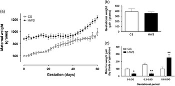
Four pregnant sows were included in the control group (CS) with a pre-gestational weight of 602±11 g and five in HWS group with 879±17 g (P<0.01). Time course of weight gain showed that total gestational weight gain was comparable between the two groups (Fig. 1a and 1b). However, CS showed a relatively constant increase in weight along gestation. In contrast, HWS had a depressed weight gain during the first two out of three of gestation and a brisk increase in the last third of gestation, relative to CS (Fig. 1a and 1c).
Maternal weight during gestation in control and obese guinea pigs. Time course of maternal weight gain (a), total gestational weight gain (b) and weight gain for each third of gestation (c) in control (CS, open circles or bars) and high weight (HWS, solid circles or bars) sows. *P>0.05, **P>0.01 v. CS, ANOVA.

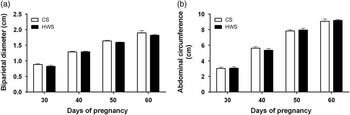
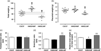
Biparietal diameter (Fig. 2a) and abdominal circumference (Fig. 2b) during gestation in HWSF were comparable with CS fetuses at every point studied. Similarly, at near term (60 days) mean fetal weight in HWS group (82.7±3.8 g) was comparable with CS fetuses (82.5±1.8 g, n=14). However, none of the HWSF showed a body weight in range of the confidence interval (78.6–86.4 g) of the control group (Fig. 3a). For further analysis HWS fetuses were classified according to their body weight as low (LWF, range 55.8–74.8 g) or high (HWF, range 88.7–107.7 g) regarding control group confidence interval. A retrospective analysis of fetal biometry during pregnancy considering this stratification in HWS showed a reduced abdominal circumference in LWF only at 40 days of gestation (Supplementary Fig. 1a) but no changes in the abdominal circumference growth rate (Supplementary Fig. 1b). LWF and HWF from HWS were present in a same litter and these characteristics were not associated to specific position in the uterus or horns. This was also observed in control sows as reported.Reference Turner and Trudinger 21
Pre-gestational weight and fetal biometry during gestation. Biparietal diameter (a) and abdominal circumference (b) in fetuses from control (CS, open bars) and high weight (HWS, solid bars) sows.

Pre-gestational weight and fetal characteristic at term. Fetal body weight (a), placental weight (b), as well as fetal to placental (c), brain to liver (d) and brain to body (e) weight ratios, in fetuses from control sows (CS/CF, open circles or bars), fetuses with high weight from obese sows (HWS/HWF, solid circles or bars) and fetuses with low weight from obese sows (HWS/LWF, grey circles or bars). *P>0.05, **P>0.01, † P<0.01 v. CS/CF, ANOVA.

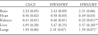
Placental weight was not altered in HWF (6.56±0.33 g) but reduced in LWF (4.60±0.27 g) compared with controls (6.14±0.23 g) (Fig. 3b), however, fetal to placental ratio was comparable among groups (Fig. 3c). Conversely, brain and heart weight were similar in the three fetal groups, whereas kidneys, liver and lungs weights were lower in LWF without significant changes in HWF compared with controls (Table 1). Furthermore the ratio between brain-to-liver weight, as well as brain-to-body weight, were increased in LWF (Fig. 3d–3e). In contrast, HWF have a decreased brain to body weight percentage relative to controls and LWF.
Fetal organ weights at near term

CS/CF, control fetus from control sows; HWS/HWF, high weight fetuses from high weight sows; HWS/LWF, low weight fetus from high weight sows.
Values expressed as mean (s.e.m.).
*P<0.05 v. CS/CF; **P<0.01 v. CS/CF, one-way ANOVA.
Umbilical artery blood flow and vascular reactivity
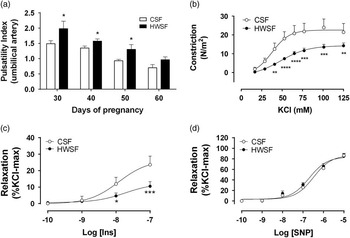
Umbilico–placental blood flow measured by Doppler velocimetry showed that pulsatility index (PI) was increased in HWSF at 30 (~1.3-fold increase), 40 (~1.2-fold increase) and 50 (~1.4-fold increase) days of gestation compared with controls (CSF) (Fig. 4a). At 60 days of gestation, there were no differences in the PI between control and HWS fetuses, however, LWF showed a substantial increase in umbilical artery PI (1.38±0.12 a.u.) compared with their HWF siblings (0.90±0.09 a.u.) and fetuses from control sows (0.70±0.10 a.u.) (Supplementary Fig. 2). Resistance index throughout gestation was comparable between HWSF and CSF with a subtle increase at 50 days (data not shown).
Pre-gestational weight and umbilico–placental reactivity. (a) Umbilical artery pulsatility index during gestation in control sows (CSF, open bars) and high weight sows (HWSF, solid bars) fetuses. Concentration dependent constriction to KCl (b) and relaxation to cumulative concentration of insulin (c) and sodium nitroprusside (SNP) (d) in umbilical arteries from fetuses from control sows (CSF, open circles) and fetuses with high weight from obese sows (HWSF, solid circles). *P>0.05, **P>0.01 v. CSF, ANOVA.

In order to determine the effects of high pre-gestational weight in the mother on umbilical arteries reactivity, analysis of vasoactive responses was carried out on arteries isolated from HWF and LWF. Ex vivo vascular reactivity of umbilical arteries was similar in HWF and LWF, therefore we decided to group these responses. Umbilical arteries from HWSF have a decreased contractile response to increasing concentration of KCl (14.3±1.0 N/m2) relative to CSF (22.6±2.2 N/m2) (Fig. 4b) without changes in the EC50. Conversely, umbilical arteries from guinea pig did not relaxed to acetylcholine (data not shown), but showed a NOS-dependent relaxing response to insulin as occurs in human umbilical arteries.Reference Krause, Prieto and Munoz-Urrutia 18 HWSF showed a reduced maximal relaxation (12.4±3.2%Kmax) without changes in the pD2 to insulin (7.72±0.45) (Fig. 4c) compared with controls fetuses (26.4±5.3%Kmax; pD2 7.92±0.33), and this response was completely inhibited by the NOS inhibitor L-NAME. In contrast, the response to exogenous NO (SNP) were comparable between the two groups (~85% Kmax) (Fig. 4d).
Femoral arteries vascular reactivity
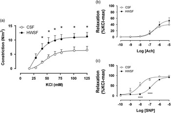
Similar to umbilical arteries, isolated femoral arteries from low and high fetal weight showed comparable ex vivo vascular responses, therefore we averaged them. HWSF showed an augmented contractile response to KCl (11.1±1.2 N/m2) (Fig. 5a) relative to CSF (6.3±0.7 N/m2) without changes in the EC50. However, the relaxation to acetylcholine in terms of maximal response (~50% Kmax) and pD2 (~6.5) was comparable between the two groups (Fig. 5b). Conversely, femoral arteries from HWSF showed a comparable maximal response (~90% Kmax), but a decreased pD2 to SNP (Fig. 5c) compared with CSF (CSF pD2=8.05±0.17, HWSF pD2=6.78±0.09, P<0.01 t-test).
Femoral artery vascular reactivity in large fetuses from obese sows. Concentration dependent constriction to KCl (a) and relaxation to cumulative concentration of acetylcholine (c) and sodium nitroprusside (SNP) (d) in femoral arteries from fetuses from control sows (CSF, open circles) and fetuses with high weight from obese sows (HWSF, solid circles). *P>0.05, **P>0.01 v. CS/CF, ANOVA.

Discussion
This study showed that overweight at conception and during gestation in guinea pigs is associated with altered fetal weight, and umbilical and systemic vascular impairment at term. In guinea pig, pre-gestational overweight induced increased risk of both small and large fetuses, with an asymmetric growth in small fetuses suggesting an IUGR. The HWS fetuses, independent of their growth, showed an increased umbilical artery PI during gestation, which normalized near term only in the large fetuses. These changes were accompanied by reduced ex vivo contractility and relaxing capacity in all the fetuses for HWS. In contrast, femoral arteries from HWSF, as examples of periphery arteries, showed increased contractile force with heterogeneous changes in the endothelial-dependent relaxation as well as the response of smooth muscle layer to NO. Altogether, this data show that maternal overweight in guinea pigs associates with altered placental blood flow that may associate with the vascular changes in systemic arteries.
In this study, increased pre-gestational weight was induced in female guinea pigs by a ~33% increase in total daily caloric intake without changes in nutrients composition, differing with other rodent models in which substantial increase in lipid or carbohydrates are applied.Reference Li, Reynolds, Sloboda, Gray and Vickers 9 – Reference Toop, Muhlhausler, O’Dea and Gentili 11 During pregnancy, total weight gain was comparable between control and HWS, however, the timing of weight gain differs among these groups. Thus, for the present model any alteration in fetal physiology would be related mainly to the pre-gestational maternal metabolic condition as occurs in human population in which controlling maternal weight gain during gestation in overweight/obese women has little effects on neonatal outcomes.Reference Yu, Han and Zhu 8 , Reference Santangeli, Sattar and Huda 22 , Reference King 23
Following the fetal growth by echography did not show differences between groups, but stratification of fetuses at term according to their weight showed that offspring from HWS had an altered fetal weight, either decreased or increased, relative to offspring from sows with normal weight before pregnancy. In the case of HWF, the differences in weight were not reflected at levels of specific organs suggesting an increased weight of the carcass, however, a further analysis is required to clarify a potential obesogenic body composition. On the other hand, in LWF from HWS, the increased brain to liver ratio and the altered umbilical artery Doppler suggest the presence of a fetal growth restriction. This effect could have been unnoticed in the fetal biometry follow-up owing to the inherent limitations of this technique,Reference Turner and Trudinger 21 but it could represent a potential early onset of IUGR as occurs in other models of maternal obesityReference Busso, Mascareno and Salas 15 in which fetal growth could be compromised at embryonic stages.Reference Luzzo, Wang and Purcell 14 Altogether, this data suggest that pre-gestational overweight in guinea pigs increases the risk of IUGR and macrosomia as occurs in human pregnancies.Reference King 23
There is no clarity about how increased pre-gestational BMI impacts on placental blood flow during gestation in human pregnancies. The most accepted view is that there are little effects on utero–placental blood flow as maternal BMI increases, particularly when associated with gestational diabetes.Reference Santolaya, Kahn, Nobles, Ramakrishnan and Warsof 24 , Reference Quintero-Prado, Bugatto and Sanchez-Martin 25 However, recent reports suggest that pre-pregnancy maternal obesity in humans associates with altered fetal cardiac function,Reference Ece, Uner and Balli 26 even at early stages of fetal development,Reference Ingul, Loras, Tegnander, Eik-Nes and Brantberg 27 which could be driven by impaired umbilico–placental perfusion. In fact, recently, it has been shown that there is a positive correlation between umbilical artery PI and maternal BMI in pregnancies.Reference Sarno, Maruotti and Saccone 28 The latter suggests that increased placental vascular resistance may be associated to maternal overweight and obesity. Similarly, in this study we found that increased maternal weight is associated with higher umbilical artery PI throughout gestation, which ultimately was manifested as a decreased ex vivo maximal relaxation to insulin. The altered umbilical PI was specially maintained in LWF up to term, clear evidence of umbilio–placental dysfunction. Furthermore, umbilical artery from HWS had a reduced contractile capacity but a normal response to exogenous NO, which could represent a compensatory mechanism to deal with an increased placental resistance resulting from endothelial dysfunction in chorionic vessels.Reference Schneider, Hernandez, Farias, Uauy, Krause and Casanello 7
Changes in the PI during gestation is a predictor of blood pressure at early infancy in a control population.Reference Khoury, Knutsen, Stray-Pedersen, Thaulow and Tonstad 29 Therefore, considering the changes in PI and vascular reactivity at umbilical level, we aimed to associate these effects in fetal femoral arteries. Femoral arteries from HWS fetuses showed an increased contractile capacity, but a lower sensitivity to NO without changes in the response to acetylcholine. Several studies have shown that maternal overweight/obesity and excessive gestational weight gain is associated with increased risk for later cardiovascular diseases in the offspring.Reference Santangeli, Sattar and Huda 22 , Reference Fraser, Tilling and Macdonald-Wallis 30 This effect has been mainly attributed to the increased adiposity and metabolic complications that subjects develop during their early infancy. In fact, non-human primates born from mothers exposed to high-fat diets have an increased intima-media thickness and impaired endothelial function in the aorta at 1 year of age. Interestingly, these aortic impairments may be substantially reversed by modifications in the post-natal diet.Reference Fan, Lindsley and Comstock 31 In contrast, a rat model of increased maternal adiposity during pregnancy shows impaired endothelial function in mesenteric arteries in the offspring, independent of the body weight at adult age,Reference Gray, Vickers, Segovia, Zhang and Reynolds 32 suggesting that the vascular dysfunction in the offspring from obese mothers has an independent origin. In this context, it has been shown that in human neonates the aortic intima-media thickness positively correlates with the maternal BMI,Reference Begg, Palma-Dias, Wang, Chin-Dusting and Skilton 33 a fact that could be related with the increased vascular risk in the offspring. At the best of our knowledge, in this study we showed for the first time that increased maternal weight before gestation induces changes in the vascular reactivity of fetal systemic vessels at term, with a heterogeneous effect on endothelial NOS and NO-dependent responses but having with a consistent increased contractile force as a hallmark. Altogether, these data support the concept that the cardiovascular risk in the offspring born from mothers with pre-gestational obesity has its origins in utero.
In conclusion, this study shows that altered fetal growth in pregnancies affected by increased pre-gestational weight take place under a reduced fetal–placental blood flow and associates with an altered endothelial and vascular function in the fetal umbilical and systemic circulation. We proposed that the increased placental vascular resistance along with the augmented nutrients supply generate the fetal hypoxic-like characteristic of these pregnancies,Reference Dollberg, Marom, Mimouni and Yeruchimovich 34 , Reference Sheffer-Mimouni, Mimouni and Dollberg 35 which is an important mechanism involved in the programming of vascular function.Reference Camm, Hansell and Kane 36 , Reference Giussani and Davidge 37
Acknowledgment
None.
Financial Support
This work was supported by Fondecyt Chile (P.C., grant number 1120928), (M.F., grant number 1121145), (B.J.K., grant number 1130801), (E.A.H., grant number 1151119).
Conflicts of Interest
None.
Ethical Standards
The procedures and animal handling were approved by the Ethics Committee of the Faculty of Medicine of the University of Chile (CBA 694 FMUCH) and the Faculty of Medicine of Pontificia Universidad Catolica de Chile (CEBA-MedUC 1130801). The studies on animals were performed according with the ARRIVE guidelines, The Guide for the Care and Use of Laboratory Animals published by the US National Institutes of Health (NIH Publication No. 85–23, revised 1996) and adheres to APS’s Guiding Principles in the Care and Use of Animals.
Supplementary Material
To view supplementary material for this article, please visit http://dx.doi.org/10.1017/S2040174415007266