INTRODUCTION
The family Gempylidae belongs to the suborder Scombroidei and consists of 16 genera and 23 species, distributed mainly in tropical and temperate seas (Nakamura & Parin, Reference Nakamura and Parin1993). The oilfish Ruvettus pretiosus (Cocco, 1829) belongs to the monotypic genus Ruvettus and is a benthopelagic species widely distributed throughout the tropical and temperate waters of the world's oceans (Nakamura & Parin, Reference Nakamura and Parin1993; Pakhorukov & Boltachev, Reference Pakhorukov and Boltachev2001). It is also the only gempylid found in the Mediterranean (Nakamura & Parin, Reference Nakamura and Parin1993). Ruvettus pretiosus is the largest representative of the family reaching 2 m in length and over 50 kg in weight; however, individuals under 150 cm and 30 kg in weight are more common (Pakhorukov & Boltachev, Reference Pakhorukov and Boltachev2001).
Oilfish individuals dwell over the continental slope and seamounts mainly at depths from 100 to 700 m (Nakamura & Parin, Reference Nakamura and Parin1993), reaching depths of more than 1000 m (Pakhorukov & Boltachev, Reference Pakhorukov and Boltachev2001). At night, parts of the species populations perform feeding vertical migrations to the epipelagic zone (Pakhorukov & Boltachev, Reference Pakhorukov and Boltachev2001).
Fisheries activity targeting R. pretiosus is performed in only a few marine areas of the Indo-Pacific mainly by small scale fishing fleets using handlines (Gudger, Reference Gudger1928; Stobbs & Bruton, Reference Stobbs and Bruton1991; Koslow et al., Reference Koslow, Boehlert, Gordon, Haedrich, Lorance and Parin2000). However, oilfish specimens also appear as an incidental by-catch in numerous drifting surface longline fisheries targeting Thunnidae and swordfish: in the Indo-Pacific (e.g. He et al., Reference He, Bigelow and Boggs1997; Francis et al., Reference Francis, Griggs and Baud2004; Bromhead et al., Reference Bromhead, Ackerman, Graham, Wight, Wise and Findlay2005), in the Atlantic (e.g. Castro et al., Reference Castro, de la Serna, Macías and Mejuto2000; Mejuto et al., Reference Mejuto, García-Cortés and de la Serna2002) and in the Mediterranean (e.g. Castro et al., Reference Castro, de la Serna, Macías and Mejuto2000; Mejuto et al., Reference Mejuto, García-Cortés and de la Serna2002; Tserpes et al., Reference Tserpes, Tatamanidis and Peristeraki2006; Peristeraki et al., Reference Peristeraki, Kypraios, Lazarakis and Tserpes2007). In the eastern Mediterranean, R. pretiosus is the second most abundant species caught by drifting surface longline fleets targeting swordfish both in terms of weight and in terms of frequency, but it is still not often caught; Peristeraki et al. (Reference Peristeraki, Kypraios, Lazarakis and Tserpes2007) report a catch per unit of effort of 1.7 individuals/1000 hooks. Moreover, the species comprises an incidental by-catch of demersal longline and gill net fisheries targeting hake in the north-eastern Atlantic (Erzini et al., Reference Erzini, Goncalves, Bentes, Lino and Ribeiro2001; Santos et al., Reference Santos, Gaspar, Monteiro and Vasconcelos2002) and it is also occasionally caught by other gears such as demersal trawls (Elbaraasi et al., Reference Elbaraasi, Elmariami, Elmeghrabi and Omar2007) and purse seines (Romanov, Reference Romanov2002).
Despite its cosmopolitan distribution and appearance as by-catch in a broad range of marine areas, published information about the biology of R. pretiosus is very scarce. The majority of the studies conducted concern biochemical analyses (e.g. Cox & Reid Reference Cox and Reid1932; Nevenzel et al., Reference Nevenzel, Rodegker and Mead1965; Nevenzel, Reference Nevenzel1970; Bone, Reference Bone1972; Ruiz-Guttierez et al., Reference Ruiz-Gutierrez, Perez-Zarza, Muriana and Bravo1997; Nichols et al., Reference Nichols, Mooney and Elliott2001) revealing very high lipid concentrations—mainly in the form of wax esters—in oilfish tissues; hence the common name of the species. As a result, oilfish flesh has purgative properties if eaten in large quantities (Nakamura & Parin, Reference Nakamura and Parin1993; Shadbolt et al., Reference Shadbolt, Kirk and Roche2002; Leask et al., Reference Leask, Yankos and Ferson2004). Other studies describe some morphological characteristics of the species (Gudger & Mowbray, Reference Gudger and Mowbray1927; Bone, Reference Bone1972; Nakamura & Parin, Reference Nakamura and Parin1993) and provide information about its spatial and vertical distribution (e.g. Gudger, Reference Gudger1928; Pakhorukov & Boltachev, Reference Pakhorukov and Boltachev2001; Tserpes et al., Reference Tserpes, Tatamanidis and Peristeraki2006; Damalas & Megalofonou, Reference Damalas and Megalofonou2007) and behaviour (Bone, Reference Bone1972; Pakhorukov & Boltachev, Reference Pakhorukov and Boltachev2001).
To our knowledge there are no systematic studies on the diet and reproduction of the species. There are a few studies where some stomach contents have been reported, including fish, cephalopods and crustaceans (Nakamura & Parin, Reference Nakamura and Parin1993; Yamamura, Reference Yamamura1997; Pakhorukov & Boltachev, Reference Pakhorukov and Boltachev2001); while sex and maturity status were identified in a few specimens by Pakhorukov & Boltachev (Reference Pakhorukov and Boltachev2001). In this study we investigate the diet and we provide for the first time data on the reproductive maturity of R. pretiosus in the eastern Mediterranean, from specimens caught by the Greek fishing fleets.
MATERIALS AND METHODS
Sample collection
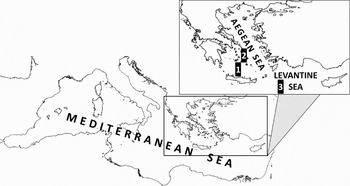
During the period from October 2007 to July 2008, 50 Ruvettus pretiosus specimens caught by Greek fishing boats targeting swordfish (Xiphias gladius) or hake (Merluccius merluccius) were collected and examined. The fishing gears used were respectively drifting surface longlines or demersal longlines. The specimens were caught in the Cretan Sea, the central Aegean Sea and the Levantine Sea (Figure 1).
Map of the eastern Mediterranean showing the marine areas from where oilfish specimens were sampled. (1) Cretan Sea; (2) Central Aegean Sea; (3) Levantine Sea.

Drifting surface longlines operated in the open sea, fishing at depths of 10–20 m from the surface during the night. This time period coincides with the vertical migrations of R. pretiosus specimens to the epipelagic zone (Pakhorukov & Boltachev, Reference Pakhorukov and Boltachev2001). Demersal longlines operated close to the sea bottom (depths 250–550 m) during the whole day, in areas with mostly muddy substrates and gentle slopes which form a suitable habitat for R. pretiosus (Pakhorukov & Boltachev, Reference Pakhorukov and Boltachev2001). In both fishing gears, the baits used were a variety of fish and cephalopod species.
The specimens of R. pretiosus were collected either on-board (eight specimens) or from landings (42 specimens). In the first case, total length (TL) and head length (HL) were measured, sex was identified, gonad samples were stored in 3.7% buffered formalin and stomachs were stored in deep freeze (–20oC) for later laboratory analysis. In the case of the specimens sampled from landings, only the head, gonads and stomachs were kept in deep freeze (–20oC) by the fishermen, since those body parts were removed on-board and they would be otherwise discarded. Deep frozen heads, gonads and stomachs supplied by the fishermen were transferred to deep freezing facilities ashore for later laboratory analysis. In the laboratory, deep frozen gonads were thawed, sex was identified and gonad samples were stored in 3.7% buffered formalin for later histological analysis. Preserving the stomachs in deep freezing conditions prior to the stomach content analysis (e.g. Chancollon et al., Reference Chancollon, Pusineri and Ridoux2006; Potier et al., Reference Potier, Marsac, Cherel, Lucas, Sabatié, Maury and Ménard2007) aimed to minimize the decrease in quality of the stomach contents and it also allowed identification and measurements of prey items to take place in a controlled environment.
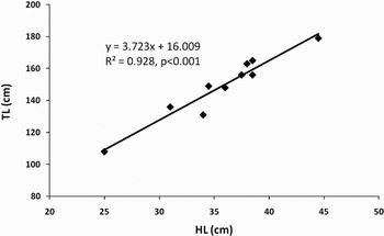
Since the majority of oilfish came from landings—hence no TLs were available—we transformed the HLs to TLs by constructing a TL–HL equation using data from the eight specimens collected on-board for this study and from two other specimens caught in the Mediterranean which were described in the literature (Bettoso & Dulcic, Reference Bettoso and Dulcić1999; Elbaraasi et al., Reference Elbaraasi, Elmariami, Elmeghrabi and Omar2007).
Sex identification was possible in 47 out of 50 specimens, as in three cases the gonads were not supplied by the fishermen. The sex-ratio of the specimens caught with surface and demersal longlines was statistically compared to a 1:1 ratio using a Chi-square (χ2) analysis.
Stomach content analysis
In order to study the diet of R. pretiosus, deep frozen stomachs were thawed in the laboratory, the number of intact empty and full (those containing at least one trophic object) stomachs was counted and stomach content analysis was performed in the latter ones. Only 30 out of the 50 specimens caught had an intact stomach; the remaining 20 stomachs were partially or fully ripped by the fishermen's handling.
Two methods of stomach content analysis were used: percentage numerical abundance and percentage occurrence (Hyslop, Reference Hyslop1980; Hernandez-Garcia, Reference Hernandez-Garcia1995; Chancollon et al., Reference Chancollon, Pusineri and Ridoux2006). A coefficient of prey numerical frequency, %No = (Ni/ Nt) × 100, and a coefficient of prey occurrence frequency, %O = (Nsi/Nsf) × 100 were calculated; where Ni was the number of prey of trophic group i, Nt was the total number of prey estimated as the smallest number from which all fragments could have originated (Cortez et al., Reference Cortez, Castro and Guerra1995), Nsi was the number of stomachs containing each group i, and Nsf was the total number of stomachs containing food, each stomach being counted as many times as the number of different types of prey it contained (Cortez et al., Reference Cortez, Castro and Guerra1995). An index of prey numerical importance (Castro & Santana del Pino, Reference Castro and Santana del Pino1995; Hernandez-Garcia, Reference Hernandez-Garcia1995) was also obtained as %I = (%No × %O)1/2 × 100.
Histological analysis
For the study of the reproduction of R. pretiosus, the gonads from 47 specimens (42 females and 5 males) were examined histologically. Pieces of gonad tissues were dehydrated, cleared in xylol and embedded in paraffin. Sections (5 µm) were made and stained with Mayer's haematoxylin and eosin Y (Clark, Reference Clark1981).
The ovaries were classified in a total of six histological stages and the testes were classified in two histological stages. Histological scoring in females was according to the maturation stage of the most advanced type of oocytes observed (West, Reference West1990). The diameters of at least 100 oocytes from each stage sectioned through their nucleus were measured in order to calculate the average oocyte diameter in each stage. Oocyte diameters were measured in sections coming from gonad samples collected on board except from the last two histological stages where only deep frozen gonads were available.
RESULTS
Total length–head length equation
The TL–HL equation constructed for Ruvettus pretiosus (Figure 2) was derived from data on a range of head lengths between 25.0 and 44.5 cm. While, as mentioned, only 10 points were used, the range of these head lengths covered the range of all specimens of this study. Based on this equation all head lengths were transformed to total lengths.
Total length (TL)–head length (HL) relationship for Ruvettus pretiosus.

Stomach fullness
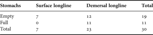
A high percentage (63.3%) of the specimens examined had empty stomachs. All the specimens caught by surface longline and 52.0% of the specimens caught by demersal longline had empty stomachs (Table 1).
Number of empty and full oilfish stomachs caught by different fishing gears.
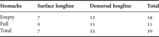
Empty, no stomach contents; full, at least one trophic object.
Stomach contents analysis
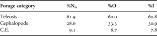
The diet of R. pretiosus consisted mainly of fish and cephalopods (Table 2). At least 13 distinct specimens of teleost fish were recorded, thus teleosts were the most important item both by number (61.9%) and occurrence (60.0%). Many fish remnants consisted of single spines, spinal columns and scales and, therefore, could not be attributed to specific species due to extended digestion. No otoliths were detected. Species that could be identified were Merluccius merluccius (Gadiformes, Merluciidae) (4 fish), Lepidopus caudatus (Perciformes, Trichiuridae), Conger conger (Anguilliformes, Congridae) and a macrourid specimen. Two chondricthyan eggs were also found in one stomach.
Comparison of the dietary importance of the major forage categories observed in oilfish stomachs. Data are given in terms of numerical frequency (%N), frequency of occurrence (%O) and index of numerical importance (%I).
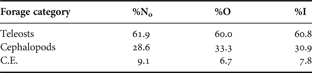
C.E., chondricthyan eggs.
At least six distinct specimens of cephalopods were detected. These were recorded in 33.3% of the stomachs sampled and represented 28.6% of the total trophic objects. In most cases only the beaks and eye lenses remained in the stomachs except in one case where the remnants allowed the identification of an individual belonging to the genus Todarodes.
Sex-ratio
Ruvettus pretiosus was found to be a gonochoristic species and no signs of intermediate or sex reversed gonads were observed. Testes had a lighter colour (white) than ovaries (pink) and had distinctive sperm ducts on both lobes.
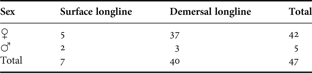
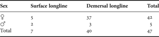
Sex was identified in 47 specimens. Of the 47 fish sexed, 42 (89.4%) were female and five (10.6%) were male. The overall ratio of males to females was 1:8.4 (Table 3). This ratio was 1:2.5 for fish caught with surface longline while it was 1:12.3 for fish caught with demersal longline (Table 3). The sex-ratio of specimens coming from the surface longline did not differ significantly from 1:1 (χ2 = 0.075; df = 1; P = 0.784), however the sample was small. On the contrary, the sex-ratio of specimens caught with demersal longline differed significantly from 1:1 (χ2 = 15.622; df = 1; P < 0.001).
Sex of the specimens sampled by each fishing gear.
Reproductive biology
FEMALES
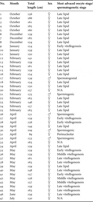
Regarding the macroscopic morphology of R. pretiosus female gonads, from October to February (Table 4) ovarian lobes were large, flaccid and bared a large cavity; hence they were characterized as ‘spent’ (West, Reference West1990). Gonads from specimens caught from April to June were more firm and robust. There was neither difference in the morphology or weight between the two lobes nor difference between the maturity stage of the oocytes coming from the posterior, middle and anterior parts of the lobe.
Sampling period, individual total length derived from head length, sex and most advanced oocyte stage (females) or spermatogenetic stage (males).

N/A, no histological analysis performed due to poor gonad condition.
At the histological sections, all the oocytes appeared positioned on ovigerous lamellae of connective tissue. Since there had been no previous histological staging of the oocytes in R. pretiosus, within this study six maturity stages were characterized.
Primary developmental stage
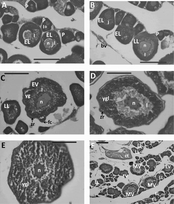
Perinucleolar stage: (oocyte diameter range: 23–9 µm, average: 44 µm) (Figure 3A, B): the perinucleolar-stage oocytes were polyedric with a high nucleus:cytoplasm ratio. During this stage large lipid droplets occurred in the cytoplasm. The nucleus displayed a small number (1–3) of nucleoli and there was intense ooplasm basophily.
Micrographs of sections (5 µm) of Ruvettus pretiosus ovaries displaying oocytes at different stages of development. Stain: haematoxylin and eosin. (A) Oocytes at perinucleolar and early lipid stage; (B) oocytes at perinucleolar, early lipid stage and late lipid stage showing differences in lipid dispersal among stages; (C) oocytes at late lipid and early vitellogenesis stage; (D) oocyte at middle vitellogenesis stage with yolk globules and a thick zona radiata; (E) oocyte at late vitellogenesis stage with large yolk globules which have covered the whole ooplasm; (F) oocytes at early lipid, late lipid, early vitellogenesis and middle vitellogenesis stage. P, perinucleolar stage; EL, early lipid stage; LL, late lipid stage; EV, early vitellogenesis stage; MV, middle vitellogenesis stage; l, lipids; n, nucleus; ct, connective tissue; bv, blood vessel; yg, yolk granules; ygl, yolk globules; zr, zona radiata; fc, follicle cells. Scale bars = 100 µm (A–E); 200 µm (F).

Early lipid stage: (oocyte diameter range: 54–97 µm, average: 70 µm) (Figure 3B): the oocytes classified at the early lipid stage exhibited a distinct area with lipids in their ooplasm close to the nucleus. At this stage the oocytes appeared larger due to an increase of the ooplasm and they had more nucleoli (5–7) adjoining the nuclear envelope.
Late lipid stage: (oocyte diameter range: 68–161 µm, average: 106 µm) (Figure 3B, C). At this stage, the lipid droplets which were previously concentrated in a distinct area close to the nucleus spread into the ooplasm encircling the nucleus. The ooplasm grew in size and became less basophilic while the size of the nucleoli reduced and their number increased.
Secondary developmental stage
Early vitellogenesis stage: (oocyte diameter range: 102–194 µm, average: 140 µm) (Figure 3C). At this stage an acidophilic zone began to emerge at the periphery of the ooplasm which was formed by small acidophilic granules (yolk granules). In addition, a thin acidophilic zona radiata appeared around the ooplasm and it was encircled by follicle cells. The lipids of the ooplasm continued to increase in size, reducing its basophily.
Middle vitellogenesis stage: (oocyte diameter range: 169–272 µm, average: 211 µm) (Figure 3D). At the middle vitellogenesis stage the acidophilic yolk globules had been formed and they appeared at the periphery of the ooplasm, while the basophily of the ooplasm was further reduced due to an increase of lipid concentration. The lipid droplets were visible only at the area around the nucleus which was the only area that had not been filled with yolk globules. At this stage, the oocyte size increased significantly and zona radiata appeared thicker.
Late vitellogenesis stage: (oocyte diameter range: 224–366 µm, average: 290 µm) (Figure 3E). The oocytes at this advanced stage of vitellogenesis displayed a great increase in both size and number of acidophilic yolk globules which covered the entire ooplasm. The oocyte size increased and zona radiata appeared thicker.
Within this study it was not possible to collect individuals possessing gonads at more advanced maturity stages (i.e. nucleus migratory stage, pre-hydrated stage and hydrated stage). Furthermore, apart from the earlier described oocytes, some atretic forms were also observed.
MALES
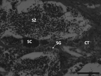
Histological examination of the five male specimens showed that the individual caught in early February had spent gonads, while the four sampled between late February and April were in the spermiogenic phase (Table 4), bearing seminiferous tubules where most stages of sperm development were visible (spermatogonia, spermatocytes and spermatozoa) (Figure 4).
Seminiferous tubules of a male Ruvettus pretiosus at spermiogenic phase (bar = 50 µm). Stain: haematoxylin and eosin. Section: 5 µm. CT, connective tissue; SG, spermatogonium; SC, spermatocytes; SZ, spermatozoa.

DISCUSSION
The frequency of empty stomachs observed in Ruvettus pretiosus specimens in this study (63.3%) is very large compared to that typically observed in other carnivorous large pelagic Scombroidei such as swordfish (Xiphias gladius) and albacore (Thunnus alalunga), in the same Mediterranean area (Peristeraki & Tserpes, Reference Peristeraki, Tserpes and Tsimenides2001) or in other marine regions (Chancollon et al., Reference Chancollon, Pusineri and Ridoux2006; Potier et al., Reference Potier, Marsac, Cherel, Lucas, Sabatié, Maury and Ménard2007). The frequency of empty stomachs in the above mentioned studies is usually less than 20%. This is probably a result of the species less active mode of life (Bone, Reference Bone1972; Pakhorukov & Boltachev, Reference Pakhorukov and Boltachev2001) or might be also related to the significant amount of stored energy (Arrington et al., Reference Arrington, Winemiller, Loftus and Akin2002) illustrated by the high concentration of lipids (especially wax esters) in oilfish tissues (Nevenzel et al., Reference Nevenzel, Rodegker and Mead1965; Bone, Reference Bone1972; Ruiz-Guttierez et al., Reference Ruiz-Gutierrez, Perez-Zarza, Muriana and Bravo1997; Nichols et al., Reference Nichols, Mooney and Elliott2001). Pakhorukov & Boltachev (Reference Pakhorukov and Boltachev2001) also have reported that only part of the species population performs nocturnal vertical trophic migrations, while the rest remains close to the sea bottom. Furthermore, they observed predatory activity in both epipelagic and benthopelagic zones. In the current study, empty stomachs were observed in all specimens caught in the epipelagic zone, while this was the case for only about half of the specimens caught by demersal longlines at depths between 250 and 550 m (Table 1). In the latter specimens, benthic and benthopelagic preys were identified in the non-empty stomachs. Consequently, it can be speculated that the individuals performing nocturnal vertical trophic migrations—which are very energetically demanding since the nocturnal feeding habitats might occur several hundred meters higher than the diurnal habitats—are generally those that were not able to cover their dietary needs in the benthopelagic zone. However, the small number of specimens sampled from the epipelagic zone does not allow us to conclude on such a hypothesis. Pakhorukov & Boltachev (Reference Pakhorukov and Boltachev2001) also reported mesopelagic prey items in the stomachs of R. pretiosus specimens caught by mesopelagic trawls during the night. In the current study such prey items were not identified, since all specimens caught by surface longlines had empty stomachs.
Stomach content analysis showed that R. pretiosus specimens in the eastern Mediterranean are mostly ichthyophagous (Table 2) consuming also cephalopods, similarly to many other gempylid species (Nakamura & Parin, Reference Nakamura and Parin1993). Those findings are in agreement with fishermen's claims that oilfish are better caught on hooks baited with fish rather than squids. Nakamura & Parin (Reference Nakamura and Parin1993) also include crustaceans among the prey items of R. pretiosus. However, no crustacean remains were identified in the stomachs analysed within the current study.
Sex has been macroscopically identified and classified as ‘male’ or ‘female’ in R. pretiosus specimens in previous studies (Gudger & Mowbray, Reference Gudger and Mowbray1927; Pakhorukov & Boltachev, Reference Pakhorukov and Boltachev2001), but this is the first time according to our knowledge that the gonochoristic reproductive strategy of the species is confirmed by histological methods. Gonochoristic reproductive strategy has been also observed in all other benthopelagic and mesopelagic gempylid species (Rowling & Reid, Reference Rowling and Reid1992; Nakamura & Parin, Reference Nakamura and Parin1993; Lorenzo & Pajuelo, Reference Lorenzo and Pajuelo1999; Griffiths, Reference Griffiths2002) investigated to date.
The higher abundance of female oilfish specimens caught by demersal longlines in depths from 250 to 550 m (Table 3) could be attributed to differences in the bathymetric distribution between males and females. Lorenzo & Pajuelo (Reference Lorenzo and Pajuelo1999) reported that the sex-ratio in roudi escolar Promethichthys prometheus—another gempylid species with similar ecology to that of the oilfish—varied between 1:6.5 to 1:5.3 in favour of females in depths of 200–500 m, while being 1:0.63 to 1:0.37 in favour of males in depths of 600–900 m. In addition, Griffiths (Reference Griffiths2002) in his study on snoek (Thyrsites atun)—another benthopelagic gempylid species—observed in many areas more females in inshore shallow waters and more males in offshore deep waters during the spawning season. Griffiths (Reference Griffiths2002) attributed this distribution pattern to metabolic processes, i.e. easier access to trophic resources for females during the energetically demanding spawning season. The above mentioned studies support the assumption that there may be different bathymetric distributions for male and female R. pretiosus specimens. This difference could also explain why the clearly unbalanced sex-ratio of specimens caught in the benthic zone does not seem to exist when considering the oilfish caught in the epipelagic zone; specimens of both genders are expected to forage close to the surface at night when they are not able to cover their dietary needs in the benthic zone. This difference in bathymetric distribution could be attributed to energetic reasons, as proposed by Griffiths (Reference Griffiths2002) for snoek. As Lorenzo & Pajuelo (Reference Lorenzo and Pajuelo1999) also proposed for roudi escolar, the observed space partitioning between sexes, which makes females more accessible to demersal longline fisheries than males, could potentially make oilfish populations more vulnerable to unrestrained fishing compared to species without this characteristic.
During the winter period (October–February) the most advanced oocytes in all but one female were at the late lipid stage, i.e. resting stage (Table 4) (Forberg, Reference Forberg1982). The first specimens with early vitellogenetic oocytes appear in April while during May and June the oocytes reach the middle and late vitellogenesis stages (Table 4). According to those results we can assume that the reproductive period occurs during the summer months, possibly peaking around July/August. A similar reproductive period has been observed for roudi escolar (Lorenzo & Pajuelo, Reference Lorenzo and Pajuelo1999).
During the period from May to September, cooperating fishermen tend to switch their fishing effort from the benthic to the epipelagic zone, thus limiting the availability of R. pretiosus specimens from their diurnal habitats. In addition, during July–August the presence of R. pretiosus specimens in surface longlines in the studied area is lower (Vasilakopoulos et al., Reference Vasilakopoulos, Tzanatos and Tserpes2009), suggesting that the species may perform migrations in specific spawning grounds. Therefore, within the current study we were unable to collect specimens from the period of July to September to identify the oocyte maturation stages.
The presence in oilfish ovaries of oocytes from many different developmental stages during May to June (Figure 3F) indicates that the species is a multiple spawner with either asynchronous or group-synchronous oocyte development pattern. The exact type of oocyte development pattern has to be confirmed in the future by checking the oocyte diameter distribution in specimens closer to the spawning time (Murua & Saborido-Rey, Reference Murua and Saborido-Rey2003). Nevertheless, histological studies on other gempylid and also trichiurid and scombrid species have shown that the asynchronous pattern generally prevails among those fish (Martins & Haimovici, Reference Martins and Haimovici2000; Griffiths, Reference Griffiths2002; Murua & Saborido-Rey, Reference Murua and Saborido-Rey2003; Abascal & Medina, Reference Abascal and Medina2005).
The oocyte maturity stages that we identified within the current study generally agree with the classification suggested by Corriero et al. (Reference Corriero, Desantis, Deflorio, Acone, Bridges, De La Serna, Megalofonou and De Metrio2003) for bluefin tuna. The only differences are in regard to the division of the lipid stage into two stages instead of one and the division of the vitellogenetic into three stages rather than two. The latter categorization has been also proposed for the amberjack (Seriola dumerili) (Grau et al., Reference Grau, Crespo, Riera, Pou and Carmen Sarasquete1996).
A distinctive morphological characteristic of the oilfish oocytes is the presence of several lipid droplets as early as the perinucleolar stage. Several perinucleolar oocytes also displayed a large lipid droplet with similar size to that of the nucleus (Figure 3A). In most other large pelagic species the accumulation of lipids in the ooplasm commences later; during the lipid/previtellogenic stage (Grau et al., Reference Grau, Crespo, Riera, Pou and Carmen Sarasquete1996; Arocha, Reference Arocha2002; Corriero et al., Reference Corriero, Desantis, Deflorio, Acone, Bridges, De La Serna, Megalofonou and De Metrio2003). The reason for this early and intense accumulation of lipids in the species oocytes is unknown but it might be related to the particularly high presence of lipids in all its tissues (Nevenzel et al., Reference Nevenzel, Rodegker and Mead1965; Bone, Reference Bone1972; Ruiz-Guttierez et al., Reference Ruiz-Gutierrez, Perez-Zarza, Muriana and Bravo1997; Nichols et al., Reference Nichols, Mooney and Elliott2001).
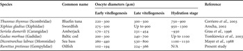
Another significant characteristic of the species oocytes is their relatively small size, indicating that oifish spawn pelagic eggs. Comparing the oocyte size calculated for R. pretiosus in our study with the oocyte size of other large pelagic or benthopelagic species that also produce pelagic eggs, the inferior size of the former is obvious (Table 5). Even if we take into account the small shrinkage of the oocytes during the preparation of the histological sections (West, Reference West1990), R. pretiosus is expected to spawn eggs with a diameter of about 1 mm at most. It is also noted that diameters of middle and late vitellogenetic oocytes were measured in histological sections of initially deep frozen gonads sampled from landings. Deep freeze tends to increase oocyte size (Klibansky & Juanes, Reference Klibansky and Juanes2007), therefore oocyte diameters of middle and late vitellogenetic oocytes might be slightly overestimated. However, no obvious differences in oocyte size were observed in the earlier maturity stages between gonads sampled on-board and from landings, therefore this could be also the case for middle and late vitellogenetic oocytes.
Oocyte diameters as measured in histological sections at early and late vitellogenesis and hydration stage for some large pelagic and benthopelagic species with pelagic eggs.

N/A, not available.
The relatively small egg size of the oilfish compared to other large pelagic and benthopelagic species could be balanced by a possible higher caloric value due to the high presence of lipids that we observed (Kamler, Reference Kamler2005). Spawning great numbers of small-sized eggs can be attributed to an evolutionary pressure such as great and stochastic egg/larvae mortality (Kolm & Ahnesjo, Reference Kolm and Ahnesjo2005) which could be related to the fact that oilfish possibly spawn in depths of several hundred metres, as observed for other gempylids with similar ecology (Lorenzo & Pajuelo, Reference Lorenzo and Pajuelo1999), and its fry occurs in the surface layers (Shcherbachev et al., Reference Shcherbachev, Parin, Pakhorukov and Piotrovskii1986 in Pakhorukov & Boltachev, Reference Pakhorukov and Boltachev2001). Therefore, small egg size probably serves the quicker dispersal and movement to the epipelagic zone and is related to the high mortality that the eggs/larvae are expected to experience. It is also noted that egg diameters of approximately 1 mm or smaller are typically observed in small bathypelagic species (Marshall, Reference Marshall1953).
Finally, the histological examination of the species male gonads showed that they resemble those of other phylogenetically related large pelagic species, such as the swordfish (Corriero et al., Reference Corriero, Desantis, Bridges, Kime, Megalofonou, Santamaria, Cirillo, Ventriglia, Di Summa, Deflorio, Campobasso and De Metrio2007), since generally spermatogenesis is highly conserved among the different fish species (Nobrega et al., Reference Nóbrega, Batlouni and Franca2009). In addition, we observed mature male specimens much earlier in the year than the expected spawning period, which is in accordance with results obtained for other Scombroidei such as swordfish (Young et al., Reference Young, Drake, Brickhill, Farley and Carter2003) and Pacific bluefin tuna (Sawada et al., Reference Sawada, Seoka, Kato, Tamura, Nakatani, Hayashi, Okada, Tose, Miyashita, Murata and Kumai2007).
In general, biological studies for fish species that are not targeted by some specialized fisheries but appear only as incidental by-catches are difficult to conduct due to the low availability and/or quality of samples. In this study, the small number of individuals caught during the on-board sampling forced us to use landed specimens for which TL was indirectly estimated from the available HL measurements. This however did not affect stomach content and gonad analysis and the information provided clarifies some basic biological aspects of a poorly studied species. Such information could potentially have a higher significance for fisheries in the future as the overexploitation of traditional fish stocks worldwide leads to increasing interest in new fisheries resources; both by profiting better from the by-catches and by switching fishing effort to deeper waters (Pieiro et al., Reference Pieiro, Casas and Baón2001). Experimental fishing targeting R. pretiosus that would focus on collecting specimens from the species diurnal and nocturnal habitats throughout the year could contribute further valuable information about the species diet and reproduction, in addition to other biological and ecological aspects such as growth and distribution.
ACKNOWLEDGEMENTS
We thank the captains and the crews of the fishing vessels ‘Fountoulakis’ and ‘Popi’ for their assistance in sample collection. Many thanks also to P. Peristeraki and Dr S. Somarakis for their advice on the stomach content analysis and oocyte staging respectively, and to L. McPherson and Dr E. Tzanatos for their helpful comments on earlier versions of this manuscript. Thanks are also due to an anonymous referee for providing valuable comments and suggestions. This study has been carried out with the financial assistance of the European Commission, in the frames of the STREP Project ‘EFIMAS’ (Contract No. SSP8-CT-2003-502516). P.V. was also supported by a scholarship from the Greek Public Welfare Foundation ‘Propondis’.