Introduction
Strongyloidiasis is caused by parasitic soil-transmitted helminths (STH) of the genus Strongyloides, of which the most common is Strongyloides stercoralis. This species is widely distributed throughout tropical and temperate regions of the world but detailed disease prevalence maps are lacking (Liu & Weller, Reference Liu and Weller1993). Today, strongyloidiasis is of growing public health importance and is now grouped with the so-called neglected tropical diseases (NTDs) (WHO, 2002, 2008). From a global perspective, and due to lack of precise epidemiological data, the exact number of people living with strongyloidiasis is not presently known, but it is estimated to be between 30 million and 100 million or higher (Siddiqui & Berk, Reference Siddiqui and Berk2001).
Many aspects of strongyloidiasis are poorly understood and comparing cross-sectional studies between geographical regions can be problematic (Olsen et al., Reference Olsen, van Lieshout, Marti, Polderman, Polman, Steinmann, Stothard, Thybo, Verweij and Magnussen2009). This is mainly due to the low sensitivity of the currently available diagnostic tests (Steinmann et al., Reference Steinmann, Zhou, Du, Jiang, Wang, Wang, Li, Marti and Utzinger2007; Stothard et al., Reference Stothard, Pleasant, Oguttu, Adriko, Galimaka, Ruggiana, Kazibwe and Kabatereine2008; Agrawal et al., Reference Agrawal, Agarwal and Ghoshal2009; Knopp et al., Reference Knopp, Mohammed, Rollinson, Stothard, Khamis, Utzinger and Marti2009) and scarcity of targeted epidemiological surveys, making strongyloidiasis one of the most poorly described NTDs from a public health perspective (Olsen et al., Reference Olsen, van Lieshout, Marti, Polderman, Polman, Steinmann, Stothard, Thybo, Verweij and Magnussen2009). Moreover, the use of the Baermann concentration and Koga agar plate methods (Koga et al., Reference Koga, Kasuya, Khamboonruang, Sukhavat, Ieda, Takatsuka, Kita and Ohtomo1991; Hernandez-Chavarria & Avendano, Reference Hernandez-Chavarria and Avendano2001) can be logistically problematic in terms of both time and expenditure, as they may each require repetitive stool sampling and agar plates should be incubated for 2–3 days before being viewed (Nielsen & Mojon, Reference Nielsen and Mojon1987).
In preventive chemotherapy for NTDs, single-dose treatments are the preferred approach for community-based treatment programmes, but their impact depends on the choice and availability of the antihelminthic drug(s), their spectrum of efficacies and suitability of co-administration (Speare & Durrheim, Reference Speare and Durrheim2004). For treatment of strongyloidiasis, Mectizan (ivermectin 0.2 mg/kg) is typically the drug of choice as it has been shown to be more effective in clearing Strongyloides infection than albendazole (i.e. 400 mg twice daily for 3 days) (Siddiqui & Berk, Reference Siddiqui and Berk2001; Keiser & Utzinger, Reference Keiser and Utzinger2008). Availability of this drug is, however, often restricted to control programmes for lymphatic filariasis and/or onchocerciasis, and is not easily obtainable outside this remit. In contrast, albendazole is more widely available and easily obtainable over the counter in developing world pharmacies and is typically co-administered with praziquantel during mass drug interventions for control of schistosomiasis and soil-transmitted helminths.
Although there is renewed interest in the control of NTDs in Uganda (Kolaczinski et al., Reference Kolaczinski, Kabatereine, Onapa, Ndyomugyenyi, Kakembo and Brooker2007), the geographical distribution of strongyloidiasis is poorly understood and it could be that the distribution of this parasite is being perturbed by ongoing preventive chemotherapy initiatives. Previously, two surveys performed in peri-urban regions of the country near Lake Victoria have reported prevalence levels of 12.3% among HIV-positive mothers (diagnosis by stool charcoal-culture) (Muhangi et al., Reference Muhangi, Woodburn, Omara, Ornoding, Kizito, Mpairwe, Nabulime, Ameke, Morison and Elliott2007) and 0.9% among schoolchildren (diagnosis by microscopy of faeces–saline solution method) (Kabatereine et al., Reference Kabatereine, Kemijumbi, Kazibwe and Onapa1997). Much higher prevalence levels were expected in rural regions of Uganda (Handzel et al., Reference Handzel, Karanja, Addiss, Hightower, Rosen, Colley, Andove, Slutsker and Secor2003); however, a recent parasitological survey performed in shoreline villages of Lake Albert, which also used a strongyloidid enzyme-linked immunosorbent assay (ELISA), found only 4.4% of mothers and 2.1% of their children to be sero-positive. The study suggested that the distribution of S. stercoralis may be broadly determined by the different ecozones within Uganda (Stothard et al., Reference Stothard, Pleasant, Oguttu, Adriko, Galimaka, Ruggiana, Kazibwe and Kabatereine2008).
The aim of this survey was first to ascertain the prevalence of strongyloidiasis among mothers and their preschool-aged children from eastern Uganda, an area with as yet unknown strongyloidid endemicity. Three different diagnostic tools were employed: Baermann concentration and Koga agar methods on collected stool samples and strongyloidid ELISA on serum from finger-prick blood. Additionally, we attempted to identify the impact of local preventive chemotherapy campaigns targeting other NTDs (namely STHs with 400 mg of albendazole and intestinal schistosomiasis with 40 mg/kg of praziquantel) on the prevalence of strongyloidiasis and other STHs and intestinal schistosomiasis 21 days and 1 year after initial treatment.
Methods
Survey area and questionnaire
The baseline study was conducted in January/February 2009 in the villages of Nakalanga (0°19′238″N, 33°29′196″E), Walumbe (0°23′608″N, 33°21′903″E), Kayanja (0°11′556″N, 33°29′837″E) and Nakirimira (0°21′459″N, 33°22′040″E), located along the shoreline of Lake Victoria in Mayuge District (Sousa-Figueiredo et al., Reference Sousa-Figueiredo, Pleasant, Day, Betson, Rollinson, Montresor, Kazibwe, Kabatereine and Stothard2010).
Each mother was asked a suite of 24 questions with regard to demographic information, general socio-economic indicators, access to health care, general self-reported health conditions and other variables (e.g. water contact behaviours and access to medications), on her behalf and on behalf of her child.
Field diagnosis of Strongyloides spp.
Three different diagnostic tools were employed for detecting Strongyloides spp. infection: ELISA on serum from finger-prick blood, Baermann concentration and Koga agar methods from stool samples.
Single finger-prick blood was collected from each participant to prepare serum for strongyloidid ELISA (IVD Research Inc., Carlsbad, California, USA). ELISA reactions were read both visually and using a portable ELISA plate reader (Labtech International Ltd., East Sussex, UK) to quantify optical density (OD) at 450 nm, following the manufacturer's instructions (samples with over 0.2 OD/450 nm were considered positive). For direct diagnosis of Strongyloides spp. infections (i.e. visual identification of larvae/eggs), a single stool sample was used to conduct the Baermann concentration method and double stool samples (collected 24 h apart) were used to prepare the Koga agar plates. Further details on all three diagnostic methods have been published previously (Stothard et al., Reference Stothard, Pleasant, Oguttu, Adriko, Galimaka, Ruggiana, Kazibwe and Kabatereine2008).
Sensitivity, specificity, positive predictive value (PPV) and negative predictive value (NPV) of strongyloidid ELISA were calculated against the proxy ‘gold standard’ of parasitological diagnosis using Baermann concentration and Koga agar plate methods (Harper & Reeves, Reference Harper and Reeves1999). The diagnostic performance of each test was calculated considering mothers and preschool-aged children as single populations, at baseline and 21 days after treatment, and (exact) 95% confidence intervals were calculated as described below.
Stool sample analysis for diagnosis of other intestinal parasites
Duplicate Kato–Katz (KK) smears were prepared from each stool sample on each day (2 days in total), and examined using a microscope at × 100 magnification, for presence of Schistosoma mansoni (intestinal schistosomiasis), hookworm, Ascaris lumbricoides and Trichuris trichiura (Katz et al., Reference Katz, Chavez and Pellegrino1972), as well as Hymenolepis nana (dwarf tapeworm) and Enterobius vermicularis (pinworm). The numbers of eggs for each helminth found were counted and recorded as eggs per gram (epg).
Follow-up surveys
Twenty-one days after the baseline survey, the initial cohort of 120 mothers and 232 preschool-aged children from the four villages were recruited for a follow-up survey (89% mothers and 84% of preschool-aged children reported back) for further diagnoses of helminths in order to assess the impact of initial chemotherapy on the prevalence and intensity of infections.
One year after the baseline survey, 81 mothers and 164 preschool-aged children were followed-up again (68% and 71% recovery, respectively) for further diagnoses of helminths, this time with duplicate Kato–Katz smears from single stool samples, in order to assess the re-infection potential in the local environment for these age groups. Insufficient funds precluded collection of second-day stool samples.
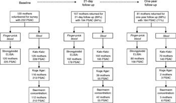
For information on how many tests were performed at baseline and the follow-ups, see fig. 1. Note that upon follow-up only individuals who were positive at baseline and reported back for follow-up were tested using Baermann concentration and the Koga agar method, due to limited resources. On the 21-day follow-up, an additional random selection of 30–40 individuals was tested as a corroborative spot-check.
Flow chart depicting the number of mothers and preschool-aged children (PSAC) surveyed at baseline and follow-ups (21-day and 1-year follow-ups), as well as information on the number of tests performed. The target for the initial baseline survey was 120 mothers and their respective two children (total of 240 children). Note that upon the 1-year follow-up, Kato–Katz smears were performed on single, not double, stool samples, unlike in previous surveys.

Statistical analyses
Data were collected from each individual using pro-forma data sheets, which were then entered using EpiDataTM and Microsoft Excel™. The data thus collated were analysed using STATA version 11 (StataCorp LP, College Station, Texas, USA) and the R statistical package® version 2.8.1 (http://www.r-project.org/). Details on the calculation of 95% confidence intervals (CI95) for prevalence values, the geometric mean of Williams (GMW), and prevalence comparisons using (one-tailed) Fisher's exact modification of the 2 × 2 chi-squared test have been published previously (Sousa-Figueiredo et al., Reference Sousa-Figueiredo, Pleasant, Day, Betson, Rollinson, Montresor, Kazibwe, Kabatereine and Stothard2010).
Ethical review, anthelminthic treatments and cure rates
Ethical permission for the study was granted by the London School of Hygiene and Tropical Medicine (LSHTM 5538·09) and the National Council of Science and Technology, Kampala, Uganda.
All mothers and children were offered treatment for schistosomiasis (40 mg/kg praziquantel) and intestinal worm infections (400 mg albendazole) at baseline as well as follow-ups if infection persisted. For younger/smaller children ( < 2 years old), the praziquantel tablets were crushed and mixed with orange juice and sugar before administration (spoon-feeding), with the help of the mother, along with a chewable orange-flavoured half-tablet of albendazole (200 mg). Lack of access to ivermectin at the local level meant that treatment of strongyloidiasis occurred with a regimen of 400 mg of albendazole, twice daily for 3 days. This regimen was undertaken by all parasite-positives at baseline at the end of the 21-day follow-up survey; ivermectin was made available for the 1 year follow-up.
Cure rates, defined as the percentage of the infected population (at baseline) found negative 21 days after treatment, were calculated for Strongyloides spp. (according to Baermann, Kogar agar plate, strongyloidid ELISA methods), hookworms and S. mansoni (according to Kato–Katz) after single-dose albendazole and praziquantel treatment at baseline (Bennett & Guyatt, Reference Bennett and Guyatt2000).
Results and discussion
In these four villages, the prevalence of strongyloidiasis was moderate, according to Baermann concentration and Koga agar plate methods combined, reaching 8.0% in mothers (aged between 16 and 70 years of age, mean age of 30) and 3.8% in preschool-aged children (aged between 5 months and 6 years of age, mean age of 3 years, female to male ratio of 1.1) before treatment. Numbers of strongyloidid larvae encountered in the faecal samples were low overall, with cases producing one (7 mothers and 6 preschool-aged children) or two larvae (2 mothers and 2 preschool-aged children) according to Baermann concentration and/or Koga agar plate methods. Importantly, the Koga agar plate method proved to be challenging to perform in the field without compromising the integrity of the cultures, especially when moving from village to village. This likely decreased the sensitivity achieved here, but could also indicate that these worms were not amenable to this culture medium.
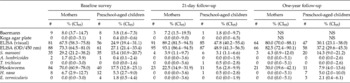
These results contrast with the local high prevalence of hookworm (70.0% in mothers and 17.5% in preschool-aged children), but imply that this eastern ecozone is within the ecological range possible for Strongyloides spp. transmission (Appleton et al., Reference Appleton, Maurihungirire and Gouws1999; Handzel et al., Reference Handzel, Karanja, Addiss, Hightower, Rosen, Colley, Andove, Slutsker and Secor2003; Brooker et al., Reference Brooker, Whawell, Kabatereine, Fenwick and Anderson2004). Nevertheless, if resources become available, further surveys should be conducted using repetitive stool samples for a more sensitive diagnosis, especially as larval excretion can vary daily (Nielsen & Mojon, Reference Nielsen and Mojon1987; Dreyer et al., Reference Dreyer, Fernandes-Silva, Alves, Rocha, Albuquerque and Addiss1996). Prevalence levels of other infections are summarized in table 1.
Number of positives (#) and prevalence (and CI95) of Strongyloides spp. according to three different diagnostic methods: Baermann concentration, Koga agar plate and strongyloidid ELISA (read visually and using a spectrophotometer at 450 nm), as well as that of other helminths as determined by two-sample duplicate Kato–Katz smears (S. mansoni, A. lumbricoides, T. trichiura, hookworms, H. nana and E. vermicularis) in mothers and preschool-aged children from villages around Lake Victoria at baseline, 21-day and 1-year follow-up surveys. NOTE: 1-year follow-up Kato–Katz smears were performed on a single stool sample (see Methods); Baermann and Koga agar plate techniques were performed on few individuals upon 1-year follow-up (see fig. 1) so prevalence levels are non-significant (NS).

From the questionnaire data, 50% of families have access to a pit-latrine, the majority wash their vegetables before cooking (96%), are aware of STHs (76%) and schistosomiasis (91%). Additionally, some reported having received albendazole (mothers: 40%; preschool-aged children: 37%) and praziquantel (mothers: 63%; preschool-aged children: 44%) prior to this survey. The latter observations indicate that NTD control measures have good outreach, even in two demographic strata not actively targeted by the national control programme. Interestingly, some mothers (44%) reported eating soil or clay, i.e. geophagia, (Woywodt & Kiss, Reference Woywodt and Kiss2002), a factor known to be highly associated with STHs (Glickman et al., Reference Glickman, Camara, Glickman and McCabe1999).
Strongyloidid diagnoses
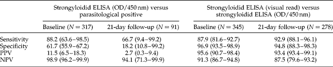
To improve diagnosis in the absence of repetitive stool sampling, we used multiple diagnostic methods simultaneously, as proposed previously (Siddiqui & Berk, Reference Siddiqui and Berk2001). However, strongyloidid diagnosis using the ELISA technique (OD/450 nm) yielded far more positives than those identified using the Baermann concentration and Koga agar methods (see table 1 for prevalence levels). When establishing the diagnostic performance of strongyloidid ELISA versus single Baermann concentration/double Koga agar methods (as ‘gold standard’), we found a sensitivity of 88.2% and a specificity of 61.7% at baseline, which declined after mass drug administration (sensitivity of 66.7% and specificity of 18.2%). Both results are much lower than those reported previously (van Doorn et al., Reference van Doorn, Koelewijn, Hofwegen, Gilis, Wetsteyn, Wismans, Sarfati, Vervoort and van Gool2007; Stothard et al., Reference Stothard, Pleasant, Oguttu, Adriko, Galimaka, Ruggiana, Kazibwe and Kabatereine2008) (see table 2 for full results). Interestingly, the majority of individuals diagnosed negative by ELISA were not excreting larvae; this point is of note as our previous survey in Lake Albert found proportionally more larva-excreting individuals who were ELISA negative, a phenomenon perhaps attributable to immunosuppression or failure to produce an antibody response (Stothard et al., Reference Stothard, Pleasant, Oguttu, Adriko, Galimaka, Ruggiana, Kazibwe and Kabatereine2008).
Diagnostic performance of strongyloidid ELISA (OD at 450 nm) using Baermann concentration results combined with Koga agar plate results as ‘gold-standard’, and diagnostic performance of visually read strongyloidid ELISA, using ELISA read by spectrophotometer as ‘gold-standard’, in mothers and their preschool-aged children from eastern Uganda, before and 21 days after a mass-drug administration campaign (see Methods). Values are expressed in % and brackets indicate CI95; PPV stands for positive predictive value, NPV stands for negative predictive value.
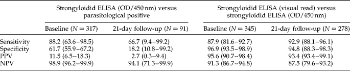
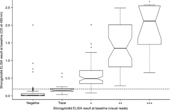
Importantly, ELISA results read visually were correlated with results gathered using a rather expensive and fragile spectrophotometer (see table 1 and fig. 2). Therefore, visual reads in the field are equally satisfactory, a significant fact when considering that most NTD surveillance field-surveys are financially and logistically constrained and field-proven ELISA methods are rather scarce.
Graphical representation of the relationships between strongyloidid ELISA results visually read (shades of yellow coded as negative, trace, positive, double-positive and triple-positive) and optical density (OD) at 450 nm at baseline. The horizontal bold bar in the middle shows the median OD for each visual code; both boxes show the interquartile range (25th to 75th percentile); the vertical dashed lines show the minimum and maximum values, or, in the presence of outliers (the circles), show 1.5 times the interquartile range (roughly two times the standard deviation); the notches at the ‘waist’ give an impression of CI95 around the medians, where boxes whose notches do not overlap have significantly different medians (P < 0.05). Horizontal dashed/dotted lines indicate the threshold over which values were considered positives (0.2 OD/450 nm). * Indicates that upper confidence interval extends over the interquartile range due to smaller sample size.

As might be expected, a ‘gold standard’ of single stool sample Baermann concentration and double stool sample Koga agar plates likely leads to an underestimation of ‘true’ prevalence (Nielsen & Mojon, Reference Nielsen and Mojon1987). However, there may be additional explanations for the discrepancy between prevalence levels resulting from direct (visual identification of larvae or eggs via Baermann concentration and Koga agar plate methods) and indirect diagnoses (identification of an immune response via the ELISA). Even though ELISA is reported to have good diagnostic sensitivity and specificity (van Doorn et al, Reference van Doorn, Koelewijn, Hofwegen, Gilis, Wetsteyn, Wismans, Sarfati, Vervoort and van Gool2007; Stothard et al., Reference Stothard, Pleasant, Oguttu, Adriko, Galimaka, Ruggiana, Kazibwe and Kabatereine2008), it is unable to distinguish between past and current infection and is proposed to cross-react with other nematode infections, e.g. filariae (Lindo et al., Reference Lindo, Atkins, Lee, Robinson and Bundy1996; Sudarshi et al., Reference Sudarshi, Stumpfle, Armstrong, Ellman, Parton, Krishnan, Chiodini and Whitty2003). The district where this study took place is predicted to be of low prevalence for lymphatic filariasis (Onapa et al., Reference Onapa, Simonsen, Baehr and Pedersen2005a), Mansonella perstans (Onapa et al., Reference Onapa, Simonsen, Baehr and Pedersen2005b) and onchocerciasis (Kolaczinski et al., Reference Kolaczinski, Kabatereine, Onapa, Ndyomugyenyi, Kakembo and Brooker2007), so ELISA results may not be confounded by co-infections with these parasites. However, one must note that prevalence levels for lymphatic filariasis and M. perstans within the district are broad estimations as no school from within its borders was selected by Onapa et al. (Reference Onapa, Simonsen, Baehr and Pedersen2005a, Reference Onapa, Simonsen, Baehr and Pedersenb) in their mapping attempts.
We hypothesize that the putative killing of nematodes/schistosomes by albendazole and praziquantel may be increasing antibody production by lymphocytes stimulated by this release of additional antigens, later detected as significantly elevated titres in the strongyloidid ELISA assay (see table 1). Thus the performance of the strongyloidid ELISA seems to be compromised by anthelminthic treatments.
To shed better light on these speculations, sensitive and unequivocal methods of diagnosis are needed and polymerase chain reaction (PCR) methods appear most promising, as a real-time PCR assay has been developed for detection of S. stercoralis DNA in stool. The assay was shown to be 100% sensitive and specific (Verweij et al., Reference Verweij, Canales, Polman, Ziem, Brienen, Polderman and van Lieshout2009); however, its use as routine diagnostic tool in the clinical setting of the developing world where co-infections are numerous is still to be proven. Nevertheless, the fact that collected faecal samples can be stored in alcohol opens up possibilities for in-country analysis where reference molecular laboratories could replace the need to perform diagnoses in situ by roving field teams.
Anthelminthic treatment at 21-day follow-up
Few samples were found to be positive at baseline or follow-up (6 negatives post-treatment of the 10 positives at baseline), consequently the confidence intervals around the ascertained cure rate are very wide (cure rate 60.0%; CI95 26.2–87.8), overlapping with the values reported by Steinmann and colleagues (2007). Importantly, to assess not only the true parasitological prevalence of strongyloidiasis, but also to ascertain the cure rate of single-dose albendazole treatment, four or more consecutive-day faecal samples per individual should have been used for Baermann concentration and Koga agar diagnostics, before and after treatment (Dreyer et al., Reference Dreyer, Fernandes-Silva, Alves, Rocha, Albuquerque and Addiss1996), as single stool sample diagnosis is likely to underestimate the ‘true’ S. stercoralis prevalence (Marti & Koella, Reference Marti and Koella1993). However, this was beyond the remit of the present study.
Due to the low number of parasitological positives identified before and after treatment, very little can be inferred about the efficacy of the treatment regimen applied in this survey against strongyloidiasis. Nevertheless, we recommend that ivermectin should be used in the future even if albendazole is more readily available. Although ivermectin is available for the treatment of filarial infections within the national control programme, it is not being offered for treatment of strongyloidiasis in local settings, nor is it easy to purchase from government or private pharmacies (Siddiqui & Berk, Reference Siddiqui and Berk2001; Keiser & Utzinger, Reference Keiser and Utzinger2008). In Uganda, if control of strongyloidiasis is to be integrated with ongoing NTD control, and patient management is to improve, availability of this drug must improve.
In this survey, praziquantel and albendazole treatment performed well against S. mansoni and common STH infections. Hookworm infections decreased significantly from 70.0 to 22.5% and from 17.5 to 5.6% in mothers and preschool-aged children, respectively, with a general cure rate of 76.2% (CI95 66.9–84.0%; 105 initial egg positives resulted in 80 egg negatives). These results concur with those reported in a recent meta-analysis of WHO-recommended anthelminthics commonly used against STH infections (Keiser & Utzinger, Reference Keiser and Utzinger2008). The impact of the chemotherapy regimen on A. lumbricoides, T. trichiura, H. nana and E. vermicularis in this survey is debatable, since very few cases were found and re-tested; nevertheless, all individuals diagnosed positive at baseline, except for one H. nana case, were negative post-treatment. Prevalence of intestinal schistosomiasis decreased significantly from 29.2 to 3.9% and from 15.4 to 3.1% in mothers and preschool-aged children, respectively, with a general cure rate of 93.3% (CI95 83.8–98.2%, 60 initial egg positives resulted in 56 egg negatives).
Prevalence patterns after annual follow-up
The prevalence of strongyloidiasis according to Baermann concentration and Koga agar plate methods was similar at follow-up to that at baseline. However, far fewer people were re-tested (see fig. 1) so results should be treated with caution. Only one larval-positive individual was found (one of the four mothers tested using the Baemann concentration method). Strongyloidid ELISA also showed similar results at baseline and at 1-year follow up. In this case the results are more reliable as a large number of individuals were surveyed at the 1-year follow-up (see table 1). In mothers, prevalence levels decreased slightly from levels at the 21-day follow-up (ELISA OD: 82.5% versus 93.1%, P = 0.034), reaching levels similar to those diagnosed at baseline (ELISA OD: 82.5% versus 73.3%, P = 0.17). In preschool-aged children, prevalence levels found 1 year after baseline were also lower than at the 21 day follow-up (ELISA OD: 37.2% versus 48.9%, P = 0.036), but still significantly higher than those identified at baseline (OD ELISA: 37.2% versus 27.1%, P = 0.043). The marked rise in strongyloidid ELISA positives following initial treatment followed by a significant decline in numbers at the 1-year follow-up is further evidence in support of the hypothesized acute triggering of Strongyloides-non-specific immunogenicity after chemotherapy and its recognition by the strongyloidid ELISA (i.e. cross-reactivity).
Hookworm prevalence levels did not change significantly between follow-ups for both mothers (22.5% versus 30.0%, P = 0.29; GMW 2.279 epg versus 3.909 epg) and preschool-aged children (5.6% versus 7.9%, P = 0.50; GMW 0.279 epg versus 0.357 epg), and were significantly lower than those recorded pre-treatment (mothers: 30.0% versus 70.0%, P < 0.00 001, GMW 3.909 epg versus 52.388 epg; preschool-aged children: 7.9% versus 17.5%, P = 0.008; GMW 0.357 epg versus 1.417 epg).
The prevalence of intestinal schistosomiasis showed different longitudinal dynamics between the two studied age groups. In mothers, prevalence levels identified 1 year after initial treatment were lower than those identified at baseline (4.3% versus 29.2%, P < 0.0001; GMW 0.164 epg versus 1.955 epg), remaining very similar to the 21-day post-treatment levels (4.3% versus 3.9%, P = 1.0; GMW 0.164 epg versus 0.121 epg). On the other hand, intestinal schistosomiasis prevalence levels in preschool-age children recovered, equalling those identified pre-treatment (14.3% versus 15.4%, P = 0.88; GMW 0.820 epg versus 0.764 epg) and being significantly higher than those identified 21 days post-treatment (14.3% versus 3.1%, P = 0.0003; GMW 0.820 epg versus 0.099 epg).
These observations identify differences in the natural history and dynamics of transmission of schistosomiasis between mothers and their young children at this location, where the potential for re-infection appears to have increased in the younger age-classes, even after successful treatment rounds, emphasizing the need for periodic treatment as children this young are at continuous risk for infection, even in areas of moderate to low transmission, such as the ones studied here. The necessity for regular treatment of preschool-aged children is discussed elsewhere (Johansen et al., Reference Johansen, Sacko, Vennervald and Kabatereine2007; Stothard & Gabrielli, Reference Stothard and Gabrielli2007a, Reference Stothard and Gabriellib; Sousa-Figueiredo et al., Reference Sousa-Figueiredo, Pleasant, Day, Betson, Rollinson, Montresor, Kazibwe, Kabatereine and Stothard2010).
In conclusion, standard dose praziquantel (40 mg/kg) and single-dose albendazole treatments were found to be very effective against intestinal schistosomiasis and common STH infections, with debatable impact on Strongyloides spp. Diagnosis of strongyloidiasis with ELISA in this region of Uganda appears to be cross-reacting with other parasitic infections. For this reason, future surveillance of strongyloidiasis should apply the Baermann concentration method or, where possible, also attempt to implement new quantitative PCR assays on stool samples preserved in alcohol. Integration of routine epidemiological screening for strongyloidiasis in mothers and preschool-aged children, especially those with HIV, and better availability of ivermectin in non-filarial endemic areas, should be encouraged.
Acknowledgements
We wish to thank the children and mothers who took part in the study. We also thank the Ministry of Health of Uganda for their support during the survey and the field staff of the Vector Control Division, Uganda. The Strongyloides and Hookworm data presented in this paper formed a part of the final dissertation for a Clinical Research intercalated BMSc (M.D.). We acknowledge financial support from the Wellcome Trust, UK (J.C.S.F., M.B., N.B.K. and J.R.S.) and Royal Society of Tropical Medicine and Hygiene, UK (M.D.).