Introduction
The vast majority of laryngeal neoplasms are epithelial in origin, comprising mainly squamous cell carcinoma, dysplasia and/or keratosis, and recurrent respiratory papillomatosis. The diagnosis of these lesions is often apparent, and a management plan can be tentatively formulated based on clinic-sited endoscopy and, at times, imaging, prior to obtaining a confirmatory tissue biopsy. Non-epithelial, submucosal laryngeal masses create more initial uncertainty among otolaryngologists attempting to make a diagnosis and develop a treatment plan. Even after excluding those submucosal lesions with a characteristic appearance (e.g. amyloidosis or intra-cordal cyst) or history of prior laryngeal intervention (e.g. Teflon® injection), the differential diagnosis still includes a variety of pathologies, and it is not always possible to predict the nature of the lesion based on examination and radiographic imaging in the clinic.
Given the relative rarity of submucosal laryngeal neoplasms, this paper presents an extensive case series together with photo-documentation, categorisation by subsite, and outcomes of treatment algorithms. This information can be useful to guide otolaryngologists in the decision-making process, prior to arrival in the operating theatre. While case series of individual types of submucosal laryngeal neoplasms exist in the literature, the examination of a broader population of multiple pathologies, as presented in this report, is helpful in order to compare differences in presentation and surgical management. Additionally, the inclusion of all patients seen with submucosal laryngeal neoplasms over an extended time period in a tertiary laryngology practice provides some indication of the relative prevalence of different types of pathology.
Materials and methods
The institutional review board of Massachusetts General Hospital approved this study.
A retrospective chart review was performed for all adult patients seen from 2002 to mid-2011 presenting with submucosal neoplasms involving the laryngeal introitus.
We excluded patients with (submucosal) squamous cell carcinoma, amyloidosis, mucosal cysts (including intra-cordal cysts, saccular cysts and laryngocoeles), Teflon granulomas, and infectious or inflammatory or autoimmune processes.
Patients' clinical presentation, operative description, histological diagnosis, and pre- and post-operative laryngeal imaging were recorded.
Results and analysis
We identified the medical records of 38 patients who met the study criteria for non-epithelial laryngeal neoplasm. The mean age at presentation was 56 years old. The study population was 59 per cent male and 41 per cent female.
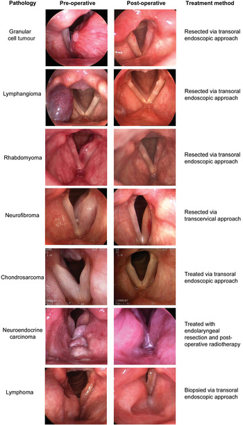
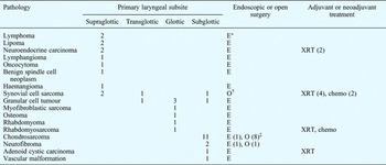
The pathologies identified in these patients are represented in Figure 1, and the laryngeal subsite distribution and treatment course are detailed in Table I. Representative pre- and post-operative images (taken when complete healing had occurred) are depicted in Figure 2. Fifty-eight per cent (22/38) of the lesions were malignant tumours.
Distribution of pathology among 38 patients presenting with submucosal laryngeal neoplasms. Malignant lesions (n = 22) are represented as detached segments on the right side, while benign lesions (n = 16) are shown as attached segments on the left.

Pre and post-operative laryngoscopic photographs of selected submucosal laryngeal neoplasms.

Laryngeal subsite and treatment modalities for submucosal neoplasms

Data represent patient numbers. *Surgery for diagnosis only. †One case was initially managed endoscopically, but eventually required a total laryngectomy for local recurrence. ‡Two patients elected to receive treatment at another institution. E = endoscopic surgery; O = open surgery; XRT = radiation therapy; chemo = chemotherapy
The primary presenting symptoms were: dysphonia (25 patients), airway restriction (six; chondrosarcoma, neuroendocrine carcinoma and granular cell tumour), dysphagia (two; lipoma and benign spindle cell neoplasm), haemoptysis (two; lipoma and synovial cell sarcoma), neck mass (two; lymphoma and synovial cell sarcoma) and neck pain (one; lymphangioma). Twenty-two of the 38 patients had undergone previous biopsy, and in some cases attempted resection, at another institution before presenting to the Massachusetts General Hospital Voice Center.
Thirty-six of the 38 patients elected to receive care at our centre. One patient with a low-grade laryngeal chondrosarcoma opted for a total laryngectomy at another institution instead of the partial laryngectomy offered at our centre. Another elderly patient presented with a long-term, low-grade, recurrent laryngeal chondrosarcoma but chose to undergo serial monitoring closer to home, since an operation would probably have worsened his functional voice and relatively patent airway.
All the remaining 36 patients were treated surgically, except for two cases of laryngeal lymphoma in which surgery was employed for diagnosis and staging only. Twenty-four patients (67 per cent) underwent a transoral, endoscopic resection, and 13 of those patients required a second endoscopic procedure to assess and potentially treat a persistent or recurrent neoplasm, of whom only one patient had unintentionally persistent disease (a benign spindle cell neoplasm). Two patients had a planned, second-stage endoscopic operation for granular cell tumours involving the anterior commissure, in order to avoid cicatrix formation. One patient with a supraglottic synovial cell sarcoma initially underwent an endoscopic resection, but ultimately required a salvage total laryngectomy for locally recurrent disease.
Eleven patients underwent initial therapeutic management with open partial laryngectomy at our institution (one for benign disease (neurofibroma) and the remainder for malignancy). Eight had no evidence of disease and an intact swallowing function. Notably, this subgroup of eight functional, disease-free patients included a young man with synovial cell chondrosarcoma who underwent induction chemoradiation followed by a transcervical partial laryngectomy for a 3 cm remnant of disease in the thyrohyoid space. At the time of writing, he had been without evidence of recurrent disease for almost five years and remained decanulated.
Three patients initially treated with an open partial laryngectomy were unable to be decanulated. One of these patients presented with recurrent laryngeal chondrosarcoma previously managed elsewhere, significant laryngeal dysfunction and tracheotomy dependence; at the time of writing, she had no evidence of disease and was taking a full oral diet, but had required a near-total laryngectomy for tumour control. The other two patients who were not decanulated were terminal cases suffering from metastatic laryngeal synovial cell sarcoma.
Radiation therapy was utilised in a substantial proportion of patients with submucosal laryngeal cancers. Eight of 10 patients with malignancies other than chondrosarcoma received radiation with or without chemotherapy, in an adjuvant or neoadjuvant manner (the other two patients had lymphoma, warranting surgery for diagnosis alone). Four of these eight patients (two with neuroendocrine carcinoma, one with rhabdomyosarcoma and one with adenoid cystic carcinoma) underwent near-total endoscopic tumour cytoreduction with preservation of key portions of laryngeal architecture, followed by planned post-operative radiation therapy; three of these four have no evidence of disease after a mean follow up of 5.9 years (range, 21–112 months), while one patient with a laryngeal neuroendocrine carcinoma expired with distant metastases (but no locoregional recurrence) more than five years after her laryngeal treatment. All four patients with synovial cell sarcoma received radiation with or without chemotherapy, either in an adjuvant or neoadjuvant manner. Radiation and/or chemotherapy have been shown to be mostly ineffective for chondrosarcoma,Reference Thompson and Gannon1 and only one of nine patients with laryngeal chondrosarcoma received adjuvant radiation therapy (due to aggressive histopathology).
Eight of the nine treated patients with laryngeal (cricoid) chondrosarcoma had no evidence of disease after a mean post-treatment follow up of 35 months (range, 10–71 months). The ninth patient was recently treated for a low grade laryngeal chondrosarcoma; a small amount of disease under the contralateral cricoarytenoid joint was intentionally not resected during an open partial laryngectomy, in order to preserve mobility of that vocal fold and permit decanulation.
Discussion
General comments
Submucosal laryngeal neoplasms present unique diagnostic and management challenges for the laryngeal surgeon. The tumour location and disease extent are important in determining the surgical options and the potential voice and swallowing outcomes. In this regard, our patients' main presenting symptoms (airway distress, dysphagia and dysphonia) roughly corresponded to tumour size and location (supraglottic, subglottic or glottic). For glottic lesions, the proximity of the tumour to the superficial layer of the lamina propria has a critical impact on the potential for post-resection voice restoration, since this vibratory layer cannot be recreated if it is destroyed by the tumour or by the surgery removing the tumour. This should be especially considered if a glottic lesion is suspected to lie deep to the superficial layer of the lamina propria, in the paraglottic space. Pre-operative radiographic imaging, as well as pre-resection endoscopic saline infusion of Reinke's space, can help to better define the location of the lesion with respect to the superficial layer of the lamina propria, thereby allowing for potential preservation of the layered microstructure of the vocal fold, within the confines of oncological safety.
Even when a prior laryngeal biopsy has been performed by a referring surgeon, the pathological diagnosis of a submucosal laryngeal neoplasm can remain in question. Because of the rarity of some of these lesions and the potential for misdiagnosis and therefore mismanagement, it is always prudent to review any previously obtained pathology slides with a pathologist experienced in mesenchymal neoplasms. Similarly, if the diagnosis is unknown a priori, the laryngeal surgeon should be cautious about aggressive resection based only on frozen section assessment (e.g. lymphoma).
Radiographic imaging was commonly used in the management of the patients in this series. Specifically, for vascular-appearing submucosal laryngeal lesions (n = 3), computed tomography (CT) angiography and/or magnetic resonance angiography were used to better characterise the haemorrhagic potential and the consequent need for pre-operative angiography with embolisation. Additionally, CT imaging is invaluable in monitoring for recurrence of laryngeal chondrosarcoma, since, in contradistinction to most epithelial laryngeal neoplasms, recurrence will often appear radiographically before it is seen endoscopically. Furthermore, imaging is helpful to characterise the possible extension of deep paraglottic lesions to neighbouring spaces (i.e. the pyriform sinus, pre-epiglottic space and cricothyroid space), giving guidance as to whether an endoscopic or transcervical approach is warranted.
While the goal of any curative surgical intervention for these neoplasms should be complete tumour eradication, voice preservation and/or restoration is also a key objective. In particular, post-resection phonatory glottic incompetence is something that can and should be treated once the tumour has been eradicated and the likelihood of neoplastic recurrence is low.Reference Zeitels, Jarboe and Franco2, Reference Zeitels3 In the same respect, benign lesions involving the anterior commissure can be treated with staged endoscopic procedures (addressing the dominant side first) in order to avoid the creation of an anterior commissure synechia or web, with resultant dysphonia and reduced ability to monitor for recurrence.
Selected benign neoplasms
Granular cell tumour
Granular cell tumours were the most common benign, submucosal laryngeal neoplasm in this series (n = 5). These neoplasms arise from undifferentiated mesenchymal cells or Schwann cells. Laryngeal involvement accounts for only 7–10 per cent of all reported cases of granular cell tumours, but approximately 50 per cent involve the true vocal fold. Typically, the lesions are well circumscribed but not encapsulated; a prior biopsy can make the distinction between tumour and normal tissue less obvious. Tumour growth is slow and recurrence rates range from 2–8 per cent. None of the five patients in this series had evidence of recurrence, with a mean follow up of 35 months after surgery. Approximately 10 per cent are multi-focal. In 2000, 206 laryngeal cases were identified in the literature, with only one case of malignant transformation.Reference Sataloff, Ressue, Portell, Harris, Ossoff and Merati4
Lipoma
Lipomas were another relatively common benign lesion in this series. To date, approximately 100 laryngeal cases have been reported in the literature, with supraglottic sites (i.e. vestibular fold, epiglottis and aryepiglottic fold) being most common.Reference De Vincentiis, Greco, Mascelli, Soldo and Zambetti5 Pre-operative imaging (either by CT or magnetic resonance imaging) can often suggest this pathology. Histologically, lipomas can be completely or partially encapsulated and recurrence can result from incomplete resection, or may be suggestive of a malignant process (e.g. liposarcoma) that was incorrectly diagnosed initially. Neither of the two patients with laryngeal lipomas in this series had a recurrence, although there was limited follow up. Lipomas usually present as a single neoplasm, but if multiple lipomas are encountered a syndrome (such as neurofibromatosis or Gardner's syndrome) should be ruled out.
Neurogenic neoplasms
Neurogenic laryngeal submucosal neoplasms were encountered in two patients in this series (neurofibromas). In the larynx, both neurofibromas and schwannomas are thought to arise from the internal branch of the superior laryngeal nerve. This may explain the reported predominance (80 per cent) in the aryepiglottic fold, while the remainder are located in the vestibular or true vocal folds.Reference Ebmeyer, Reineke, Gehl, Hamberger, Mlynski and Essing6 Neurofibromas arise from perineural fibrocytes and therefore are not usually grossly encapsulated or easily separated from the underlying nerve; in contrast, schwannomas arise from perineural Schwann cells, are well encapsulated, and can in theory be peeled away from the nerve of origin. Endoscopic treatment of laryngeal neurofibromas is still possible, as evidenced by the fact that one of our two patients with this lesion underwent endolaryngeal management with preservation of vocal function (albeit with limited follow up); the other patient had a transcervical resection due to submucosal extension of the lesion into the pyriform sinus, followed by a staged reconstructive medialisation laryngoplasty (with no evidence of recurrent disease after 17 months). Another consequence of the encapsulation of schwannomas (versus neurofibromas) is the possibility of a non-diagnostic incisional biopsy as a result of the capsule not being penetrated. It is rare for a schwannoma to become cancerous, but neurofibromas have a 10 per cent malignant transformation rate and can be encountered alone or in the context of neurofibromatosis.Reference Ebmeyer, Reineke, Gehl, Hamberger, Mlynski and Essing6
Lymphatic and vascular neoplasms
Lymphatic and vascular anomalies were found in three patients. Among these was a haemangioma; while these lesions are much more common in the paediatric population and tend to resolve spontaneously, a second type of haemangioma can occur in adults of any age and typically does not spontaneously involute.Reference Lomeo, McDonald and Finneman7 Although we have previously reported on the utility of the 532 nm KTP laser for photoangiolysis and/or photoablation of laryngeal lesions with significant microvasculature (e.g. squamous cell carcinomaReference Zeitels, Burns, Lopez-Guerra, Anderson and Hillman8 and papillomaReference Franco, Zeitels, Farinelli and Anderson9), as well as for glottic ectasias and varices,Reference Zeitels, Akst, Bums, Hillman, Broadhurst and Anderson10 the KTP laser was used in only one of the three patients (in a continuous mode, with simultaneous cooling) to excise the vascular lesion en bloc. The other two lesions were also resected en bloc with a fibre-based thulium laser, which in our experience has superior coagulative abilities compared with the KTP.Reference Zeitels, Burns, Akst, Hillman, Broadhurst and Anderson11 Intra-operative blood loss was minimal for all three patients. While some have reported a technique of repeated photocoagulation, ablating through vascular laryngeal tumours rather than excising them,Reference Kishimoto, Hirano, Kato, Suehiro, Kanemaru and Ito12 this probably imparts greater thermal injury to the larynx and risks damage to nearby glottic surfaces, with the potential for post-operative dysphonia.
Selected malignant neoplasms
Chondrosarcoma
Chondrosarcomas comprised the majority of malignant submucosal lesions, both in our current series (n = 11) as well as those of others,Reference Cai, Wang and Shi13, Reference Fernandez-Acenero, Larach and Ortega-Fernandez14 and may represent the most common type of submucosal neoplasm (benign or malignant) in the larynx overall. Despite the fact that they are malignancies, their radiographic appearance (most commonly involving the cricoid cartilage) should not be correlated with the management strategies for squamous cell carcinoma presenting with similar radiographic involvement. Thus, a laryngeal function preserving resection with narrow margins has been reported to be oncologically adequate for low-grade tumoursReference Thompson and Gannon1 (as opposed to a total laryngectomy). Even a local recurrence can potentially be treated with a salvage partial laryngectomy. Along these lines, laryngeal chondrosarcomas have a low propensity for metastasis, with only 1.9 per cent (two patients out of 111) in a Mayo series metastasising to distant sites, and none to regional lymph nodes.Reference Thompson and Gannon1 (Both of these patients had previously undergone a total laryngectomy.) Given these limited data, a total laryngectomy should also not be performed on the premise of reducing the risk of metastasis.
The histological grade of chondrosarcoma can play a role in surgical planning. Depending on the circumstances, large, low-grade lesions (i.e. grade one of three) in individuals with reasonably functional voice, airway and/or swallowing may best be managed by endoscopic debulking, as opposed to a partial laryngectomy. However, patients opting for this strategy must be aware of the potential need for future procedures. Higher grade lesions (grades two to three) typically mandate total tumour removal (but not always total laryngectomy) to minimise the chance of recurrence.
Open partial laryngectomy for treatment of laryngeal chondrosarcoma is often eschewed due to a perceived lack of reconstructive options, since the majority of these lesions involve the cricoid cartilage. Yet, several reconstructive techniques proved useful in our series, including secondary healing, repair of the laryngeal defect with a thyrotracheal anastomosis, and re-establishment of the laryngeal lumen with free tissue grafts. In essence, as long as a single, functional cricoarytenoid joint can be preserved, it is unlikely that a patient with a laryngeal chondrosarcoma will require a total laryngectomy due to lack of reconstructive options.
Synovial cell sarcoma
Laryngeal synovial cell sarcomas represented a relatively large proportion of malignant submucosal tumours in this series (four out of 22), but only a handful of cases are discussed in the literature.Reference Bilgic, Mete, Ozturk, Demiryont, Keles and Basaran15, Reference Harb, Luna, Patel, Ballo, Roberts and Sturgis16 These aggressive malignancies arise from pluripotent mesenchymal progenitor synovial cells (which exist in the soft tissues of the anterior larynx, particularly the thyrohyoid ligament) and much less frequently from the synovial lining of joints within the head and neck. Around 200 synovial cell sarcomas involving various sites within the head and neck (with deep neck soft tissues slightly more commonly involved than aerodigestive tract) have been described since the first report in 1954,Reference Harb, Luna, Patel, Ballo, Roberts and Sturgis16 but a literature search revealed only 15 previously described cases in the larynx.Reference Fernandez-Acenero, Larach and Ortega-Fernandez14, Reference Harb, Luna, Patel, Ballo, Roberts and Sturgis16 While partial laryngectomy may be successful,Reference Quinn17 local recurrence is common, and thus wide resection with negative margins (e.g. total laryngectomy for sarcomas of laryngeal origin) is the recommendation for the generalised head and neck population with this cancer.Reference Harb, Luna, Patel, Ballo, Roberts and Sturgis16 Of the four patients in this series, one presented with advanced, recurrent, locoregional disease and subsequently expired. One patient was initially managed endoscopically, followed by an open partial laryngectomy, but ultimately required a total laryngectomy and developed lung metastases. Another patient underwent an open partial laryngectomy but at the time of writing had mediastinal disease. One patient who underwent neo-adjuvant chemoradiation therapy followed by open partial laryngectomy was disease-free after nearly five years of post-surgical follow up.
• Endolaryngeal, submucosal neoplasms have an extensive differential diagnosis
• In the presented 38 cases, chondrosarcomas were commonest
• Most submucosal laryngeal lesions can be treated surgically
• The decision to operate endoscopically versus trans-cervically is based on tumour pathology and location and laryngeal function
Given the paucity of data on laryngeal synovial cell sarcoma, several broader points from the overall head and neck experience are relevant. Local recurrence tends to be the most common failure pattern, and post-operative irradiation may improve local control.Reference Harb, Luna, Patel, Ballo, Roberts and Sturgis16 Regional metastases are very rare. Distant metastases are typically to the lung, often occurring in tandem with a local recurrence, and the role of chemotherapy is not clear from the existing data.Reference Harb, Luna, Patel, Ballo, Roberts and Sturgis16, Reference Lee and Shin18 The disease-specific survival in a recent, large series of head and neck synovial cell sarcomas was 72 per cent at five years.Reference Harb, Luna, Patel, Ballo, Roberts and Sturgis16
Conclusion
Subepithelial laryngeal neoplasms represent a broad spectrum of possible pathologies, but generalised laryngeal surgical management principles still apply. Some of the issues of diagnostic uncertainty are unique to the mesenchymal origin of these tumours, and surgical informed consent should be appropriately broad to reflect this and allow for flexibility in intra-operative decision-making. The literature typically organises these lesions by pathology, while the information herein gives a portrayal of how these lesions are encountered prior to the diagnostic biopsy. The relative distribution of tumours presented here, and their pre-treatment photo-documentation, should be helpful, since few surgeons will have substantial experience with any individual pathology. Most submucosal laryngeal neoplasms are managed surgically, with the addition of adjuvant radiation and/or chemotherapy for selected malignancies.
Acknowledgements
This work was supported in part by the Eugene B Casey Foundation and the Institute of Laryngology and Voice Restoration.