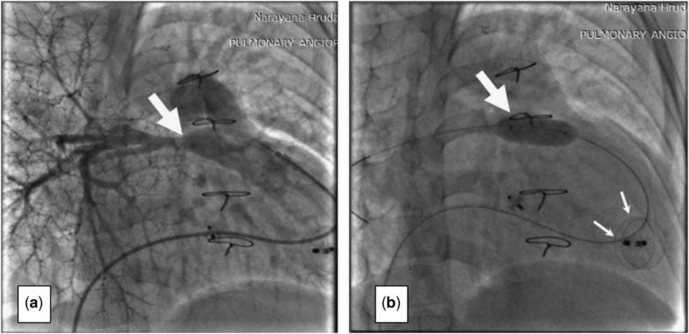
Amale child, with d-transposition of great arteries, large perimembranous ventricular septal defect, multiple additional ventricular septal defects, small muscle-bound right ventricle, and severe pulmonary stenosis with confluent, moderate-sized branch pulmonary arteries, underwent an emergency right modified Blalock–Taussig shunt on day 15 of life. In view of the cardiac anatomy, he was planned for a univentricular palliation; however, on re-evaluation at 2.5 years of age, the tricuspid valve on transthoracic echocardiography was seen to be 11 mm (−2.9 Z scores). A diagnostic cardiac catheterisation was carried out preoperatively to evaluate the right ventricle as well as obtain haemodynamic data for backup plan of single ventricle palliation. On visual impression of the angiographic images, the right ventricle appeared borderline adequate, and thereby we decided to attempt a biventricular repair with closure of the ventricular septal defect and re-construction of the right ventricular outflow tract. The parents were explained about the risk of residual ventricular septal defects, obstruction at various anastomotic sites, requirement of repeat surgeries, and possible ventricular dysfunction following extensive surgery. Owing to the vast experience of our centre with CHD surgeries, the right ventricle intraoperatively was seen to be tripartite and adequate, and hence a réparation à l’étageventriculaire procedure with closure of ventricular septal defects and takedown of the Blalock–Taussig shunt was carried out. On follow-up, he was noted to be symptomatic with effort intolerance, and transthoracic echocardiography showed a moderate residual upper ventricular septal defect and multiple apical ventricular septal defects, mild mid-right pulmonary artery stenosis, free pulmonary regurgitation, and right ventricular dysfunction. In view of these multiple abnormalities and earlier two surgeries, surgical re-intervention was deemed extremely difficult and risky. Therefore, after discussion at the multidisciplinary meeting and with the parents, it was decided to close the ventricular septal defects through the percutaneous approach in the cardiac catheterisation laboratory. Through a femoral approach, the upper muscular ventricular septal defect was closed using an 8-mm Amplatzer Muscular Ventricular Septal Defect Occluder Device (Fig 1), and an 18-mm Amplatzer Multi-Fenestrated Septal Occluder – Cribriform was used to close the multiple apical muscular ventricular septal defects (Fig 1) (AGA Medical Corporation, Nathan Ln N, Minneapolis, United States of America). On follow-up at 1 year after the procedure, the patient was asymptomatic, and his echocardiography showed improved right ventricular function and no significant residual ventricular septal defects; however, increasing right pulmonary artery stenosis was noted (Fig 2), necessitating right pulmonary artery angioplasty using an 8×20 mm cutting balloon followed by a 10×20 mm Tyshak II balloon (NuMED, Inc., Hopkinton, New York, United States of America) (Fig 2). On subsequent follow-ups, he has been asymptomatic and carrying out his activities normally.
( a ) Left ventricular angiogram showing upper muscular ventricular septal defect (VSD) and multiple apical VSDs (arrows). ( b ) VSD occlude device deployed in the upper muscular VSD (arrow). ( c ) Cribriform septal occlude device deployed in apical VSDs (arrow). ( d ) Two occlude devices in situ at the upper muscular and apical VSDs (arrow).

( a ) Right pulmonary artery (RPA) angiogram showing middle stenosis (bold arrow) ( b ) balloon angioplasty of RPA (bold arrow); the ventricular septal defect device is also seen (thin arrows).

Discussion
In this case, neither the surgical closure of multiple ventricular septal defects nor the postoperative device closure of residual ventricular septal defects made it unique. Our case was unique on two counts: first, a cardiac anomaly that was most likely to go the univentricular pathway due to borderline anatomy; however, owing to the surgical expertise, an intraoperative decision for a biventricular repair was taken and executed remarkably; and, second, the use of cribriform septal occluder device for multiple apical ventricular septal defects makes it the only case reported in the paediatric age group. The incidence of residual ventricular septal defects around the accessible areas such as the upper muscular septum and in inaccessible portions of the apical interventricular septum that require re-intervention after réparation à l’étageventriculaire procedure with closure of ventricular septal defects has been reported to the tune of 6%.Reference Carloa, Lecompteb, Tomascoc, Cohenb and Vouhéd 1 These residual ventricular septal defects, pulmonary regurgitation, and right pulmonary artery stenosis made the child symptomatic, thus necessitating intervention. Device closure of the upper muscular ventricular septal defect with a single muscular septal defect occluder device was a straightforward procedure; however, closing the multiple, closely placed apical muscular ventricular septal defects was technically difficult and was carried out with a cribriform septal occluder device. There have been reports of using cribriform septal occluder devices in adults with ventricular septal defects following acute myocardial infarction.Reference Szkutnik, Kusa and Bialkowski 2 It has been used in a child with post-traumatic multiple ventricular septal defects but not in apical ventricular septal defects.Reference Kasem, Kanthimathinathan, Mehta, Neal and Stumper 3 The occlusion of these shunts reduced the volume overload on the right ventricle; however, the worsening right pulmonary artery stenosis would have hastened the right ventricular dysfunction in the long term. Therefore, balloon angioplasty of the right pulmonary artery was carried out. Thus, the surgically uncorrectable residual shunts and obstructions were corrected by skilled, staged, and timely percutaneous interventions by the cardiologists. The close coordination and planning by the multidisciplinary team at every step made the successful outcome possible.
Acknowledgements
None.
Financial Support
This research received no specific grant from any funding agency, commercial, or not-for-profit sectors.
Conflicts of Interest
None.
Supplementary material
To view supplementary material for this article, please visit http://dx.doi.org/10.1017/S1047951116000925