Introduction
Psychopathy is a developmental disorder (Lynam et al. Reference Lynam, Caspi, Moffitt, Loeber and Stouthamer-Loeber2007) that has long been linked to attentional abnormalities (Hare & Jutai, Reference Hare, Jutai, Van Dusen and Mednick1983; Kosson & Newman, Reference Kosson and Newman1986). The aim of the current paper is to review the literature on attention and psychopathy from a cognitive neuroscience perspective. We (i) briefly describe psychopathy; (ii) consider the cognitive neuroscience of attention; and (iii) consider the literature on attention in psychopathy with respect to the cognitive neuroscience literature.
What is psychopathy?
The disorder of psychopathy involves two core components: emotional dysfunction [Callous and Unemotional (CU) traits] and antisocial behavior (Hare, Reference Hare1991; Frick, Reference Frick1995). The emotional dysfunction involves reduced guilt, empathy and attachment to significant others. The antisocial behavior component involves a predisposition to antisocial behavior from an early age. It is typically identified in adults with the Hare Psychopathy Checklist-Revised (PCL-R; Hare, Reference Hare1991, Reference Hare2003). In children and adolescents, psychopathic traits can be indexed through the Antisocial Process Screening Device (ASPD; Frick & Hare, Reference Frick and Hare2001), the Psychopathy Checklist – Youth Version (PCL-YV; Forth et al. Reference Forth, Kosson and Hare2003) or the Youth Psychopathy traits Inventory (YPI; Andershed et al. Reference Andershed, Kerr, Stattin, Levander, Blaauw and Sheridan2002). The disorder is developmental, with recent work confirming its stability from childhood into adulthood (Lynam et al. Reference Lynam, Caspi, Moffitt, Loeber and Stouthamer-Loeber2007).
One of the major strengths of the classification of psychopathy has been its utility in risk assessment. Considerable work has shown the predictive power of the PCL-R with respect to recidivism (Hart et al. Reference Hart, Kropp and Hare1988; Hare et al. Reference Hare, Clark, Grann and Thornton2000; Kawasaki et al. Reference Kawasaki, Kaufman, Damasio, Damasio, Granner, Bakken, Hori, Howard and Adolphs2001). The classification is also important with respect to treatment. Hawes & Dadds (Reference Hawes and Dadds2005) found that boys with high CU traits were less responsive to treatment than boys with conduct problems but low CU traits.
What is attention?
Attention can be considered the process by which stimuli are selected for further processing and control over behavior. Stimulus selection is biased by bottom-up sensory-driven mechanisms (e.g. visual salience) and top-down influences generated outside of sensory cortices (e.g. executive attention) (Desimone & Duncan, Reference Desimone and Duncan1995). Rather than being processed additively, stimuli are processed in a mutually inhibitory fashion whereby a gain in activity of the neurons representing one object occurs at the expense of activity of the neurons representing another (Desimone & Duncan, Reference Desimone and Duncan1995).
Bottom-up and top-down attentional control
The current dominant model of attention remains the biased competition model (Desimone & Duncan, Reference Desimone and Duncan1995). This model views attention as a result of the competition for neural representation that occurs when multiple stimuli are present in the visual field (Desimone & Duncan, Reference Desimone and Duncan1995; Duncan, Reference Duncan1998). Which stimuli win this competition and are ‘attended to’ is a product of both bottom-up and top-down functional processes. Bottom-up sensory processes include an enduring bias to objects that are moving, bright and large (Jonides & Yantis, Reference Jonides and Yantis1988; Triesman & Gormican, Reference Triesman and Gormican1988) and are considered consequences of the intrinsic functional properties of the visual system. Emotional attention, the augmentation of emotional representations within the temporal cortex by input from the amygdala, is considered to be another form of attention-based processing bias (Pessoa & Ungerleider, Reference Pessoa and Ungerleider2004).
In contrast to bottom-up processes, top-down influences on stimulus selection occur as a function of task demands (Desimone & Duncan, Reference Desimone and Duncan1995). For example, if a person is told to search for objects of a particular color, units within the sensory cortex responding to that color will be primed in one or more systems within which color is coded. Objects of the desired color will then gain a competitive advantage in the primed system (Duncan et al. Reference Duncan, Humphreys and Ward1997). In this way, objects that are not physically salient may win the competition and influence ongoing behavior. This ‘biased competition’ view of attention has been demonstrated in the visual system at the neuronal level (Reynolds et al. Reference Reynolds, Chelazzi and Desimone1999), and at the systems level using functional magnetic resonance imaging (fMRI; Kastner & Ungerleider, Reference Kastner and Ungerleider2000). Although most of the empirical work concerned competition within the visual system, it is assumed that stimulus competition occurs at multiple levels between stimulus input and behavioral output (Allport, Reference Allport1980; Duncan et al. Reference Duncan, Humphreys and Ward1997). Thus, although stimuli that acquire dominance at early stages of input exert greater control over processing downstream, competition continues to occur at each subsequent level.
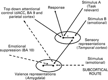
Fig. 1 represents the position that we have developed on the basis of the Desimone & Duncan (Reference Desimone and Duncan1995) model of attention and Pessoa & Ungerleider's (Reference Pessoa and Ungerleider2004) extension of this model to emotion. In this simplified representation (see also Mitchell et al. Reference Mitchell, Richell, Leonard and Blair2006), only two stimuli can be attended to: A and B. Following Desimone & Duncan (Reference Desimone and Duncan1995), in the absence of task demand information, the stimulus (or stimuli) that are attended to will be determined by bottom-up processes. If stimulus A is moving and bright, for example, and stimulus B is not, neurons representing stimulus A will be highly active and suppress, through the inhibitory connections, those units representing stimulus B. However, if stimulus A is a task-relevant stimulus and task demands stipulate a search for it, the neurons representing stimulus A will be primed by input from the frontal cortex and their activity boosted such that the neurons representing the target stimulus ‘A’ will even more effectively suppress those representing stimulus B. Considerable data demonstrate that top-down attentional control is mediated by the anterior cingulate cortex, lateral regions of the prefrontal cortex and the parietal cortex (Moran & Desimone, Reference Moran and Desimone1985; Kastner & Ungerleider, Reference Kastner and Ungerleider2000; Marois et al. Reference Marois, Chun and Gore2004).
A schematic of the attentional framework. Within this schematic only two stimuli can be attended to: A and B. A and B are representational competitors: stronger representation of A leads to weaker representation of B (and vice versa) due to the depicted inhibitory connections between these representations. In this figure, A is a task-relevant stimulus. Thus, goal-directed top-down attentional control will lead to priming of this representation, increasing its activity and its probability of suppressing the representation of B. In this figure, B is an emotional stimulus. Prior stimulus-reinforcement learning has led to an association between B and an emotional reaction. Thus B leads to activation of amygdala-dependent ‘valence representations’ that, because of their reciprocal connection with representational cortex, increase the representational strength of B.

Top-down attentional networks
Not depicted in Fig. 1, but worthy of note, is the suggestion that there are two top-down attentional networks that carry out the functions of alerting and executive attention respectively and that are innervated by different neurotransmitter systems (Posner & Rothbart, Reference Posner and Rothbart2007). Alerting involves monitoring incoming stimuli for particular targets. It is associated with right frontal and parietal regions of the cortex (Fan et al. Reference Fan, McCandliss, Fossella, Flombaum and Posner2005) and can be indexed by measures of continuous performance and vigilance tasks, that is tasks where the participant responds irregularly to a target stimulus presented rarely among non-target stimuli (Fossella et al. Reference Fossella, Sommer, Fan, Wu, Swanson, Pfaff and Posner2002).
Executive attention involves mechanisms for monitoring and resolving conflict among thoughts, feelings and responses. It is associated with the dorsomedial (including dorsal anterior cingulate) and lateral prefrontal cortex (Posner & Rothbart, Reference Posner and Rothbart2007) and can be indexed by tasks such as the Stroop tasks (Botvinick et al. Reference Botvinick, Braver, Barch, Carter and Cohen2001; Fan et al. Reference Fan, McCandliss, Fossella, Flombaum and Posner2005). It has also been suggested that this system is recruited during emotional regulation tasks, where the participant is encouraged to ‘reappraise’ the visual image; in such cases, it can be considered that the individual's attentional focus on the image and its semantic associations are altered (Posner & Rothbart, Reference Posner and Rothbart2007). Functional imaging studies examining this form of emotional regulation do indeed identify regions associated with the executive attention network; that is the dorsomedial [Brodmann Area (BA) 32, medial BA 8] frontal cortex and lateral prefrontal cortex (e.g. BA 9, 44, 46 and 47) (Ochsner et al. Reference Ochsner, Bunge, Gross and Gabrieli2002; Ochsner & Gross, Reference Ochsner and Gross2005; Phan et al. Reference Phan, Fitzgerald, Nathan, Moore, Uhde and Tancer2005).
Orienting is also worth noting. This involves selecting relevant information from sensory input and aligning attention with a source of sensory signals. The orienting system for visual events has been associated with posterior brain areas, including the superior parietal lobe and temporal parietal junction, as well as the frontal eye fields (Corbetta & Shulman Reference Corbetta and Shulman2002; Fan et al. Reference Fan, McCandliss, Fossella, Flombaum and Posner2005). It can be indexed by cued reaction time tasks (see below).
Emotional attention (for reviews of this literature, see Pessoa & Ungerleider, Reference Pessoa and Ungerleider2004; Vuilleumier, Reference Vuilleumier2005; Bishop, Reference Bishop2007)
Following Pessoa & Ungerleider (Reference Pessoa and Ungerleider2004), emotional attention can be understood as a function of the interaction between the temporal cortex and the amygdala. Emotional stimuli are unconditioned and conditioned stimuli (see Rolls, Reference Rolls1999); that is, stimuli that are either intrinsically aversive or appetitive (loud noises/food) or learnt to be aversive or appetitive (guns/money). Aversive and appetitive conditioning involves the interaction of the temporal cortex and the amygdala (LeDoux, Reference LeDoux1996); stimuli represented within the temporal cortex that become associated with valence information represented within the amygdala come to activate the amygdala. As the connections between the temporal cortex and the amygdala are reciprocal (Amaral et al. Reference Amaral, Price, Pitkanen, Carmichael and Aggleton1992), the activity of neurons representing emotional stimuli in the temporal cortex is further augmented by reciprocal feedback from the amygdala. With respect to Fig. 1, in the absence of top-down attentional control, stimulus B is more likely to be attended over stimulus A because only stimulus B (the emotional stimulus) receives additional feedback from the amygdala.
This means that if the emotional stimulus is a distracter to the stimulus determining task performance (as depicted in Fig. 1), then representational interference will be greater than if this distracter stimulus was neutral. Indeed, a variety of studies have shown that emotional stimuli cause greater interference than neutral stimuli (Vuilleumier et al. Reference Vuilleumier, Armony, Driver and Dolan2001; Harris & Pashler, Reference Harris and Pashler2004; Erthal et al. Reference Erthal, de Oliveira, Mocaiber, Pereira, Machado-Pinheiro, Volchan and Pessoa2005; Mitchell et al. Reference Mitchell, Richell, Leonard and Blair2006, Reference Mitchell, Nakic, Fridberg, Kamel, Pine and Blair2007; Blair et al. Reference Blair, Smith, Mitchell, Morton, Vythilingam, Pessoa, Fridberg, Zametkin, Sturman, Nelson, Drevets, Pine, Martin and Blair2007). Conversely, if the emotional stimulus is relevant to task performance, then there will be facilitation of performance, induced by the emotional nature of the stimulus (due to reciprocal activation from the amygdala), relative to neutral stimuli. This can be seen in emotional lexical decision paradigms, where healthy volunteers are usually significantly faster/more accurate to judge that emotional letter strings (e.g. ‘murder’) are words rather than neutral letter strings (e.g. ‘table’) (Graves et al. Reference Graves, Landis and Goodglass1981; Lorenz & Newman, Reference Lorenz and Newman2002). Lexical decisions are thought to be based on the familiarity of the semantic patterns activated by the words (Atkinson & Juola, Reference Atkinson, Juola and Kornblum1973; Plaut & Booth, Reference Plaut and Booth2000). If, as is argued (Nakic et al. Reference Nakic, Smith, Busis, Vythilingam and Blair2006), the semantic representations of emotional words both activate and receive reciprocal feedback from the amygdala, then they should be faster to recognize than neutral words. Of note, emotional words are associated with amygdala activity (Kuchinke et al. Reference Kuchinke, Jacobs, Grubich, Vo, Conrad and Herrmann2005; Nakic et al. Reference Nakic, Smith, Busis, Vythilingam and Blair2006) and, in the context of lexical decision, this activity is correlated significantly with activity in regions of the middle temporal cortex implicated in semantic processing (Nakic et al. Reference Nakic, Smith, Busis, Vythilingam and Blair2006).
The degree of automaticity of amygdala activation
The model depicted in Fig. 1 assumes that top-down attentional control can influence the response of the amygdala to emotional stimuli (cf. Pessoa & Ungerleider, Reference Pessoa and Ungerleider2004). If the neurons representing stimulus A are primed by the frontal–parietal network implicated in top-down attentional control, they will be more successful in suppressing the activity of neurons representing the emotional stimulus B, which will thus be associated with reduced amygdala activity. However, this issue remains debated. Alternative claims made include suggestions that: (i) amygdala activation, at least by fearful facial expressions, is automatic and not influenced by attention to the expression (Dolan & Vuilleumier, Reference Dolan and Vuilleumier2003); (ii) directing attention away from fearful expressions in fact increases amygdala activity to these expressions (Williams et al. Reference Williams, McGlone, Abbott and Mattingley2005); and (iii) increasing attentional load results in reduced specificity of the amygdala response so that it responds to disgusted facial expressions (but decreases insula activity to this expression) (Anderson et al. Reference Anderson, Christoff, Panitz, De Rosa and Gabrieli2003).
There have been some studies that have indicated that attentional manipulations do not alter the level of amygdala responding to fearful expressions; that is, that the amygdala response to these expressions is automatic (Vuilleumier et al. Reference Vuilleumier, Armony, Driver and Dolan2001; Anderson et al. Reference Anderson, Christoff, Panitz, De Rosa and Gabrieli2003). However, several other studies have shown that attentional manipulations considerably alter the response of the amygdala to fearful expressions (Pessoa et al. Reference Pessoa, McKenna, Gutierrez and Ungerleider2002, Reference Pessoa, Padmala and Morland2005; Bishop et al. Reference Bishop, Jenkins and Lawrence2007; Mitchell et al. Reference Mitchell, Nakic, Fridberg, Kamel, Pine and Blair2007) and other emotional stimuli (Ochsner & Gross, Reference Ochsner and Gross2005; Blair et al. Reference Blair, Smith, Mitchell, Morton, Vythilingam, Pessoa, Fridberg, Zametkin, Sturman, Nelson, Drevets, Pine, Martin and Blair2007). Moreover, it remains unclear why a particular type of stimulus, if cortically represented, would be immune from the impact of attention. It could be suggested (cf. Dolan & Vuilleumier, Reference Dolan and Vuilleumier2003) that the amygdala response to fearful expressions is in fact driven by activity through a subcortical route (which might be relatively immune to attentional load). However, it would be surprising if this route activated the amygdala to such a degree that input from the cortical representation of the expression could not further increase neural activity (see Mitchell et al. Reference Mitchell, Nakic, Fridberg, Kamel, Pine and Blair2007).
The subcortical route
A subcortical route to the amygdala is considered a necessary condition for automatic threat-processing. Fig. 1 depicts an assumed subcortical route to the amygdala for some emotional stimuli. There is evidence of two dissociable routes in other mammalian species at least for auditory stimuli (Armony et al. Reference Armony, Servan-Schreiber, Romanski, Cohen and LeDoux1997; LeDoux, Reference LeDoux and Aggleton2000). There have been analogous suggestions that information on the emotional expressions of others can be conveyed either by a subcortical pathway (retinocollicular–pulvinar–amygdalar) or by a cortical pathway (retinogeniculostriate–extrastriate–fusiform) (de Gelder et al. Reference de Gelder, Vroomen, Pourtois and Weiskrantz1999; Morris et al. Reference Morris, Ohman and Dolan1999; Adolphs, Reference Adolphs2002). Support for the suggestion of a subcortical pathway was provided by work with G. Y., a patient with a long-standing right-sided hemianopia after occipital lobe damage at the age of 8 years (de Gelder et al. Reference de Gelder, Vroomen, Pourtois and Weiskrantz1999). This ‘blindsight’ patient showed some ability to discriminate (by guessing) between different facial expressions in his blind hemifield. Later neuroimaging work with G. Y. demonstrated differential amygdala responses to fearful versus happy expressions when these were presented to both the blind and seeing hemifields. However, striate and fusiform activity only occurred in response to stimuli presented to the seeing hemifield (Morris et al. Reference Morris, DeGelder, Weiskrantz and Dolan2001).
The idea of a subcortical visual route to the amygdala is not without its critics, however (Pessoa, Reference Pessoa2005). The theoretical basis for the existence of the subcortical visual processing route is based on work involving the rodent auditory system (Quirk et al. Reference Quirk, Repa and LeDoux1995; LeDoux, Reference LeDoux1996). Evidence for a subcortical visual route has also been provided for rats (Shi & Davis, Reference Shi and Davis2001) but not yet for primates (Pessoa, Reference Pessoa2005). Moreover, the animal studies showed that the subcortical auditory processing route could not support differential conditioning (Armony et al. Reference Armony, Servan-Schreiber, Romanski, Cohen and LeDoux1997; LeDoux, Reference LeDoux and Aggleton2000). By contrast, early neuroimaging work suggested that the putative visual subcortical pathway in humans might be able to provide a detailed enough representation to allow differential conditioning (Morris et al. Reference Morris, Ohman and Dolan1999). However, later data examining the perceptual capacity of the subcortical route through the manipulation of the spatial frequency of face stimuli indicated that the pulvinar and superior colliculus carried coarse information (Vuilleumier et al. Reference Vuilleumier, Armony, Driver and Dolan2003). This would suggest that the visual subcortical pathway would not provide sufficient detail to support differential conditioning.
Frontal regulation of emotional responding
In addition to a subcortical system for increasing the salience of threat-related stimuli, the brain is thought to have regulatory mechanisms that reduce the impact of emotional processing when the emotional stimuli are insignificant or interfere with adaptive behavior. We have already discussed briefly one route, executive attention, by which the frontal cortex can influence emotional responding. However, there seem to be reasonable data indicating that specific regions of the frontal cortex may in fact exert an inhibitory influence on the amygdala directly. The medial prefrontal cortex projects to the basolateral amygdala (Ongur & Price, Reference Ongur and Price2000), and is thought to regulate emotion by selecting or inhibiting amygdala outputs (Rosenkranz & Grace, Reference Rosenkranz and Grace2001).
There have been suggestions that attentional control of emotional distracters recruits the rostral anterior cingulate cortex (rACC) (Whalen et al. Reference Whalen, McInerney, McNally, Wilhelm, Jenike and Rauch1998; Shin et al. Reference Shin, Whalen, Pitman, Bush, Macklin, Lasko, Orr, McInerney and Rauch2001; Vuilleumier et al. Reference Vuilleumier, Armony, Driver and Dolan2001; Bishop et al. Reference Bishop, Duncan, Brett and Lawrence2004; Etkin et al. Reference Etkin, Egner, Peraza, Kandel and Hirsch2006). However, some studies report dorsal rather than rostral ACC activity in response to emotional distracters (Davis et al. Reference Davis, Taylor, Hutchison, Dostrovsky, McAndrews, Richter and Lozano2005; Haas et al. Reference Haas, Omura, Constable and Canli2006; Blair et al. Reference Blair, Smith, Mitchell, Morton, Vythilingam, Pessoa, Fridberg, Zametkin, Sturman, Nelson, Drevets, Pine, Martin and Blair2007; Mitchell et al. Reference Mitchell, Nakic, Fridberg, Kamel, Pine and Blair2007). Moreover, work has shown that the rACC shows increased responding to emotional stimuli even in contexts where attentional control is not required (Breiter et al. Reference Breiter, Etcoff, Whalen, Kennedy, Rauch, Buckner, Strauss, Hyman and Rosen1996; Elliott et al. Reference Elliott, Dolan and Frith2000; Kosson et al. Reference Kosson, Budhani, Nakic, Chen, Saad, Vythilingam, Pine and Blair2006; Nakic et al. Reference Nakic, Smith, Busis, Vythilingam and Blair2006). It is therefore possible that some of the results that were suggested to indicate control over the amygdala's response in fact represented excitatory activation from the amygdala. Importantly, very few studies have manipulated distracter salience through emotional and non-emotional manipulation in the same study. Those that have, have indicated that emotional distracters, like neutral comparable distracters, lead to the recruitment of the same ‘executive attention’ neural systems: dorsal anterior cingulate, dorsolateral prefrontal and parietal cortices (Compton et al. Reference Compton, Banich, Mohanty, Milham, Herrington, Miller, Scalf, Webb and Heller2003; Luo et al. Reference Luo, Mitchell, Jones, Mondillo, Vythilingam and Blair2007).
In addition to the emotion regulation tasks that have concentrated on the regulation of emotion through reappraisal (i.e. the attention-based activation of different, or less negative, semantic representations of the stimuli; see Ochsner & Gross, Reference Ochsner and Gross2005), other studies have examined emotional regulation through suppression (i.e. the distancing of the self from the stimulus/the inhibition of any emotional reaction to the stimulus) (e.g. Beauregard et al. Reference Beauregard, Levesque and Bourgouin2001; Levesque et al. Reference Levesque, Eugene, Joanette, Paquette, Mensour, Beaudoin, Leroux, Bourgouin and Beauregard2003). Of interest, this form of emotional regulation has activated regions of the anterior orbital frontal cortex not typically seen during reappraisal paradigm; that is BA 10/11 (Beauregard et al. Reference Beauregard, Levesque and Bourgouin2001; Levesque et al. Reference Levesque, Eugene, Joanette, Paquette, Mensour, Beaudoin, Leroux, Bourgouin and Beauregard2003). Animal data suggest that this region has at least some interconnectivity with the amygdala (Ongur & Price, Reference Ongur and Price2000). Moreover, this region is also recruited during ‘automatic’ emotional suppressions, that is in the context of tasks where emotional distracters significantly interfere with task performance (e.g. the Attentional Stroop and Emotional Interrupt tasks; Blair et al. Reference Blair, Smith, Mitchell, Morton, Vythilingam, Pessoa, Fridberg, Zametkin, Sturman, Nelson, Drevets, Pine, Martin and Blair2007). This region may be suppressing amygdala activity by activating inhibitory interneurons within the amygdala (Rosenkranz & Grace, Reference Rosenkranz and Grace2001).
Theoretical accounts of psychopathy
Two main viewpoints can be distinguished in current theories on psychopathy. One view suggests that psychopathy reflects an attentional impairment (MacCoon et al. Reference MacCoon, Wallace, Newman, Baumeister and Vohs2004; Hiatt & Newman, Reference Hiatt, Newman and Patrick2006). An alternative view suggests that psychopathy reflects a specific form of emotional dysfunction (Patrick, Reference Patrick1994; Lykken, Reference Lykken1995; Blair et al. Reference Blair, Mitchell and Blair2005; Frick & Marsee, Reference Frick, Marsee and Patrick2006; Kiehl, Reference Kiehl2006). More detailed evaluations of these positions have been presented elsewhere (Blair, Reference Blair2006) and so are only summarized here.
The attentional theoretical perspective particularly reflects the work of Joe Newman and colleagues, who have provided an influential, attention-based model of psychopathy: the response modulation (RM) hypothesis (Newman, Reference Newman, Cooke, Forth and Hare1998; Lorenz & Newman, Reference Lorenz and Newman2002; Newman et al. Reference Newman, Brinkley, Lorenz, Hiatt, MacCoon, Herve and Yuille2007). This is a cognitive model that suggests that RM involves ‘a rapid and relatively automatic (i.e. non-effortful or involuntary) shift of attention from the effortful organization and implementation of goal-directed behavior to its evaluation’ (Newman et al. Reference Newman, Schmitt and Voss1997). This ‘brief and highly automatic shift of attention … enables individuals to monitor and, if relevant, use information that is peripheral to their dominant response set (i.e. deliberate focus of attention)’ (Lorenz & Newman, Reference Lorenz and Newman2002). Within this view, the core impairment with respect to psychopathy is thought to relate to reduced response modulation. According to this view, ‘the impulsivity, poor passive avoidance, and emotion-processing deficits of individuals with psychopathy may all be understood as a failure to process the meaning of information that is peripheral or incidental to their deliberate focus of attention’ (Lorenz & Newman, Reference Lorenz and Newman2002).
Within the emotion theoretical perspective, there is relative consensus that the amygdala is dysfunctional (Patrick, Reference Patrick1994; Blair et al. Reference Blair, Mitchell and Blair2005; Kiehl, Reference Kiehl2006). However, the extent to which other neural systems are also implicated remains debated. Blair et al. (Reference Blair, Mitchell and Blair2005) implicate the ventromedial prefrontal cortex as well as the amygdala whereas Kiehl (Reference Kiehl2006) implicates a considerably greater variety of structures including the anterior and posterior cingulate, superior temporal cortex and the hippocampus. With respect to the nature of the impairment, all positions agree that the processing of punishment information is disrupted (Patrick, Reference Patrick1994; Lykken, Reference Lykken1995; Blair et al. Reference Blair, Mitchell and Blair2005; Frick & Marsee, Reference Frick, Marsee and Patrick2006; Kiehl, Reference Kiehl2006), although Blair et al. (Reference Blair, Mitchell and Blair2005) have specified that this seems to involve the use of punishment information during stimulus-reinforcement rather than stimulus-response learning. There is some debate regarding the extent to which reward processing is disrupted. Some authors have argued that the deficit only implicates punishment information (Lykken, Reference Lykken1995; Frick & Marsee, Reference Frick, Marsee and Patrick2006). However, Blair et al. (Reference Blair, Mitchell and Blair2005) hold that stimulus–reward association formation and use is also dysfunctional, just to a lesser extent than stimulus–punishment association formation and use. This view is consistent with that of Verona et al. (Reference Verona, Patrick, Curtin, Bradley and Lang2004).
Attention and psychopathy
Bottom-up and top-down attentional control
Newman and colleagues have suggested that psychopathy may be associated with dysfunction in bottom-up and top-down attentional control. Thus, according to Hiatt & Newman (Reference Hiatt, Newman and Patrick2006, p. 346), ‘psychopaths proposed difficulty accommodating secondary information can be understood as difficulty accommodating bottom-up, stimulus-driven information, especially when the bottom-up information is inconsistent with or unrelated to the current top-down, effortful focus of attention’. Additionally, they have considered that psychopathy might reflect ‘a deficit in automatically allocating top-down attention’ (MacCoon et al. Reference MacCoon, Wallace, Newman, Baumeister and Vohs2004, p. 429). According to their position, in psychopathy top-down attention is less able to shift ‘automatically to nondominant cues that suggest a more adaptive response’ (MacCoon et al. Reference MacCoon, Wallace, Newman, Baumeister and Vohs2004, p. 429).
It is important to note that Newman and colleagues have proposed a rather different model of top-down attentional control from that assumed by Desimone & Duncan (Reference Desimone and Duncan1995) and other theorists (Allport, Reference Allport1980; Lavie, Reference Lavie1995; Corbetta & Shulman, Reference Corbetta and Shulman2002; Posner & Rothbart, Reference Posner and Rothbart2007). Rather than (or in addition to) top-down attention being involved in the priming of task-relevant representations, Newman and colleagues assume that it has a more general function in ‘automatically shifting to nondominant cues that might suggest a more adaptive response’. Indeed, they argue that ‘psychopathic individuals have a deficit in automatically allocating top-down resources to nondominant cues’ (MacCoon et al. Reference MacCoon, Wallace, Newman, Baumeister and Vohs2004, p. 429). However, from the perspectives of the models of top-down attention (Desimone & Duncan, Reference Desimone and Duncan1995; Posner & Rothbart, Reference Posner and Rothbart2007), it is unclear that top-down resources are automatically allocated to non-dominant cues. Moreover, it is unclear how a top-down attentional system could determine which non-dominant cues might suggest a more adaptive response; the computations driving this automatic shifting have yet to be specified. In addition, in at least one class of paradigms, individuals with psychopathy have been found to be unimpaired in shifting attention to non-dominant cues. These are the attentional set-shifting tasks; for example the Wisconsin Card Sorting Task (WCST) and the extradimensional set-shifting component of the Intradimensional/Extradimensional Discrimination (ID/ED) task. Individuals with psychopathy show no impairment on these tasks (LaPierre et al. Reference LaPierre, Braun and Hodgins1995; Blair et al. Reference Blair, Colledge and Mitchell2001; Mitchell et al. Reference Mitchell, Colledge, Leonard and Blair2002).
Top-down attentional networks
As noted above, alerting involves monitoring incoming stimuli for particular targets. It can be indexed by continuous performance and vigilance tasks (Posner & Rothbart, Reference Posner and Rothbart2007). Relatively little work has examined the ability of individuals with psychopathy to perform such tasks. Raine & Venables (Reference Raine and Venables1988) found no behavioral impairment in individuals with higher scores on the PCL relative to comparison individuals on a continuous performance task. Yet they did report larger and more prolonged P300 event-related potentials (ERPs) at parietal sites in individuals with psychopathy relative to comparison individuals and argued that this suggested at least short-term enhanced attention capacities in individuals with psychopathy. However, this has been questioned by data from Kiehl and colleagues. They observed reduced P300 ERPs in individuals with psychopathy during two different oddball tasks that involved an infrequent target (i.e. continuous performance) as well as task-irrelevant novel stimuli (Kiehl et al. Reference Kiehl, Hare, Liddle and McDonald1999, Reference Kiehl, Bates, Laurens, Hare and Liddle2006). Both these studies reported larger late ERP negativities in the individuals with psychopathy. Kiehl et al. (Reference Kiehl, Smith, Hare, Mendrek, Forster, Brink and Liddle2001, Reference Kosson, Budhani, Nakic, Chen, Saad, Vythilingam, Pine and Blair2006) point out that patients with temporal lobe damage, relative to controls, or patients with parietal lobe damage as well as epileptic patients following resection of the amygdala and anterior superior temporal gyrus show very similar results (Johnson, Reference Johnson1993; Yamaguchi & Knight, Reference Yamaguchi, Knight, Haschke, Roitbak and Speckmann1993). Kiehl et al. (Reference Kiehl, Bates, Laurens, Hare and Liddle2006) suggest that these data might reflect ‘abnormal attention and orienting processes’ in individuals with psychopathy. However, if their anatomical analysis proves correct, it would suggest that the abnormal attention relates to emotional attention (see below) rather than any form of top-down attentional control. Neither the amygdala nor the temporal cortex is implicated in any of the putative top-down attentional systems (Corbetta & Shulman, Reference Corbetta and Shulman2002; Posner & Rothbart, Reference Posner and Rothbart2007). As such, these data cannot be used as evidence for positions suggesting enhanced selective attention through top-down attentional systems (see below).
Executive attention involves mechanisms for monitoring and resolving response conflict and can be indexed by response conflict paradigms such as the Stroop task and its variants. A series of studies have examined executive attention in individuals with psychopathy (Newman et al. Reference Newman, Schmitt and Voss1997; Mitchell et al. Reference Mitchell, Colledge, Leonard and Blair2002; Hiatt et al. Reference Hiatt, Schmitt and Newman2004; Blair et al. Reference Blair, Newman, Mitchell, Richell, Leonard, Morton and Blair2006; Dvorak-Bertsch et al. Reference Dvorak-Bertsch, Sadeh, Glass, Thornton and Newman2007; Vitale et al. Reference Vitale, Brinkley, Hiatt and Newman2007). These studies have indicated that executive attention is either intact or even possibly superior in individuals with psychopathy. Thus, individuals with psychopathy show no indications of the increased interference on the classic color-word Stroop task (Hiatt et al. Reference Hiatt, Schmitt and Newman2004; Dvorak-Bertsch et al. Reference Dvorak-Bertsch, Sadeh, Glass, Thornton and Newman2007) or counting Number Stroop tasks (Blair et al. Reference Blair, Newman, Mitchell, Richell, Leonard, Morton and Blair2006), and may show decreased interference on some color-word Stroop variants and reading Number Stroop tasks (see below). These data suggest that regions of the dorsomedial (including the dorsal anterior cingulate cortex) and dorsolateral prefrontal cortex necessary for executive attention are not dysfunctional in psychopathy.
There have also been reports of reduced interference on color word Stroop tasks, where the target and distracter information are spatially separated (e.g. the word ‘blue’ is written in white ink and surrounded by a green triangle; Hiatt et al. Reference Hiatt, Schmitt and Newman2004). Individuals with psychopathy have also been found to show slightly reduced interference on a reading Number Stroop task (Blair et al. Reference Blair, Newman, Mitchell, Richell, Leonard, Morton and Blair2006) and in a picture-word interference task (Newman et al. Reference Newman, Schmitt and Voss1997; Vitale et al. Reference Vitale, Brinkley, Hiatt and Newman2007). In this context it is worth noting the results of Hare & Jutai (Reference Hare, Jutai, Van Dusen and Mednick1983), who presented distracter tones to participants while they engaged in a video game. ERPs to the tone indexed representation of the task-irrelevant tone stimulus. The individuals with psychopathy showed a significantly reduced N100 amplitude to the tone relative to the comparison individuals only while playing the video game, not if the tone was presented alone (Hare & Jutai, Reference Hare, Jutai, Van Dusen and Mednick1983). In short, these last data indicate the possibility of enhanced executive attention in individuals with psychopathy.
There have been suggestions that data from passive avoidance paradigms can be considered evidence for an attentional viewpoint (Newman et al. Reference Newman, Brinkley, Lorenz, Hiatt, MacCoon, Herve and Yuille2007; MacCoon et al. Reference MacCoon, Wallace, Newman, Baumeister and Vohs2004; Hiatt & Newman, Reference Hiatt, Newman and Patrick2006). In passive avoidance paradigms, participants must learn to approach those stimuli that become associated with reward, and avoid those stimuli that become associated with punishment (Newman & Kosson, Reference Newman and Kosson1986). Such paradigms are functionally identical to go/no-go paradigms used in animal work (Schoenbaum et al. Reference Schoenbaum, Chiba and Gallagher1999, Reference Schoenbaum, Nugent, Saddoris and Setlow2002) although rather different from the go/no-go paradigm used in human work (where no stimulus-reinforcement value learning is necessary, the participant is told to respond to one type of stimulus and avoid another). There are considerable data indicating that individuals with psychopathy are impaired in passive avoidance learning; specifically, they fail to learn to avoid the stimuli associated with punishment (Newman & Kosson, Reference Newman and Kosson1986; Newman & Schmitt, Reference Newman and Schmitt1998; Blair et al. Reference Blair, Mitchell, Leonard, Budhani, Peschardt and Newman2004). However, it is unclear how such data are compatible with classic models of top-down attentional control. On trials in which the participant receives punishment, there are no representational competitors to this punishment information that could suppress its processing; reward information may have been delivered but this would have occurred on a previous trial at least several seconds before. Moreover, if top-down attentional control was necessary for performance on the passive avoidance paradigm, it would suggest that lesions of regions implicated in top-down attentional control (e.g. dorsal anterior cingulate, dorsolateral prefrontal cortex and parietal cortex) would disrupt passive avoidance learning. However, there is no evidence in support of this, although there are considerable data that lesions of the amygdala disrupt performance on this task (Ambrogi Lorenzini et al. Reference Ambrogi Lorenzini, Baldi, Bucherelli, Sacchetti and Tassoni1999; Gallagher & Schoenbaum, Reference Gallagher and Schoenbaum1999; Schoenbaum et al. Reference Schoenbaum, Chiba and Gallagher1999). In short, although data from passive avoidance studies have been cited in support of attention-based accounts of psychopathy, the data currently suggest that such tasks index stimulus-reinforcement (reward and punishment) learning rather than attentional mechanisms.
Orienting involves selecting relevant information from sensory input and aligning attention with a source of sensory signals and can be indexed by cued reaction time tasks. Again, very little work has investigated orienting in individuals with psychopathy. However, Howland et al. (Reference Howland, Kosson, Patterson and Newman1993) used a cued reaction time task. In this task, participants responded to target stimuli presented on either side of the screen with left and right button presses respectively. Preceding the presentation of each target stimulus, a cue appeared on the left, right, or center of the screen. On valid trials the cue and target stimulus were on the same side, for invalid trials on opposite sides, and in the center of the screen for neutral trials. In this task, individuals with psychopathy made more errors than comparison individuals on invalidly cued trials (although the effect was significant only for left-side target stimuli) and neutral cues (here the effect was significant only for right-side target stimuli). These data, although not robust (given the alternating laterality of the effects), suggest a strengthened orienting response in individuals with psychopathy that is more difficult to override. The specific neurocognitive mechanism for this difficulty remains unclear.
Emotional attention
As stated earlier, emotional attention can be understood as a function of the interaction between the temporal cortex and the amygdala; the activity of neurons representing emotional stimuli in the temporal cortex is further augmented by reciprocal feedback from the amygdala (cf. Pessoa & Ungerleider, Reference Pessoa and Ungerleider2004; Mitchell et al. Reference Mitchell, Richell, Leonard and Blair2006). This means that if the emotional stimulus is a distracter to the task-relevant stimulus, then representational interference will be greater in healthy individuals than if this distracter stimulus was neutral. If psychopathy is associated with amygdala dysfunction, as has been suggested (Patrick, Reference Patrick1994; Blair et al. Reference Blair, Mitchell and Blair2005; Kiehl, Reference Kiehl2006), then significantly less interference by emotional distracters should be seen. Two studies have examined this issue (Christianson et al. Reference Christianson, Forth, Hare, Strachan, Lidberg and Thorell1996; Mitchell et al. Reference Mitchell, Richell, Leonard and Blair2006). Mitchell et al. (Reference Mitchell, Richell, Leonard and Blair2006) examined the extent to which an emotional distracter presented immediately before and after very rapidly presented target stimuli interfered with simple motor responses to these target stimuli. Although the comparison individuals showed significant interference to both positive and negative relative to neutral distracters, the individuals with psychopathy showed no significant interference to these distracters. Christianson et al. (Reference Christianson, Forth, Hare, Strachan, Lidberg and Thorell1996) examined the extent to which emotional, relative to neutral, content in images interfered with the recollection of non-emotional, peripheral content. The comparison individuals showed significantly reduced recall of peripheral content for emotional slides whereas the individuals with psychopathy did not.
For situations in which the task-relevant stimulus is emotionally salient, healthy individuals generally show enhanced task performance. This facilitation is thought to occur because of reciprocal activation from the amygdala that enhances the competitive strength of the task-relevant emotional stimulus. By contrast, if, as is proposed for psychopathy, there is amygdala dysfunction, individuals with this disorder will not benefit from reciprocal activation and will not show facilitation of performance by emotional task-relevant material. As noted earlier, this can be indexed by emotional lexical decision paradigms (Nakic et al. Reference Nakic, Smith, Busis, Vythilingam and Blair2006), where individuals with psychopathy show significantly reduced facilitation relative to comparison individuals (Williamson et al. Reference Williamson, Harpur and Hare1991; Lorenz & Newman, Reference Lorenz and Newman2002).
It is interesting to note data from patients with a disorder that in some functional respects represents an inverse to psychopathy: post-traumatic stress disorder (PTSD). Considerable neuroimaging data demonstrate that individuals with PTSD show elevated amygdala responses to threat stimuli, even threat stimuli such as fearful expressions that are not part of the original trauma (Bremner, Reference Bremner2004; Liberzon & Martis, Reference Liberzon and Martis2006; Rauch et al. Reference Rauch, Shin and Phelps2006). On the basis of the above analysis, patients with PTSD should show heightened interference by emotional distracters (rather than the reduced interference seen in psychopathy) and heightened facilitation by emotional targets (rather than the reduced facilitation seen in psychopathy). In line with this suggestion, considerable data demonstrate that patients with PTSD show heightened interference by emotional distracters (Williams et al. Reference Williams, Mathews and MacLeod1996; Bar-Haim et al. Reference Bar-Haim, Lamy, Pergamin, Bakermans-Kranenburg and van2007). Recent data also demonstrate that patients with PTSD show heightened facilitation by emotional targets (Vythilingam et al. Reference Vythilingam, Blair, McCaffrey, Scaramozza, Jones, Nakic, Mondillo, Hadd, Bonne, Mitchell, Pine, Charney and Blair2007).
Functional imaging data can also reveal the priming of emotional representations in the temporal cortex by reciprocal amygdala activation. Thus, several studies have shown heightened fusiform/temporal cortex responses to emotional relative to neutral expressions (Vuilleumier et al. Reference Vuilleumier, Armony, Driver and Dolan2001; Mitchell et al. Reference Mitchell, Nakic, Fridberg, Kamel, Pine and Blair2007). This is thought to represent, in part, the reciprocal feedback of the amygdala to this region of the temporal cortex. Studies examining expression processing in both adults with psychopathy (Deeley et al. Reference Deeley, Daly, Surguladze, Tunstall, Mezey, Beer, Ambikapathy, Robertson, Giampietro, Brammer, Clarke, Dowsett, Fahy, Phillips and Murphy2006) and children with psychopathic traits (Marsh et al. Reference Marsh, Finger, Mitchell, Reid, Sims, Kosson, Towbin, Leibenluft, Pine and Blair2008) have shown a reduced differential response with the fusiform cortex to fearful relative to neutral expressions. These data are consistent with reduced priming of emotion-relevant representations in the temporal cortex by reciprocal amygdala activation in individuals with psychopathy (although only Marsh et al. Reference Marsh, Finger, Mitchell, Reid, Sims, Kosson, Towbin, Leibenluft, Pine and Blair2008 observed reduced amygdala activity).
Implications of the attention data for theoretical views on psychopathy
The RM hypothesis
As noted earlier, the RM hypothesis is an influential, attention-based model of psychopathy (Newman, Reference Newman, Cooke, Forth and Hare1998; Lorenz & Newman, Reference Lorenz and Newman2002; Newman et al. Reference Newman, Brinkley, Lorenz, Hiatt, MacCoon, Herve and Yuille2007). However, the degree to which the model's implicit conception of attention in healthy individuals is consistent with dominant cognitive neuroscience views on attention is unclear (Allport, Reference Allport1980; Desimone & Duncan, Reference Desimone and Duncan1995; Corbetta & Shulman, Reference Corbetta and Shulman2002; Posner & Rothbart, Reference Posner and Rothbart2007). In particular, it is unclear how the frontal cortex begins ‘automatically shifting to nondominant cues that might suggest a more adaptive response’. Moreover, individuals with psychopathy are unimpaired in shifting attention to nondominant cues in attentional set-shifting tasks such as the WCST and the extra-dimensional set-shifting component of the ID/ED task (LaPierre et al. Reference LaPierre, Braun and Hodgins1995; Blair et al. Reference Blair, Colledge and Mitchell2001; Mitchell et al. Reference Mitchell, Colledge, Leonard and Blair2002). In addition, individuals with psychopathy appear sensitive to non-task relevant information in classic color-word Stroop and counting Number Stroop paradigms (Hiatt et al. Reference Hiatt, Schmitt and Newman2004; Blair et al. Reference Blair, Newman, Mitchell, Richell, Leonard, Morton and Blair2006; Dvorak-Bertsch et al. Reference Dvorak-Bertsch, Sadeh, Glass, Thornton and Newman2007).
It could be argued that Raine & Venables' (Reference Raine and Venables1988) finding of larger and more prolonged P300 ERPs during continuous performance tasks in individuals with psychopathy suggests enhanced attention capacities. The suggestion might be of over-priming of task-relevant stimuli leading to the suppressed representation of other stimuli in the environment (cf. Desimone & Duncan, Reference Desimone and Duncan1995). By this account, the considerable emotional impairment seen in individuals with psychopathy would reflect enhanced attention of non-emotional environmental features (cf. Hiatt & Newman, Reference Hiatt, Newman and Patrick2006). In essence, individuals with psychopathy would be showing enhanced attention-based emotional regulation (cf. Ochsner & Gross, Reference Ochsner and Gross2005). Consistent with such an ‘exaggerated selective attention’ version of the RM hypothesis are reports of reduced interference on some Stroop tasks variants (Newman et al. Reference Newman, Schmitt and Voss1997; Hiatt et al. Reference Hiatt, Schmitt and Newman2004; Blair et al. Reference Blair, Newman, Mitchell, Richell, Leonard, Morton and Blair2006; Vitale et al. Reference Vitale, Brinkley, Hiatt and Newman2007) and reduced processing of distracter tones while engaged in a video game (Hare & Jutai, Reference Hare, Jutai, Van Dusen and Mednick1983). However, such an account does face difficulties in dealing with the selective nature of the reduction in interference seen in Stroop-type variants (see Hiatt et al. Reference Hiatt, Schmitt and Newman2004; Blair et al. Reference Blair, Newman, Mitchell, Richell, Leonard, Morton and Blair2006; Dvorak-Bertsch et al. Reference Dvorak-Bertsch, Sadeh, Glass, Thornton and Newman2007). In short, an attention-based account for psychopathy cannot be considered supported as yet.
Emotion dysfunction accounts
Accounts suggesting that the functioning of the amygdala is dysfunctional in psychopathy are supported by data indicating reduced emotional attention in psychopathy. This reduced priming of emotional representations is seen as reduced interference by emotional distracters (Christianson et al. Reference Christianson, Forth, Hare, Strachan, Lidberg and Thorell1996; Mitchell et al. Reference Mitchell, Richell, Leonard and Blair2006), reduced facilitation of emotional targets (Williamson et al. Reference Williamson, Harpur and Hare1991; Lorenz & Newman, Reference Lorenz and Newman2002) and reduced utilization of relevant stimulus features (Dadds et al. Reference Dadds, Perry, Hawes, Merz, Riddell, Haines, Solak and Abeygunawardane2006). Functional imaging data also indicate reduced activation of emotional representations in the temporal cortex potentially because of reduced reciprocal amygdala activation (Deeley et al. Reference Deeley, Daly, Surguladze, Tunstall, Mezey, Beer, Ambikapathy, Robertson, Giampietro, Brammer, Clarke, Dowsett, Fahy, Phillips and Murphy2006; Marsh et al. Reference Marsh, Finger, Mitchell, Reid, Sims, Kosson, Towbin, Leibenluft, Pine and Blair2008). Moreover, it is worth remembering Kiehl and colleagues' anatomical analysis of their findings of larger late ERP negativities in the individuals with psychopathy as indicative of amygdala/temporal cortex damage (Kiehl et al. Reference Kiehl, Smith, Hare, Mendrek, Forster, Brink and Liddle2001, Reference Kiehl, Bates, Laurens, Hare and Liddle2006). In addition, recent work has shown that patients with amygdala lesions show reduced eye gaze to the eye region, that this is related to their impairment in fearful face recognition and that attentional instructions to focus on the eye region could abolish the recognition impairment (Adolphs et al. Reference Adolphs, Gosselin, Buchanan, Tranel, Schyns and Damasio2005). Importantly, recent work by Dadds et al. (Reference Dadds, Perry, Hawes, Merz, Riddell, Haines, Solak and Abeygunawardane2006) in children with psychopathic tendencies has reported reduced eye gaze to the eye region and that attentional instructions to focus on the eye region abolish the recognition impairment. In short, the emotion attention literature in psychopathy is supportive of emotion dysfunction-based accounts of the disorder (Patrick, Reference Patrick1994; Lykken, Reference Lykken1995; Blair et al. Reference Blair, Mitchell and Blair2005; Frick & Marsee, Reference Frick, Marsee and Patrick2006; Kiehl, Reference Kiehl2006).
It is worth noting the emotion dysfunction accounts provide no explanation of the reduced interference seen on some Stroop task variants (Newman et al. Reference Newman, Schmitt and Voss1997; Hiatt et al. Reference Hiatt, Schmitt and Newman2004; Blair et al. Reference Blair, Newman, Mitchell, Richell, Leonard, Morton and Blair2006; Vitale et al. Reference Vitale, Brinkley, Hiatt and Newman2007). It is possible that the reduced interference reflects weaker representation of the distracter information (i.e. reading is less of a prepotent response), possibly due to the reduced education/greater truancy of individuals with psychopathy (Blair et al. Reference Blair, Mitchell and Blair2005). However, this would not account for the reduced interference on the spatially distributed color-word Stroop tasks (Hiatt et al. Reference Hiatt, Schmitt and Newman2004). Importantly, it does provide a clear prediction: individuals with psychopathy would show weaker representation of [reduced blood oxygen level dependent (BOLD) responses to] similar information in non-conflict situations. In other words, they would show reduced neural activity to word stimuli presented as non-Stroop stimuli (generally reduced representation of these stimuli would mean less interference from their representation in Stroop contexts). This prediction remains to be examined.
Conclusions
In this review, we relate the cognitive neuroscience of attention to relevant data from work with individuals with psychopathy. Considerable progress has been made in understanding the neural systems involved in attention and their functional contributions. This enables a more detailed analysis of any potential attention deficit in psychopathy.
There are hints of enhanced top-down attentional systems in psychopathy, particularly in some aspects of executive attention. These would be consistent with an ‘exaggerated selective attention’ version of Newman's RM hypothesis. However, it remains, for example, unclear why changes in the spatial location of the distracter information on Stroop tasks should have such striking effects on the performance of individuals with psychopathy. Such data are not easily explained within dominant cognitive neuroscience views of attention.
The data do suggest reduced emotional attention in psychopathy; that is reduced priming of emotion-relevant representations in the temporal cortex by the amygdala. These data are consistent with models of psychopathy stressing amygdala dysfunction (Patrick, Reference Patrick1994; Blair et al. Reference Blair, Mitchell and Blair2005; Kiehl, Reference Kiehl2006) and emotion-based models of the disorder more generally (Lykken, Reference Lykken1995; Frick & Marsee, Reference Frick, Marsee and Patrick2006). It is to be hoped that greater understanding of the neurocognitive basis of psychopathy will be translated into more efficacious treatments of this disorder in the near future.
Acknowledgments
R. J. R. B. was supported by the Intramural Research Program of the NIH:NIMH. D. G. V. M. was supported by funding from the Natural Sciences and Engineering Research Council of Canada, and the Ontario Mental Health Foundation New Investigator Fellowship.
Declaration of Interest
None.