Introduction
Conduction system pacing has emerged as a more physiologically driven alternative to apical right ventricular pacing and is now being utilised in selected cases for cardiac resynchronisation therapy. Reference Vijayaraman, Chelu and Curila1 Pacing from the right ventricular apex results in slow myocyte-to-myocyte propagation of the electrical activation wavefront throughout both ventricles. Consequently, surface electrocardiograms exhibit a wide QRS complex and left bundle branch block pattern, indicative of electrical dyssynchrony. Reference Paton and Witte2 The main concern in that most adult patients studies compare LBA pacing only right ventricular apical pacing. The concept of selective-site pacing was developed with the aim of preventing the negative continuum associated with ventricular apical pacing, and includes pacing from the septum, from the outflow tract, and from selective His bundle pacing. Reference Cantù, De Filippo and Gabbarini3 Children will be exposed to pacing for a longer period than adults, making conduction system pacing crucial. The proximal interventricular septum has been demonstrated in studies as a better alternative pacing site compared to the apical region. Reference Karpawich, Singh and Zelin4 Permanent His bundle pacing (selective or non-selective) has been found to be the most physiological as it does not induce ventricular dyssynchrony (electrical or mechanical). Reference Mar, Devabhaktuni and Dandamudi5 Left bundle branch area pacing is a more recent technique gaining rapid acceptance due to its broader target area and excellent electrical parameters. Reference Dai, Dai and Guo6 We would like to share the short-term results of our case series on left bundle branch area pacing in paediatric patients.
Material and method
Patient population
Between December 2021 and April 2024, a total of 19 patients who underwent left bundle branch area pacing were included in the study. All procedures were conducted at a single centre and approved by the local ethics committee. Written informed consent was obtained from all patients before the procedure. Clinical and periprocedural data were collected from electronic medical records and the electrophysiology laboratory database. Ten patients were male, and nine were female. All patients had complete atrioventricular block and met a class I or IIa indication for permanent pacemaker implantation according to the European Society of Cardiology guidelines. Reference Glikson, Nielsen and Kronborg7 Seven patients had congenital complete atrioventricular block, while 12 had postoperative complete atrioventricular block. In some CHDs, particularly in complete atrioventricular septal defects, the location of the AV node and His bundle is often more posterior–inferior. Therefore, special attention should be paid during implantation, and the thickness of the basal interventricular septum and the presence of a septal patch in the heart were evaluated before the procedure. The left ventricular end-diastolic diameter and interventricular septum thickness were measured using parasternal long-axis M-mode echocardiography. synthetic tissue, the presence of a patch, and asynchronous contraction render M-mode cardiac function assessment inadequate. A left ventricular ejection fraction was measured using modified Simpson’s method, and a value below 50% was accepted as indicative of reduced systolic function. A left ventricular end-diastolic diameter z-score above two was considered increased. The measurements were performed by the same operator. To minimise the margin of error, the average of at least three measurements was taken.
Implantation technique
All procedures were conducted under deep sedation without intubation by a single paediatric electrophysiologist. There is no universally recognised method for the implantation of leads in congenital heart disease. The preoperative assessment involves a thorough examination of the patient’s anatomical and surgical history, along with a review of original operative reports whenever available. Reference Moore and Dalal8 Prior to each implantation, venography was performed from an ipsilateral peripheral intravenous line. Endocardial leads were inserted using the percutaneous subclavian vein technique in all cases. Furthermore, the pacemaker generator was consistently positioned in a prepectoral subfascial location for all patients. The SelectSecure lead (model 3830, Medtronic Inc., Minneapolis, USA) and Select Site C315 His or C304 His sheaths (Medtronic Inc., Minneapolis, USA) are used in operation. The 12-lead electrocardiography recordings and the intracardiac electrograms from the pacing lead were simultaneously displayed and continuously recorded on an electrophysiology recording system. First of all, a sheath was placed in the right femoral vein, and from there, the His catheter was advanced to the His region to identify the His area. The delivery sheath was inserted via the left subclavian vein or axillary vein. Next, the Select Secure pacing lead was advanced through the sheath, positioning its tip just beyond the distal part of the sheath for pacing and local activation potential recording. The sheath, along with the pacing lead, was then carefully advanced towards the His region, specifically inferior to the septal leaflet of the tricuspid valve, approximately 1–1,5 cm below it according to the patient’s age and weight under fluoroscopic guidance in the right anterior oblique 30° view. The lead made contact with the septal region (the right side of the septum). Unipolar pacing was performed from here. Criterias indicating right ventricular septal pacing are the presence of a “W” morphology in lead V1, discordance between leads aVR (negative QRS) and aVL (positive QRS), and lead II exhibiting a more positive R wave than lead III. During the procedure, the lead is carefully inserted into the septum with the aid of fluoroscopy, using a 30° left and right anterior oblique view. The sheath is positioned counterclockwise on the septum, with the tip pointing towards approximately two o'clock in the left anterior oblique view. If there is a patch in the septal region, slightly more inferiorly, or if there is scar tissue, we performed fixation in a location that would pace the left bundle branch in proximity. Then the lead is securely advanced into the septum by rotating (Figure 1). Subsequently, impedance sensing and post-pacing QRS morphology were evaluated. As the lead progresses further into the septum, the impedance increases. A unipolar impedance drop of 50–100 ohms suggests that the tip electrode has engaged the left ventricular endocardium. If left bundle capture is not achieved, the lead is advanced while taking care to avoid perforating the septum. Consistent left ventricular activation time of less than 80 milliseconds, measured as the duration from the stimulation artieact to the peak of the R wave in lead V6 (Figure 2), indicates successful left bundle capture. Reference Padala and Ellenbogen9 It should be noted that recording the left bundle potential during the procedure is optional and not essential for achieving left bundle capture. Caution should be exercised to avoid excessive rotations beyond this point, as it may cause the lead to cross into the left ventricular cavity. After achieving an appropriate position, the sheath was removed using a specialised slitting tool. To prevent lead stretching during the growth of smaller paediatric patients, a redundant lead loop was created in the right atrium, and an unwinding test was performed for both ventricular and atrial leads by pulling the leads from the pacemaker pocket, following previously described procedures. Reference Cantu, De Filippo and Gabbarini10 In smaller patients, a loop measuring approximately 60–80 mm was created, allowing for growth over approximately 6–8 years in the paediatric age group (with 10 mm of loop length considered sufficient to accommodate body growth per year). Reference Gheissari, Hordof and Spotnitz11 Great care was taken to ensure that the loop did not induce ectopic beats, was not overbent to the point of being prone to fracture, and did not pose a risk of protruding into the right ventricle. Lead adhesions over time can often negate any pre-determined looping. For older patients over eight years of age, the lead length was determined by calculating the target height and estimating the yearly increase in the patients' height. The delivery sheath was then gently withdrawn, and pacing parameters were rechecked. Left bundle branch area pacing was confirmed by previously published criteria Reference Chen, Wu, Su, Su and Huang12
( a ) In the radiographic image in the RAO position, the catheter at the his position is indicated with a star, and the lead near the left bundle is indicated with an arrow ( b ) the radiological image of the injection in the septum and lead position is shown in the LAO view before lead implantation ( c ) AP radiography after pacemaker implantation of a different patient who developed complete AV block after coarctation surgery.

( a ) It has been shown that the left ventricular activation time is very short ( time to peak = 68 msec). ECG paper speed: 50 mm/second, sensitivity: 1 mv/10 mm ( b ) post-procedure ECG of the same patient. ECG paper speed: 25 mm/second, sensitivity: 1 mv/10 mm. In a 16-year-old patient who developed permanent atrioventricular block due to traumatic effects of the guide wire in the left ventricular outflow tract during stent implantation for aortic coarctation, we initially performed left bundle pacing. We have been monitoring the patient for 2 years. The pacing parameters are quite good, and we are following the cardiac functions as normal.

Statistical analysis
Statistical analysis was performed using SPSS 22.0 (IBM Corporation, Armonk, New York, United States). Quantitative data were expressed as mean ± standard deviation or median range (minimum-maximum) values. Categorical values were presented as n (number) and % (percentage). Differences between two groups were compared using Wilcoxon’s signed-rank test. The p-value < 0.05 was considered statisticallly significant.
Results
Out of the patients, 10 were male, and nine were female. The median age and weight were seven Reference Paton and Witte2–Reference Ponnusamy, Basil and Vijayaraman18 and 38 kg (13–56), respectively. All patients had complete atrioventricular block. Seven patients had congenital complete atrioventricular block, while 12 patients had postoperative complete atrioventricular block (five ventricular septal defect, four atrioventricular septal defect, one atrial septal defect, one double outlet right ventricle, and one coarctation of aorta) (Table 1). Five patients who had undergone congenital heart surgery and four patients with isolated complete atrioventricular block were previously subjected to epicardial right ventricular pacing. Among these patients, those in VVIR mode were set to a lower rate of 60. Atrioventricular (DDD mode) leads were implanted in 10 patients, and ventricular leads (VVIR mode) were implanted in nine patients. In five of our patients, ventricular dysfunction developed due to prior right ventricular epicardial pacing. Two of these patients had isolated atrioventricular block, while the rest developed after congenital heart surgery. The median time for the development of ventricular dysfunction due to right ventricular pacing was 62 months (12–112). Of these patients in whom cardiac dysfunction developed, two were undergoing pacing in VVIR mode, while the remainings were in DDD mode. Left bundle branch pacing were applied in the remaining ten patients as the initial procedure. Three of these patients with isolated atrioventricular block had normal ejection fraction values; however, their left ventricular end-diastolic diameter z-scores were above two. The other seven patients developed atrioventricular block after surgery. We observed left bundle branch potential in only six of our patients.
Patients characteristics

Values are n (number), median (min-max), M male, F female, kg kilogram, AV = atrioventricular; AVSD = atrioventricular septal defect; VSD = ventricular septal defect; ASD = atrial septal defect; DORV = double outlet right ventricle.
Before the procedure, the measurement of median interventricular septal thickness was 7.2 mm (5.7–10.2) in M-mode echocardiography performed in the parasternal long axis. The Z score for interventricular septum according to BSA was a median of + 1.2 with a range of 0.9 to 1.5.
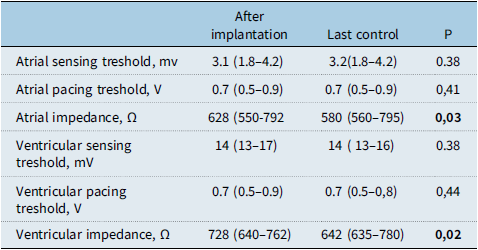
The paced QRS duration after left bundle branch area pacing was 92 msec (80–117). In one patient, left bundle branch area pacing could not be performed, and implantation was done in a septal position close to the left bundle. The median R wave amplitude, threshold, and impedance values were 14.7 mV (13.3–16.8), 0.7 mV (0.5–1.1), and 728 ohms (640–762), respectively (Table 2). The median procedure duration was 145 minutes (80–218). The median fluoroscopy dose was 18.7 mGy (13.5–34.2).
Lead parameters during implantation time and last control

Values are given as median ( minimum-maximum). MV = milivolt; V = Volt; Ω = ohm.
Postoperative echocardiography revealed that the electrodes were located near the endocardium of the left ventricular septum. No complications such as myocardial perforation and pericardial effusion occurred.
Follow-up
After the procedure, patients were monitored at one month post-procedure and then at three months intervals. The mean follow-up duration was 16.8 months (ranging from 3 to 29 months). There were no significant changes in pacing parameters such as R wave amplitude and threshold. Although the changes in impedance values were statistically significant, these changes remained within normal limits. In all patients who experienced cardiac dysfunction from right ventricular epicardial pacing, the left ventricular ejection fraction measured by the modified Simpson method increased above 65% within six months at most. In three of these patients, ejection fraction and left ventricular enddiastolic diameter z scores returned to normal at three months, while in the remaining two patients, this occurred at six months. During the follow-up period, no infections, lead dislocations, pocket haematomas, pneumothorax, or pericardial/pleural effusions were observed. Additionally, tricuspid valve issues were not detected in any patient during echocardiographic checks.
Discussion
The short duration of the QRS complex is important as it helps preserve cardiac function in the long term. While left bundle branch pacing shows progress in achieving ventricular synchronisation compared to non-conduction system pacing, it does not result in physiological interventricular synchronisation because it can cause a right bundle branch block. Especially in paediatric patients, the risk of developing ventricular dysfunction is higher due to prolonged exposure to pacing compared to adults. A number of studies have shown that septal pacing can recreate a more synchronised contraction and a narrow QRS complex while maintaining LV function. However, in pacing performed from the septal region of the right ventricle, there can be different effects on cardiac contraction depending on the location. Reference Peschar, de Swart, Michels, Reneman and Prinzen13 Therefore, conduction system pacing is crucial in today’s practice. In studies related to His bundle pacing, high threshold values, the need for early replacement, and an increase in threshold values are commonly observed issues associated with the development of blockages below the His bundle. Reference Vijayaraman, Chung and Dandamudi14 Consequently, it has become less preferred. Although there is no clear evidence showing definitive indications for left bundle branch area pacing in paediatric patients, it has become a more preferred modality today due to its ability to provide appropriate pacing parameters and physiological pacing. The noticification of cases in children is limited. Reference Wenlong, Baojing, Chencheng and Jianzeng15 To the best of our knowledge, our study is the largest series conducted in children.
The success criteria for left bundle branch pacing area include the following: 1) The QRS pattern of pacing should display a right bundle branch block or an R wave in lead V1; 2) the presence of a left bundle branch potential should be recorded during the intrinsic rhythm; 3) the stim-left ventricular activation times (the duration from stimulus to the peak of the R wave in leads V4–V6) should be consistently short at both high and low pacing outputs. 4) The lead should be positioned in the left ventricular septal subendocardium, which can be confirmed by angiogram or echocardiogram. The first, third, and fourth criteria are necessary for a successful left bundle branch area pacing procedure, while the second criterion directly indicates left bundle branch capture. If the third criterion is not met, the implantation is considered as left ventricular septal pacing. Reference Chen, Wu, Su, Su and Huang12
During unipolar left bundle branch area pacing, the QRS morphology often shows a pattern that resembles right bundle branch block in lead V1, with a qR or Qr morphology. This pattern can potentially help improve left bundle conduction in patients with left bundle branch block. The observed right bundle branch block pattern is typically incomplete and can be influenced by factors such as the extent of capture of the distal His bundle or proximal left bundle, distal conduction system disease, and septal-Purkinje connections. However, relying solely on QRS morphology is not a reliable indicator of left bundle capture. The right bundle branch block pattern may not be present if the pacing site is located in the superior septum or near the distal His bundle or proximal left bundle. Additionally, left ventricular septal pacing without capturing the left bundle can also produce a right bundle branch block pattern. One key difference is that left ventricular septal pacing leads to prolonged activation of the left ventricular free wall compared to left bundle branch area pacing. Reference Liu, Wang, Sun, Qin and Zheng16 In accordance with previous studies; the presence of R wave in V1 was not accepted as a sufficient evidence for left bundle capturing in our study. It was also supported by seeing the left bundle branch potential and the stim to peak left ventricular activation time below 80 ms. While paced QRS duration is one parameter that can be utilised, it does not consistently provide an accurate representation of left ventricular mechanical activation. Reference Peschar, de Swart, Michels, Reneman and Prinzen13 Additionally, the fundamental echocardiographic parameter of ejection fraction is often, and mistakenly, regarded as a precise measure of myocardial contractility.
The peak left ventricular activation time, which is measured from the onset of the pacing spike to the peak of the R wave in lead V5–6, is used to assess the speed of left ventricular free-wall activation. It is a valuable indicator for identifying pacing lead depth and left bundle branch capture. In cases where left bundle branch capture is achieved, the peak left ventricular activation time typically remains < 80 ms and remains consistent across different output levels. Reference Wu, Chen and Wang17 An increase in peak left ventricular activation time, such as from 10 V to 12 V, suggests that the lead is positioned away from the left bundle region, indicating the need for careful advancement of the lead. However, it is important to consider that peak left ventricular activation time may be influenced by factors like intraventricular conduction defects and ischaemic cardiomyopathy, which may require further refinement of cut-off values. Recording the left bundle branch potential is important, particularly in patients without complete heart block or complete left bundle branch block. The left bundle branch potential typically appears as a sharp, high-frequency deflection approximately 15–30 ms before the onset of the surface QRS. Left bundle branch potential was observed in only six of our patients. This potential plays a significant role in confirming the lead depth and assessing the extent of conduction block within the left bundle branch.
Performing left bundle branch area pacing in children presents challenges due to the thinner ventricular septum compared to adults, which can affect lead placement and pose a risk of septal perforation. In a study, a drop in lead impedance below 450 ohms during lead insertion into the septum was defined as the cut-off value for septal perforation. Reference Ponnusamy, Basil and Vijayaraman18 The need for a pacemaker in children often arises after congenital heart surgery, and the presence of a septal patch from the surgery can complicate or prevent the procedure. Prior to the procedure, the interventricular septum thickness in all patients should be evaluated, and if they have undergone surgery, the location of the septal patch should also be assessed. We were able to perform the procedure on a patient with an interventricular septum measuring as small as 5.7 mm in diameter. We easily evaluated the septum in our patients using transthoracic echocardiography. In cases where these measurements and patch localisations cannot be clearly assessed, transesophageal echocardiography may be considered. In our patients, transthoracic echocardiography was sufficient. We did not encounter any implantation problems due to interventricular septum thickness.
The SelectSecure lead (model 3830, Medtronic Inc., Minneapolis, USA) and Select Site C315 His or C304 His sheaths (Medtronic Inc., Minneapolis, USA) are used in operation, while an electrophysiological multichannel recorder is used to simultaneously document intracardiac electrograms and 12-lead electrocardiography. Moreover, the Pacing System Analyzer is used to test the pacing parameters and record intracardiac electrograms via the pacing lead. We successfully performed left bundle area pacing in 18 out of 19 patients. In fact, in the remaining patient, the values were close to the left bundle pacing range, even if they did not match exactly; however, due to the slightly prolonged stim-to-peak ventricular activation time, it has been indicated as close to the left bundle. During the follow-up period, no significant changes in pacing parameters were observed. Another important consideration is the potential for lead position changes as children grow and develop, which could impact pacing. Thus far, no adverse changes in pacing parameters have been noted due to lead position changes.
Left bundle branch area pacing is an emerging pacing technique, and currently, only the short-term safety profile is known. Among 530 published cases, six (1%) lead dislodgements and nine (1.7%) septal perforations have been reported. Reference Chen, Wei and Bai19 No major complications like transient ischaemic attack/stroke or pericardial effusion have been described. Lead-related complications such as sensing issues and delayed threshold rise have not been reported. However, potential complications like intramural haematoma, coronary artery injury, tricuspid regurgitation, risk of septal contractile stress fracture of the lead, and risk of lead extraction require investigation in future clinical studies. Reference Chen, Wei and Bai19 There are case reports documenting issues with transvenous pacing leads prolapsing into the pulmonary artery. However, we have not encountered such problems in our practice, as we ensure to leave an appropriate amount of slack tailored to the patient’s age and weight Reference Berul, Villafane and Atkins20 Since the interventricular septum is thinner in paediatric age group, attention should be paid to septal perforation during the implantation procedure. No major complications were seen in our patients.
In a study conducted by Jinghao Li and colleagues involving 48 paediatric patients, it was found that left bundle branch area pacing results in a narrower QRS duration and improved ventricular synchrony compared to right ventricular septal pacing. The study demonstrated that the pacing parameters, including capture threshold and interventricular mechanical delay, were more favourable in the left bundle branch area pacing group. These findings suggest that left bundle branch area pacing may be a more effective approach for achieving optimal cardiac function in paediatric patients requiring pacemaker implantation. Reference Li, Jiang and Cui21 In our cases, patients who developed cardiac dysfunction due to right ventricular pacing, the systolic functions returned to normal within maximum of six months. Unfortunately, the long-standing emphasis on ejection fraction is misguided. Ejection fraction is erroneously assumed to be a measure of myocardial contractility. Ejection fraction does not provide any specific information on causation or underlying mechanisms. Reference Konstam and Abboud22 It has also been noted that relying solely on ejection fraction measurements will be inadequate in the era of strain.
Due to the longer exposure to pacing in children compared to adults, there is a higher probability of ventricular dysfunction associated with conventional pacing methods. Therefore, pacing utilising the conduction system as much as possible holds promise for the future. From this perspective, we anticipate a more widespread use of left bundle branch area pacing in the future.
Study limitations
This is a single-centre retrospective study in a limited number of patients including short-term results. As a tertiary care centre, our patient population may not represent the general population. Little is known about the long-term effects of left bundle branch area pacing on right ventricular function. Further research, including multicentre randomised controlled clinical studies, is essential to comprehensively evaluate the benefits and risks of this procedure.
Conclusion
Left bundle branch area pacing is safe, feasible, and reliable for providing physiological pacing in paediatric patients. Left bundle branch area pacing was observed to be successful in paediatric patients with narrow QRS duration and stable pacing parameters. This procedure has also been shown to be effective in correcting cardiac dysfunction in patients exposed to long-term right ventricular apical pacing. While improvements are needed, the short learning curve, high success rate, and low pacing threshold of left bundle branch pacing indicate its potential for widespread clinical application, offering benefits to more children.
Acknowledgements
None.
Financial support
This research received no specific grant from any funding agency, commercial, or not-for-profit sectors.
Competing interests
None.