Introduction
Nasopharyngeal cancer is common amongst the Chinese population in South-East Asia. In Singapore, its prevalence is estimated at 10.8 per 100 000, and it is the sixth commonest cancer affecting males.Reference Seow, Koh, Chia, Shi, Lee and Shanmugaratnam1 Patients frequently present late with advanced tumour. The commonest presentation is neck swelling from lymph node metastasis. Early tumours are often asymptomatic and discovered incidentally. Targeted examination of the postnasal space is performed in patients with a suspicious clinical history. Large tumours are easy to identify with conventional white light endoscopy. Early tumours, however, may appear similar to adenoidal tissue or even normal nasopharyngeal mucosa.
The efficacy of narrow-band imaging via video-endoscopy in the early detection of pre-malignant and malignant lesions has been shown in regions such as the lung and gastrointestinal tract.Reference Kumagai, Inoue, Nagai, Kawano and Iwai2, Reference East, Tan, Bergman, Saunders and Tekkis3 Studies on applications of narrow-band imaging in the head and neck region are scarce in comparison. There have been five publications to date on the use of narrow-band imaging in the nasopharynx for the detection of primary or recurrent nasopharyngeal carcinoma.Reference Lin and Wang4–Reference Wang, Lin, Lee and Weng8 The criteria for suspicion of nasopharyngeal carcinoma vary between reports and are not yet clearly defined.
In this study, nasopharyngeal masses were examined using both white light and narrow-band imaging. The features of both white light and narrow-band imaging as regards to normal adenoidal tissue and nasopharyngeal carcinoma were then compared.
Method
Ethics approval was sought and obtained from the institutional review body (DSRB Domain D/10/267).
Patients attending the ENT clinic who required nasopharyngeal examination due to their presenting complaint, and who were found to have a postnasal mass, were invited to participate in this prospective study. Informed consent was obtained.
Patients underwent nasendoscopic evaluation using both conventional white light and narrow-band light. Photodocumentation of the nasopharynx was performed. Biopsies of the nasopharyngeal mass were taken for histological confirmation of diagnosis. Serological analysis for Epstein–Barr virus, a tumour marker for nasopharyngeal carcinoma, was performed for all patients.
Exclusion criteria included an age younger than 21 years, pregnancy, lack of consent, cognitive impairment, coagulopathy and previous Epstein–Barr virus serological analysis.
Results
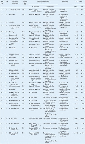
We recruited 30 consecutive patients who were found to have a nasopharyngeal mass on routine nasendoscopic examination and who consented to participate in the study. Patients’ symptoms and examination findings for white light and narrow-band imaging are shown in Table I. Twenty-one of the 30 patients had no evidence of malignancy on either histological or serological testing. Eight patients had nasopharyngeal carcinoma. One patient with papillary adenocarcinoma of the nasopharynx was excluded, as our aim was to compare nasopharyngeal carcinoma with non-neoplastic nasopharyngeal tissue.
Patient characteristics and results

Y = years; NPC Hx = history of nasopharyngeal carcinoma; EBV = Epstein–Barr virus serology; VCA = viral capsid antigen (immunoglobulin (Ig) A); Ea = early antigen (IgA); M = male; PNS = postnasal space; L = left; FOR = fossa of Rosenmuller; R = right; F = female
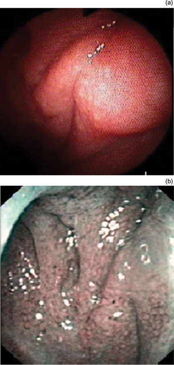
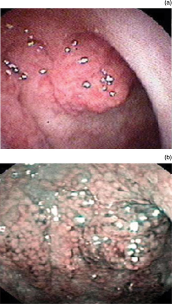
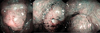
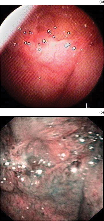
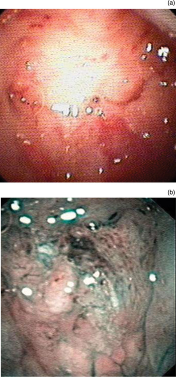
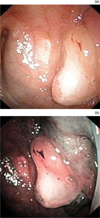
Of the 21 patients with normal adenoidal tissue, 17 were found to have a central nasopharyngeal mass, 2 had a unilateral nasopharyngeal mass and 2 had fullness at the fossa of Rosenmuller. Narrow-band imaging features identified in all these patients were as follows: (1) a regularly arranged, follicular pattern, and (2) each ‘follicle’ being composed of a pale centre with a surrounding dark periphery (Figures 1 to 3).
Endoscopic appearance of normal nasopharynx, comparing (a) conventional white light and (b) narrow-band light. Regularly arranged follicular patterns with pale-centred follicles and dark peripheries are clearly visualised with narrow-band imaging.

Endoscopic appearance of adenoidal tissue, comparing (a) conventional white light and (b) narrow-band light.

Endoscopic appearance of adenoidal tissue seen with narrow-band light.

Of the eight patients with nasopharyngeal carcinoma, one presented with a central nasopharyngeal mass, three had a unilateral nasopharyngeal mass and four had a mass at the fossa of Rosenmuller. The features of narrow-band imaging of these cases of nasopharyngeal carcinoma were as follows: (1) absence of surface patterns (n = 7), and/or (2) ‘reverse’, haphazard follicular patterns, with each follicle being composed of a dark brown centre with a surrounding pale periphery (n = 3) (Figures 4 to 6). Brown speckling was seen both in patients with adenoids and those with nasopharyngeal carcinoma (19 per cent of adenoids cases, 25 per cent of nasopharyngeal carcinoma cases).
Endoscopic appearance of nasopharyngeal carcinoma, comparing (a) conventional white light and (b) narrow-band light. The latter clearly shows haphazardly arranged follicular patterns and an increase in brown areas on the surface of the suspicious lesion.

Endoscopic appearance of nasopharyngeal carcinoma, comparing (a) conventional white light and (b) narrow-band light. The latter shows irregular follicular patterns with reversal of the usual follicular pattern, i.e. showing a dark brown centre with a pale periphery.

Endoscopic appearance of nasopharyngeal carcinoma, comparing (a) conventional white light and (b) narrow-band light. The latter shows absence of surface patterns.

Discussion
Narrow-band imaging via video-endoscopy is an innovative technology which uses optical interference filters to create narrow-band light capable of highlighting superficial mucosal microvasculature, an important characteristic which has been proposed to differentiate neoplastic and non-neoplastic lesions.Reference Seow, Koh, Chia, Shi, Lee and Shanmugaratnam1 Narrow-band imaging is available in many recent video-endoscope models, activated from the monitor console or the endoscope itself. This imaging modality has been increasingly recognised as a highly sensitive and accurate tool for the early detection of superficial mucosal neoplastic lesions. Much work has been done evaluating the diagnostic precision of narrow-band imaging versus conventional white light imaging, for the detection of early tumours in regions such as the lung and gastrointestinal tract.Reference Kumagai, Inoue, Nagai, Kawano and Iwai2, Reference East, Tan, Bergman, Saunders and Tekkis3
Studies on applications of narrow-band imaging in the head and neck region are scarce by comparison. In a recently published, multi-centre, randomised, controlled trial from Japan assessing narrow-band imaging in the early detection of synchronous and metachronous superficial oropharyngeal and hypopharyngeal squamous cell carcinoma in patients with oesophageal cancer, the sensitivity of narrow-band imaging was reported to be 100 per cent, compared with 8 per cent for conventional white light endoscopic imaging.Reference Muto, Minashi, Yano, Saito, Oda and Nonaka9 Kumagai et al. have proposed criteria for narrow-band imaging features suspicious of malignancy in regions other than the nasopharynx, as follows: (1) a well demarcated, brown lesion, with (2) scattered, brown dots within the lesion.Reference Kumagai, Inoue, Nagai, Kawano and Iwai2 Brown speckling represents microvascular proliferation, which is reported to be a characteristic feature of neoplastic and pre-neoplastic lesions on narrow-band imaging.
To date, there have been five publications on the use of narrow-band imaging in the nasopharynx for the detection of primary or recurrent nasopharyngeal carcinoma, and the criteria for suspicion of malignancy in this region have not yet been clearly defined.Reference Lin and Wang4–Reference Wang, Lin, Lee and Weng8
Lin et al. reported the successful detection of early nasopharyngeal carcinoma recurrence using narrow-band imaging, following the criteria proposed by Kumagai et al. Reference Lin and Wang4 A well demarcated, brownish area with brownish spots was found in an area of the nasopharynx otherwise noted to be smooth and symmetrical when viewed with conventional endoscopy.
Vlantis et al., on the other hand, did not notice these features in their patient with nasopharyngeal carcinoma.Reference Vlantis, Chan, Chan, Woo, Tong and van Hasselt5 They observed that, in adults, mucosal lymphoid tissue had a regular follicular pattern on narrow-band imaging, with pale follicles surrounded by a thin, dark border, and a ratio of pale follicle to dark border (‘pale-to-dark’ ratio) of roughly 90 per cent.Reference Vlantis, Chan, Chan, Woo, Tong and van Hasselt5 In their patient with nasopharyngeal carcinoma, they described a reversal in the pale-to-dark ratio of the follicles. Therefore, they concluded that this abnormal vascular pattern may signal an early event in the evolution of nasopharyngeal carcinoma.Reference Vlantis, Chan, Chan, Woo, Tong and van Hasselt5
Ho et al. examined narrow-band imaging appearances in nasopharyngeal carcinoma and benign nasopharyngeal lesions.Reference Ho, Lee and Chu7 They too observed that benign lymphoid hyperplasia demonstrated a regular, cobblestone appearance on narrow-band imaging. Irregular, engorged vascular patterns and/or microvascular proliferative patterns were seen in 32 of 41 nasopharyngeal carcinoma cases (78 per cent) in their study.
In our study, narrow-band imaging of normal adenoids revealed features consistent with descriptions given by other authors. Regularly arranged follicular patterns, with each follicle having a pale centre and surrounding dark periphery, were seen in all patients with normal adenoidal tissue. In patients with nasopharyngeal carcinoma, regularly arranged follicular patterns were not seen. Patterns were either absent (88 per cent) or, if present, tended to be haphazard and irregular (38 per cent) with reversal of the follicular pattern described by Vlantis et al. Reference Vlantis, Chan, Chan, Woo, Tong and van Hasselt5 Increased microvascularity, presenting as brown speckling on narrow-band imaging, was seen in 19 per cent of our patients with benign adenoidal tissue and in 25 per cent of our patients with nasopharyngeal carcinoma.
• Endoscopic narrow-band imaging can detect early pre-malignant and malignant lesions in the lung and gastrointestinal tract
• Nasopharyngeal applicability is poorly researched, and diagnostic criteria are ill-defined
• This study reports narrow-band imaging characteristics of normal adenoids and nasopharyngeal carcinoma
• This modality may enable earlier, more accurate detection of nasopharyngeal malignancy
Wang et al. found that scattered brown spots were seen in recurrent nasopharyngeal carcinoma post-irradiation, as well as in the nasopharynx of patients with no recurrence, presumably due to non-specific inflammatory changes due to irradiation.Reference Wang, Lin, Lee and Weng8 Studies not involving the head and neck region have reported that increased microvascular density due to inflammation can mimic the narrow-band imaging appearance of dysplasia and carcinoma; indeed, narrow-band imaging has been reported to have a high incidence of false positive results in patients with colitis.Reference Dekker, van den Broek, Reitsma, Hardwick, Offerhaus and van Deventer10 Brown speckling, a criteria proposed by Kumagai et al.,Reference Lin and Wang4 may therefore not be as specific a finding in the nasopharynx as the other features described.
Conclusion
This study found that normal adenoidal tissue consistently demonstrated narrow-band imaging features previously described by other authors. Nasopharyngeal carcinoma, on the other hand, may vary widely in appearance; however, our study found that loss or irregularity of follicular patterns, and reversal of follicular patterns, were characteristic features.
From the limited literature thus far, narrow-band imaging appears to be a promising and useful adjunct enabling earlier and more accurate detection of malignancy in the nasopharynx. However, much work remains to be done. Further studies into the diagnostic precision of narrow-band imaging are in progress.
Acknowledgement
This study was funded by a National Medical Research Council Enabling Grant from the Ministry of Health, Singapore.