Introduction
Several studies have shown that antidepressants can modulate the processing of emotional stimuli in healthy volunteers, without affecting mood or anxiety (Harmer, Reference Harmer2008). For example, repeated antidepressant administration reduced the perception of aversive facial expressions (Harmer et al. Reference Harmer, Shelley, Cowen and Goodwin2004) and modified amygdala response to emotional faces (Harmer et al. Reference Harmer, Mackay, Reid, Cowen and Goodwin2006; Murphy et al. Reference Murphy, Norbury, O'Sullivan, Cowen and Harmer2009). Initial evidence suggests that antidepressants given to clinical samples could have the same effect on emotional processing shown in healthy volunteers and this could be important for symptom remission. In one study a single dose of reboxetine compared to placebo reversed the negative biases in facial emotion recognition present in depressed subjects without any effects on mood or anxiety (Harmer et al. Reference Harmer, Goodwin and Cowen2009). Furthermore, increased recognition of happy facial expressions following 2 weeks of citalopram was predictive of therapeutic response after 6 weeks of treatment (Tranter et al. Reference Tranter, Bell, Gutting, Harmer, Healy and Anderson2009). Similarly, 7 days of escitalopram administration in depressed subjects reduced the amygdala response to fearful faces compared to placebo, in the absence of any effect on mood or other clinical symptoms (Godlewska et al. Reference Godlewska, Norbury, Selvaraj, Cowen and Harmer2012).
The translation from pre-clinical to clinical samples is vital for understanding how early changes in emotional processing may be involved in clinical response to antidepressant treatment. However, clinical studies are limited by the slow recruitment rate of unmedicated patients, general cognitive confounds and significant heterogeneity between patients. Hence, there may be significant value in the identification of a model of depression that can be used to test and refine hypotheses before complex clinical studies are initiated. One approach has been to use populations that model some of the pathological processes seen during illness without meeting criteria for depressive or anxiety disorders themselves. Neuroticism (N) is a personality trait characterized by an enduring tendency to experience both negative affect and significant levels of anxiety, widely recognized as a risk factor for psychiatric disorders (Lahey, Reference Lahey2009). Studies have shown that subjects with high N present biases in emotion processing such as impaired recognition of happy faces, similar to those shown by clinically depressed samples (Chan et al. Reference Chan, Goodwin and Harmer2007), and also biases in attentional processes, more reminiscent of anxiety-related alterations (Di Simplicio et al. Reference Di Simplicio, Norbury and Harmer2012). At a neural level, high N has been associated with altered processing of affective information in the amygdala, anterior cingulate and medial prefrontal cortex (PFC) (Haas et al. Reference Haas, Omura, Constable and Canli2007, Reference Haas, Constable and Canli2008; Chan et al. Reference Chan, Norbury, Goodwin and Harmer2009). Given that anxious and depressive facets are present equally in subjects with high N, investigating the early effects of antidepressants on emotional processing in highly neurotic participants may provide a model for exploring antidepressants effects on neural circuitry characterized by negative biases relevant to both anxious and depressive psychopathology.
Randomized clinical trial data suggest, however, that N is associated with poor remission rate to antidepressant treatment (Katon et al. Reference Katon, Unutzer and Russo2010) and this personality phenotype may also be a risk factor for unwanted adverse increases in agitation and anxiety early in treatment. There have been few experimental medicine studies exploring the effects of antidepressant treatment on psychological processes relevant to depression in a highly neurotic sample although we previously reported 7 days of selective serotonin reuptake inhibitor (SSRI) administration was able to decrease medial PFC activity during the categorization of negative self-descriptors, in highly neurotic subjects. Such effects are suggestive of an early remediation of negative affective bias that may be related to rumination tendencies and depression proneness in a highly vulnerable group (Di Simplicio, Reference Di Simplicio, Norbury and Harmer2012). By contrast, early effects of SSRI treatment on response to threat stimuli have not been assessed in a high-N sample but could be relevant to the induction of unwanted anxiety and agitation. Amygdala response to negative versus positive facial expressions is typically decreased after single-dose (Del-Ben et al. Reference Del-Ben, Deakin, McKie, Delvai, Williams, Elliott, Dolan and Anderson2005; Bigos et al. Reference Bigos, Pollock, Aizenstein, Fisher, Bies and Hariri2008; Murphy et al. Reference Murphy, Norbury, O'Sullivan, Cowen and Harmer2009; Rawlings et al. Reference Rawlings, Norbury, Cowen and Harmer2010) or 7-day (Harmer et al. Reference Harmer, Mackay, Reid, Cowen and Goodwin2006; Norbury et al. Reference Norbury, Taylor, Selvaraj, Murphy, Harmer and Cowen2009) antidepressant administration in unselected healthy volunteers. The failure to detect early anxiogenic-like effects of SSRI treatment in these models may occur because of the use of particularly low-anxious subjects in these functional magnetic resonance imaging (fMRI) studies or because fMRI cannot detect absolute differences in brain activity, which can confound relative differences identified by this technique (Murphy, Reference Murphy2010). This can be partly overcome by evaluating measures associated with absolute brain activity in the absence of stimuli presentation with the use of haemoperfusion techniques such as arterial spin labelling (ASL) and this approach was therefore included in the current study.
In this study we investigated the effects of 7 days of SSRI versus placebo administration on neural responses to threat-relevant stimuli (affective facial expressions) using fMRI and whole-brain haemoperfusion in a vulnerable population (volunteers with high-N scores). Given the complex literature on threat-relevant processing, an additional control group of subjects with low N was also tested to confirm previous results on neural differences between low- and high-risk groups (Chan et al. Reference Chan, Norbury, Goodwin and Harmer2009), and to help to clarify possible antidepressants effects on subjects with high N. We wanted to test the hypothesis that subjects with high-N scores could provide a model for early unwanted effects of SSRIs on anxiety and would therefore show paradoxical neural responses early in the treatment with an SSRI, manifest as increased amygdala responses to threat-relevant stimuli.
Method
Volunteers and design
Thirty-four healthy volunteers (18 females, mean age 25 ± 5.02 years) with a score above 16/24 on the N scale of the Eysenck Personality Questionnaire (EPQ; Eysenck & Eysenck, Reference Eysenck and Eysenck1975) (mean = 17.74 ± 2.1) and 14 healthy volunteers (11 females, mean age 33.53 ± 10.63 years) with a score below 5/24 on the N scale of the EPQ were recruited from the general population. These threshold scores represent approximately 1 standard deviation (s.d.) below and above the mean N scores of the general population aged 20–30 years (Eysenck et al. Reference Eysenck, Eysenck and Barret1985), and were evidence that a 1 s.d. increase in N scores conveys a 50–60% increase in lifetime risk for developing depression (Kendler et al. Reference Kendler, Neale, Kessler, Heath and Eaves1993). Written informed consent was provided as approved by the local ethics committee. All subjects were screened with the SCID (First et al. Reference First, Spitzer, Gibbon and Williams1998) for current or past Axis I disorders, were right-handed, in good physical health and free of any medication, excluding the contraceptive pill, and had no contraindications for fMRI scanning. The State and Trait Anxiety Inventory (STAI; Spielberger et al. Reference Spielberger, Gorsuch and Lushene1983) and the Hamilton Depression Rating Scale (HAMD; Hamilton, Reference Hamilton1960) were evaluated at baseline.
A double-blind placebo-controlled randomized design was used, randomly allocating subjects with high-N scores to 7 days of citalopram 20 mg/day (n = 18) or placebo (n = 16). A low-N group was also selected to represent a low-risk sample at the opposite end of the specific risk dimension represented by this particular personality trait factor. We have previously characterized healthy subjects with low-risk and high-risk personality profiles in terms of emotion processing both at a neuropsychological and at a neural response level (Chan et al. Reference Chan, Goodwin and Harmer2007, Reference Chan, Norbury, Goodwin and Harmer2009). Hence, the comparison between a high-risk group on placebo and a low-risk group was introduced to aid clarification of whether any drug effect would be specific to the neural responses characteristic of the N risk dimension. Subjects with low-N scores (n = 14) completed the same fMRI protocol but without receiving treatment.
During treatment, ratings for mood, anxiety and side-effects were collected daily with the Befindlichskeit Scale (BFS; von Zerssen & Schwarz, Reference von Zerssen and Schwarz1974), visual analogue scales (VAS) and a side-effects questionnaire.
Experimental task
On the last day of treatment subjects participated in a 16-min experiment using rapid event-related fMRI (Chan et al. Reference Chan, Norbury, Goodwin and Harmer2009). Subjects indicated the gender of faces showing expressions of fear and happiness at low intensity (30%), medium intensity (60%) and high intensity (100%), and neutral faces (Ekman, Reference Ekman1976; Young et al. Reference Young, Rowland, Calder, Etcoff, Seth and Perrett1997), by pressing two keys on an MRI-compatible keypad (eight trials per face type and 24 presentations of a fixation cross as baseline). Stimuli were presented in a random order for 500 ms each, and the intertrial interval varied according to a Poisson distribution (mean = 5000 ms) using E-Prime version 1.0 (Psychology Software Tools Inc., USA) and projected onto an opaque screen, viewed using angled mirrors. Accuracy and reaction times were recorded by E-Prime.
All subjects also underwent a 12-min resting state acquisition during which cerebral blood flow (CBF) levels were measured using a single-shot three-dimensional (3D) gradient and spin echo (GRASE)-ASL technique. Participants were instructed to watch a black screen, remain awake and refrain from focusing their thoughts.
Data acquisition and analysis
All subjects were scanned on a 3-T TIM Trio scanner at the Oxford Centre for Clinical and Magnetic Resonance Research (OCMR). fMRI data were acquired with a voxel resolution of 3 × 3 × 3 mm3, repetition time (TR) = 3 s, echo time (TE) = 30 ms, and flip angle = 87°. Field maps were acquired using a dual echo two-dimensional (2D) gradient echo sequence with echos at 5.19 and 7.65 ms, and TR = 444 ms. Data were acquired on a 64 × 64 × 40 grid, with a voxel resolution of 3 mm isotropic. T1-weighted structural images were acquired for subject alignment using a magnetization prepared rapid gradient echo (MPRAGE) sequence with the following parameters: voxel resolution 1 × 1 × 1 mm3 on a 176 × 192 × 192 grid, TE = 4.53 ms, inversion time (TI) = 900 ms, TR = 2200 ms. For ASL acquisition parameters see MacIntosh et al. (Reference MacIntosh, Pattinson, Gallichan, Ahmad, Miller, Feinberg, Wise and Jezzard2008).
Because of rapid movement artefacts the sample was left with 32 subjects (17 citalopram treated) for final analysis, performed using FSL version 4.1 (Smith et al. Reference Smith, Jenkinson, Woolrich, Beckmann, Behrens, Johansen-Berg, Bannister, De Luca, Drobnjak, Flitney, Niazy, Saunders, Vickers, Zhang, De Stefano, Brady and Matthews2004). Pre-processing included slice acquisition time correction, within-subject image realignment (Jenkinson et al. Reference Jenkinson, Brady and Smith2002), non-brain removal using a mesh deformation approach (Smith, Reference Smith2002), spatial normalization [to a Montreal Neurological Institute (MNI) 152 stereotactic template] and spatial smoothing, and high-pass temporal filtering was applied using a Gaussian-weighted running lines filter, with a 3-dB cut-off of 100 s. Susceptibility-related distortions were corrected as far as possible using FSL field-map correction routines (Woolrich et al. Reference Woolrich, Jbabdi, Patenaude, Chappell, Makni, Behrens, Beckmann, Jenkinson and Smith2009). An additional pre-processing procedure was used to eliminate motion artefacts by applying the FSL Independent Component Analysis tool to each individual subject separately and removing components that represented obvious scanner-related or physiological artefacts based on visual inspection of each subject's individual data (Beckmann & Smith, Reference Beckmann and Smith2004).
In the first-level analysis, individual activation maps were computed using the general linear model with local autocorrelation correction (Woolrich et al. Reference Woolrich, Ripley, Brady and Smith2001). Eight explanatory variables were modelled, including intensity (low, medium, high) of fear and happy and also neutral and fixation. The main contrasts of interest were fear versus happy expressions (and vice versa) for each intensity level. All variables were modelled by convolving the onset of each stimulus with a haemodynamic response function, using a variant of a gamma function (i.e. a normalization of the probability density function of the gamma function) with s.d. = 3 s and a mean lag of 6 s.
In the second-level analysis, whole-brain individual data were combined at the group level (citalopram versus placebo) using a mixed-effects group cluster analysis corrected for multiple comparisons (Woolrich et al. Reference Woolrich, Behrens, Beckmann, Jenkinson and Smith2004), which accounts for intra-subject variability and allows general population inferences to be drawn. Significant activations were identified using a cluster-based threshold of statistical images [height threshold of z = 2.3 and a (corrected) spatial extent threshold of p < 0.05] (Friston et al. Reference Friston, Worsley, Farckowiak, Mazziotta and Evans1994). To check for potential confounding effects of subthreshold differences in blood perfusion levels, demeaned CBF values obtained during resting-state GRASE-ASL were entered in the analysis as a voxel-wise covariate of no interest. Significant interactions were further explored by extracting the percentage blood oxygen level-dependent (BOLD) signal change within the areas of significant difference, analysed using repeated-measures ANOVA (between-subjects variable = group; within-subjects variable = valence) followed by appropriate post-hoc t tests (SPSS version 14.0; SPSS Inc., USA). Corresponding Brodmann areas (BAs) were identified by transforming MNI coordinates into Talairach space (Talaraich & Tournoux, Reference Talaraich and Tournoux1988). Our primary aim was to establish the effect of SSRI repeated administration on the responses to fearful versus happy facial expressions at each intensity level. An additional objective was to compare the neural correlates of processing fearful versus happy facial expressions at each intensity level between subjects with low-N scores and subjects with high-N scores who did not receive the active medication, using the same analysis design, to replicate previous data and help with the interpretation of the primary outcome analysis. For completeness, the results from the same analysis without correcting for blood perfusion levels are reported in the online Supplementary Material (Table S4).
In view of strong a priori evidence implicating the amygdala in facial expressions processing (Sheline et al. Reference Sheline, Barch, Donnelly, Ollinger, Snyder and Mintun2001; Surguladze et al. Reference Surguladze, Brammer, Keedwell, Giampietro, Young, Travis, Williams and Phillips2005) and showing modulation of amygdala response by repeated SSRI administration (Harmer et al. Reference Harmer, Mackay, Reid, Cowen and Goodwin2006) and high N (Chan et al. Reference Chan, Norbury, Goodwin and Harmer2009), we performed a region-of-interest (ROI) analysis. The mean BOLD percentage signal change was extracted from standard anatomical right and left amygdala masks (Harvard-Oxford anatomical atlas) and the interaction between treatment group (drug versus placebo), facial expression (happy versus fearful versus neutral) and hemisphere (right versus left) was modelled and tested using a repeated-measures ANOVA.
Perfusion data were analysed based on the method described in Chappell et al. (Reference Chappell, Okell, Jezzard and Woolrich2009). A non-parametric permutation-based (5000 iterations), two-sample (low N versus high N on placebo and placebo versus citalopram) unpaired t test approach was used to test first for whole-brain, voxel-wise, between-group differences in CBF perfusion levels (Nichols & Holmes, Reference Nichols and Holmes2002) and, second, after applying standard right and left amygdala anatomical masks.
For the behavioural and affect ratings data, one-way ANOVAs were used to examine group differences.
Results
Behavioural results and affect ratings
The two treatment groups of subjects with high-N scores were similar in terms of demographics (age and gender) and state and trait anxiety and depressive symptoms, whereas they both differed significantly from the low-N group on age and on the affect measures. Citalopram did not affect subjective ratings of state or mood as measured by VAS and BFS (all time × group ANOVAs, p's >0.05). Subjects receiving citalopram reported a trend for lower levels of calmness on the day of testing (t 31 = 2.02, p = 0.05) (Supplementary Material, Table S1).
Subjects with high-N scores on citalopram reported significantly more side-effects of nausea (time × group ANOVA, main group effect: F 31 = 10.85, p = 0.002) and these were correlated with anxiety levels on day 3 of treatment (p = 492, p = 0.038). There were no significant differences between the groups in accuracy and reaction time to all facial expressions during the gender discrimination task (Supplementary Material, Table S2).
fMRI results: low N versus high N on placebo
A detailed description of these results is presented in the online Supplementary Material. The BOLD fMRI data from the whole-brain analysis revealed greater response in the low-N group to fearful versus happy faces of high intensity compared to the high-N group in a network of brain areas including lateral and medial prefrontal and temporo-parieto-occipital cortical areas (Figs S1 and S2).
fMRI results: high N, citalopram versus placebo
Whole-brain analysis
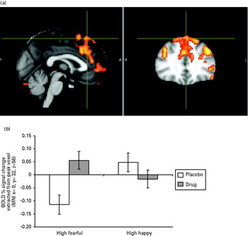
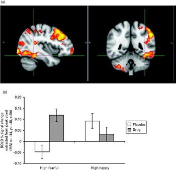
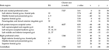
We found no significant between-group differences (citalopram versus placebo) in resting perfusion. Whole-brain analysis of BOLD data revealed four significant clusters for the interaction between group and high-intensity emotion condition: high-intensity fearful versus high-intensity happy faces (Table 1). Two clusters were located in prefrontal cortical areas bilaterally, including the superior frontal gyrus (BA 8), precentral and middle frontal gyri (BA 6, BA 9), inferior frontal gyrus and frontal pole (BA 10) and the paracingulate and dorsal anterior cingulate cortex (dACC) (BA 32) (Fig. 1 a). A third cluster included left temporo-parieto-occipital areas going from the supramarginal and angular gyrus (BA 40, BA 39) to the lateral occipital cortex from the superior (BA 39) to the inferior division (BA 19), to the fusiform gyrus (BA 19, BA 18) and inferior and middle temporal gyri (BA 37, BA 21) (Fig. 2 a). Finally, a significant difference in activation was found in the cerebellum. Post-hoc analyses revealed that these activations were driven by a greater response under citalopram to fearful compared to happy faces of high intensity (Figs 1 b and 2 b).
(a) Significant cluster of activation depicting greater blood oxygen level-dependent (BOLD) signal response in the dorsomedial prefrontal cortex (DMPFC) and left dorsolateral prefrontal cortex (DLPFC), including the dorsal anterior cingulate (dACC), in the high fearful versus high happy faces contrast in the citalopram-treated versus placebo-treated group during a gender discrimination task. Images thresholded at z > 2.3, p < 0.05, corrected. Montreal Neurological Institute (MNI) coordinates: x = 0, y = 32, z = 58. Images are in radiological format. (b) The group × emotion interaction in the PFC displaying increased activation to high fearful faces compared to high happy faces under citalopram. The percentage signal change plotted on the y axis is extracted from the mean activation voxel taken from the whole-brain analysis significant cluster.

(a) Significant cluster of activation depicting greater blood oxygen level-dependent (BOLD) signal response in the left temporo-parieto-occipital cortex, including the fusiform gyrus, in the high fearful versus high happy faces contrast in the citalopram-treated versus placebo-treated group during a gender discrimination task. Images thresholded at z > 2.3, p < 0.05, corrected. Montreal Neurological Institute (MNI) coordinates: x = − 38, y = − 46, z = − 18. Images are in radiological format. (b) The group × emotion interaction in the left temporo-parieto-occipital cortex displaying increased activation to high fearful compared to high happy faces under citalopram. The percentage signal change plotted on the y axis is extracted from the mean activation voxel taken from the whole-brain analysis significant cluster.

fMRI results of citalopram versus placebo treatment comparison: regions showing increased activation in the citalopram versus placebo group in the group × emotion interaction in the contrast of fearful faces of high intensity versus happy faces of high intensity

fMRI, Functional magnetic resonance imaging; BA, Brodmann area.
Given the trend for lower calmness ratings on the experiment day in the citalopram-treated group, a correlation between calmness scores and mean BOLD percentage signal change extracted from the clusters of significant activation was run to check whether neural responses in the citalopram group could be related to the marginally higher anxiety levels. These exploratory analyses showed significant negative correlations between calmness ratings and neural signal in the left temporo-parieto-occipital cortex in response to high fearful and high happy faces (r = − 0.553, p = 0.026) and in the medial PFC to high fearful faces (r = − 0.505, p = 0.046) within the drug-treated group.
Amygdala ROI analysis
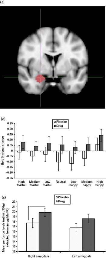
Mean perfusion levels extracted from the amygdalae revealed a significant difference in CBF in the right amygdala, with citalopram increasing baseline blood perfusion levels compared to placebo (Fig. 3 c).
(a) Amygdala region of interest (ROI). (b) The group × emotion × intensity × hemisphere interaction in the amygdala displaying a significant main group effect (F 29 = 4.63, p = 0.04). Subjects on citalopram showed a higher activation bilaterally in the amygdala to happy and fearful facial expressions of all intensities compared to subjects on placebo. Percentage signal change plotted on the y axis is extracted from right amygdala standard anatomical mask. (c) Differences in cerebral blood flow (CBF) (ml/min per 100 g) in the amygdala, with citalopram significantly increasing baseline blood perfusion levels compared to placebo in the right amygdala.

In the group comparison of percentage BOLD signal change extracted from right and left amygdala masks, no significant interaction between group, emotion and hemisphere was found, but a significant main effect of group (F 31 = 5.52, p = 0.025) was present. This effect was driven by subjects on citalopram showing a higher activation bilaterally in the amygdala to all faces regardless of emotional expression compared to subjects on placebo (Fig. 3 a, b).
A correlation between calmness ratings and mean BOLD percentage signal change extracted from the amygdala was run to check whether neural responses in the citalopram group could be related to the marginally higher anxiety levels after drug treatment. This showed a significant negative correlation between calmness ratings and mean activation in the right amygdala in response to all faces (r = − 0.504, p = 0.046). No correlation was found between calmness ratings and amygdalae perfusion levels (all p's >0.05).
Discussion
The aim of our study was to investigate the neural effects of repeated antidepressant administration on a sample of subjects who presented neural and behavioural biases in emotional processing related to increased risk for psychopathology. Our results show that 7 days of SSRI treatment in subjects with high N was associated with: (1) elevated resting perfusion in the right amygdala; (2) increased bilateral amygdalae activation to all facial expressions regardless of emotional valence; and (3) increased activation to fearful versus happy facial expressions in occipital, parietal, temporal and prefrontal cortical areas. The high-N group treated with placebo also showed decreased functional response to fearful compared to happy facial expressions in overlapping brain areas when compared to a group with low-N scores. Hence, 7 days of SSRIs seemed to shift the neural response in processing fearful facial expressions present in subjects vulnerable to psychopathology towards the response present in low-risk samples, albeit in the opposite direction to the effects typically reported in unselected healthy volunteers (Harmer et al. Reference Harmer, Mackay, Reid, Cowen and Goodwin2006; Murphy et al. Reference Murphy, Norbury, O'Sullivan, Cowen and Harmer2009; Norbury et al. Reference Norbury, Taylor, Selvaraj, Murphy, Harmer and Cowen2009; Rawlings et al. Reference Rawlings, Norbury, Cowen and Harmer2010).
Effects of N
The high-N volunteers receiving placebo in the current study showed reduced responses to fearful faces in the fusiform gyri and parieto-temporal cortices compared to the low-N control group. These areas belong to secondary sensory associative structures and have all been involved previously in visual and attentional processing of emotional facial expressions (Phan et al. Reference Phan, Wager, Taylor and Liberzon2002; Pourtois et al. Reference Pourtois, Schettino and Vuilleumier2013). Such a pattern of activation is opposite to the results of previous work using the same task (Chan et al. Reference Chan, Norbury, Goodwin and Harmer2009). As N is a multifaceted trait, comprising elements of increased reactivity, depressive mood and anxiety, it is possible that differences between the high-N groups' response to emotional faces between the two studies are secondary to subtle neuropsychological and temperamental variations between different high-N samples. In fact, the present sample reported higher levels of trait anxiety (STAI, 48.41 ± 9.4) and higher Beck Depression Inventory (BDI) scores (9.15 ± 6.1) compared to our previous neuroimaging study (STAI = 39 ± 8, BDI = 2.50 ± 1.9; Chan et al. Reference Chan, Norbury, Goodwin and Harmer2009). The current sample may therefore represent a more vulnerable group of subjects with high neurotic personality traits who responds differently to emotional facial expressions. Of note, one study has shown that N is associated with two different behavioural tendencies influencing response to danger or ambiguity: ‘volatility’, involving hypervigilance to negative cues, and ‘withdrawal’, involving avoidant behaviour towards any environmental novelty before the actual danger can be disambiguated (DeYoung et al. Reference DeYoung, Quilty and Peterson2007). The relative prevalence of these predispositions can produce differences in behavioural strategies towards threat-related stimuli: ‘volatility’ has been correlated to increased amygdala response to negative pictures and ‘withdrawal’ to reduced amygdala signal during avoidance versus approach responses irrespective of valence (Cunningham et al. Reference Cunningham, Arbuckle, Jahn, Mowrer and Abduljalil2011). These data could explain contrasting findings within high-N populations and seem to be in line with our results. In particular, the reduced pattern of neural response to threatening stimuli in our sample could reflect greater avoidant processing strategies (i.e. shifting attention away from emotional faces), which have been shown to be a function of perceived threat intensity (Bishop, Reference Bishop2007; Mogg et al. Reference Mogg, Garner and Bradley2007; Straube et al. Reference Straube, Schmidt, Weiss, Mentzel and Miltner2009). Notably, this explanation is supported by eye movement data showing that extremely high-N subjects avoid exploration of facial expression and of the eye region irrespective of expression type during the same gender discrimination task (Di Simplicio et al. 2012). If this was the case, the increased response to fearful faces with antidepressant administration may reflect decreased avoidance of socially salient and threat-relevant stimuli and lead to a normalized pattern of emotional processing.
Effects of SSRI administration
In the current study citalopram increased resting perfusion of the amygdala. A similar effect on the right amygdala was also found after single-dose citalopram administration in healthy volunteers (Chen et al. Reference Chen, Wan, O'Reardon, Wang, Wang, Korczykowski and Detre2011). Citalopram also increased functional responses to emotional faces in the same area in high-N volunteers. The amygdala is involved in salience and ambiguity detection (Davis & Whalen, Reference Davis and Whalen2001) and is possibly particularly responsive to social stimuli such as faces (Adolphs, Reference Adolphs2008). Higher levels of amygdala activity have been associated with higher negative affect, anxiety and depression (Morris et al. Reference Morris, Friston, Büchel, Frith, Young, Calder and Dolan1998; Adolphs et al. Reference Adolphs, Gosselin, Buchanan, Tranel, Schyns and Damasio2005) and decreased responses in the same area have been reported following anxiolytic drug administration (Paulus et al. Reference Paulus, Feinstein, Castillo, Simmons and Stein2005). It may therefore be relevant that high-N volunteers tended to feel less calm after SSRI compared to placebo treatment and this subjective rating was associated with amygdala responsivity in the fMRI paradigm.
The current effects seem to contrast with previous evidence showing decreased amygdala responses to fearful faces in unselected healthy volunteers with antidepressant administration (Norbury et al. Reference Norbury, Taylor, Selvaraj, Murphy, Harmer and Cowen2009; Rawlings et al. Reference Rawlings, Norbury, Cowen and Harmer2010), and this evidence has been replicated recently after 7 days of escitalopram administration in depressed patients (Godlewska et al. Reference Godlewska, Norbury, Selvaraj, Cowen and Harmer2012). Our opposite result of increased haemoperfusion and increased non-emotion specific activation in a sample characterized by high levels of anxiety and dysphoria suggests that the early actions of SSRIs on neural responses to threat may be affected by baseline characteristics of the sample and may be related to early unwanted increases in anxiety, which can be seen in some patients early in treatment (Sinclair et al. Reference Sinclair, Christmas, Hood, Potokar, Robertson, Isaac, Srivastava, Nutt and Davies2009).
However, it is notable that, although increased reactivity to threat would be regarded as aversive and unwanted, this effect acted to reverse the hypo-response seen in the placebo group. Consistent with this we have found that 7 days of citalopram treatment was able to increase occular exploration of faces in a high-N group (Di Simplicio et al. Reference Di Simplicio, Norbury and Harmer2012). Taken together, these data suggest that increased threat reactivity with SSRI treatment may be seen in this vulnerable group through a process of decreasing emotional avoidance. Although such effects would be adversive in the short term, they would be predicted to reduce anxiety in the longer term as patients gain greater exposure to benign ‘threat’ cues (such as faces) in the absence of avoidant safety behaviours, perhaps mimicking some key processes typically targeted in psychological therapy for anxiety disorders (Butler et al. Reference Butler, Fennell and Hackman2008). Future studies that combine eye-tracking methodologies and fMRI are required to validate this hypothesis. An alternative interpretation could be that the increased response to fearful facial expressions produced by citalopram in highly neurotic subjects could represent a dysfunctional effect of serotonergic manipulation, which could explain the poor clinical response to antidepressant treatment sometimes shown by these subjects when they present with a full-blown depressive disorder. This seems less likely given that the direction of the neural response after citalopram treatment resembles that of the low risk group, more suggestive of a shift towards resilience to psychopathology. Longer treatment studies with a longitudinal follow-up design on samples with high-N traits that develop depressive illness are needed to clarify what neurofunctional response correlates to the presence or absence of subjective mood change in this particular population.
In addition to the effects in the amygdala, an extended network of increased activation to fearful stimuli was also found in the PFC, from medial regions such as the dorsal ACC (BA 32, BA 9) to orbital regions such as the left ventrolateral PFC and frontal pole (BA 10, BA 47) and dorsolateral PFC (DLPFC) areas (BA 9 and BA 8) following SSRI administration. Dorsomedial PFC (DMPFC) areas have been linked to appraisal of emotional stimuli (Etkin et al. Reference Etkin, Egner and Kalisch2011) and increased DMPFC and amygdala coupling has been related to threat detection proportional to trait anxiety scores (Robinson et al. Reference Robinson, Charney, Overstreet, Vytal and Grillon2011). Orbital PFC has been implicated in regulatory function over the limbic system (Etkin et al. Reference Etkin, Egner and Kalisch2011), with the additional recruitment of DLPFC when an explicit cognitive demand is needed to moderate the distracting impact of emotional content on task performance (Ochsner & Gross, Reference Ochsner and Gross2005). Increased DMPFC and DLPFC response to fearful versus happy facial expressions in our study implies both greater neural recruitment for threat appraisal and increased requirement of neural resources to engage control systems during threat processing in high-N volunteers receiving SSRI treatment, consistent with the non-specific greater limbic signalling also seen in this group. It is unknown whether this effect on lateral PFC regions also translates into long-term better cognitive control over threatening stimuli manifest at the behavioural level, as suggested by longer treatment studies in clinical samples (Davidson et al. Reference Davidson, Irwin, Anderle and Kalin2003; Fu et al. Reference Fu, Williams, Cleare, Brammer, Walsh, Kim, Andrew, Pich, Williams, Reed, Mitterschiffthaler, Suckling and Bullmore2004).
Of note, the same volunteers were also found to show reduced responses in medial PFC to negative affective stimuli in a self-referential word categorization task (Di Simplicio, 2011). This adds to evidence of a dissociation between early effects of SSRI treatment on threat- and anxiety-related stimuli and the early induction of positive emotional bias, more relevant to therapeutic responses in depression (Browning et al. Reference Browning, Reid, Cowen, Goodwin and Harmer2007; Harmer, Reference Harmer2008). Hence, volunteers vulnerable to depression may show early positive biasing effects of antidepressants together with the anxiogenic-like responses seen here, both representing a mechanism of antidepressant drug response (Harmer et al. Reference Harmer, Goodwin and Cowen2009).
Limitations
The results from this study are limited by the absence of a low-N control group also receiving SSRI treatment to verify that the effects seen here are specific to a highly neurotic sample. However, early effects of SSRI treatment on unselected (typically low-anxious) populations have been well replicated, with one exception using high-dose citalopram (20 mg i.v. compared to 20 mg oral administration). We also did not take a measure of drug plasma levels to ensure compliance and it is possible that the volunteers in this study did not all take citalopram as instructed, even though clear differences were apparent compared to placebo. Finally, in the absence of post-scan questionnaires verifying whether subjects were able to refrain from focusing on their thoughts during the ASL resting sequence, it possible that the two treatment groups engaged in different mental activities that might have been reflected in the amygdala perfusion differences.
Future studies
Further investigations are required to assess the effects of longer-term SSRI administration in high-risk volunteers and in particular the prediction that the effects seen here after 1 week would reverse over time. We speculate that antidepressants could initially promote exposure to social stimuli, including potentially threatening stimuli, and by facilitating the experience of non-dangerous contacts with emotional stimuli, their action could lead to resetting the threat-appraisal sensitivity of the system (Carver et al. Reference Carver, Johnson and Joormann2008). To validate this hypothesis, it remains a key challenge to replicate in larger-scale clinical studies whether these initial SSRI effects on emotional processing circuitry are related to remediating an avoidant attentional pattern and to assess whether the same neural response would also be seen in the early treatment of anxiety disorders (Clark & Beck, Reference Clark and Beck2010).
Conclusions
The results from this study suggest that 7 days of SSRI administration can increase neural markers of fear reactivity in subjects at the high end of the N dimension. Such effects could be related to early increases in fear and agitation seen with antidepressant treatment in some patients. However, it is hypothesized that these effects could reflect decreased avoidance and be related to beneficial therapeutic effects seen in the longer term. Further studies are required to provide a functional test of this prediction.
Supplementary material
For supplementary material accompanying this paper visit http://dx.doi.org/10.1017/S0033291713000731.
Acknowledgements
We thank all participants in the study and the University of Siena, Italy, doctoral research program for supporting Dr M. Di Simplicio. The study was carried out as part of M. Di Simplicio's doctoral research at the University Department of Psychiatry, Oxford and supported by a Medical Research Council (MRC) grant to Dr C. Harmer.
Declaration of Interest
Dr C. Harmer has served as a consultant for P1vital, GlaxoSmithKline, Servier, Astra Zeneca, Johnson & Johnson and Lundbeck and is on the advisory board and holds shares of P1vital.