Introduction
Primary oocytes in the mammalian ovary are always at the arrested stage of first meiosis, in which their nuclei are completely covered by caryotheca and appear to have a bubble appearance; at this stage they are usually termed germinal vesicle oocytes. Following further maturation of the follicles, the oocytes in developing follicles resume meiosis, finally completing the first meiosis phase of maturation by way of metaphase (MI), anaphase (AI) and telophase (TI) stages, and extrude Pb I before ovulation (Ortiz et al., Reference Ortiz, Lucero and Croxatto1983). Following the first meiosis phase, oocytes immediately enter the second phase of meiosis, going through prophase (PII) to metaphase (MII). Subsequently, oocytes in the MII stage enter into another arrested stage, and the extruded Pb I gradually degenerates and deteriorates until fertilization. During the period of in vitro maturation of an oocyte, extrusion of Pb I has usually been used as a sign to distinguish whether the nucleus of the oocyte is mature or not. The morphologic characteristic of Pb I has also been used to determine whether an embryo should be transferred into a recipient or not in relation to research about adjunctive reproductive techniques in human beings. In general, a favourable morphology in Pb I implies that the fertilization ability of its oocyte will be good (Ebner et al., Reference Ebner, Yaman, Moser, Sommergruber, Feichtinger and Tews2000). The reproductive function of a mouse polar body was demonstrated by Wakayama and colleagues (Reference Wakayama, Hayashi and Ogura1997) and Wakayama and Yanagimachi (Reference Wakayama and Yanagimachi1998), who documented the birth of normal mouse offspring derived from Pb I and Pb II. After fertilization, the mouse Pb II and the female pronucleus in a zygote were successfully frozen in liquid nitrogen. The thawed Pb II and female pronucleus were used to reconstruct zygotes, and offspring possessing normal reproductive ability were born from the Pb II and female pronucleus (He et al., Reference He, Liu and Zev2003). Currently, there have been no other reports of similar research in other animal polar bodies.
At present, successful preservation of porcine germplasm mainly depends on the state of the living body. Cryopreservation techniques of porcine oocytes and embryos are still in a developmental stage, with great expense and effort being expended by many companies. Research regarding mouse reproduction from polar bodies has suggested a solution to the problems in preserving other animal germplasms through enabling cryopreservation of polar bodies and recombination of oocytes or zygotes (Fan, Reference Fan2000). In view of this, the present study aims to identify some of the biological characteristics of Pb I, including extrusion processes, morphology, and its isolation and preservation under different conditions, in order to provide some reference theories and techniques for further research into recombination of the oocyte polar body.
Materials and methods
Collection of porcine oocytes and preparation of porcine follicle liquid
Fresh porcine ovaries were recovered from a nearby abattoir, washed and placed into vacuum bottles containing 0.8% NaCl solution supplemented with 3200 IU/ml penicillin and 1600 IU/ml streptomycin. During treatment of the ovaries and transfer to the laboratory, the temperature was kept at between 25 and 30°C and experiments performed within 3 h. Recovery of cumulus oocyte complexes (COCs) from the follicles was carried using the method of Tan et al. (Reference Tan, Wang, Zhou and Xu2008), and porcine follicle fluid (pFF) prepared during collection of the COCs.
In vitro culture of porcine COCs
In vitro culture of COCs was performed using modified TCM 199 medium supplemented with 10% newborn calf serum (NCS), 69 μg/ml L-cysteine, 10 IU/ml pregnant mare serum gonadotropin (PMSG), 10 IU/ml human chorionic gonadotropin (hCG) and 1 μg/ml 17 β-oestradiol in a four-well culture plate, into each of which about 50 COCs were transferred in 1 ml medium and covered with mineral oil. After culture of the COCs in an incubator for at least 2 h under conditions of 39°C and 5% CO2 with saturation humidity, those of the COCs that developed oocytes with normal morphology, uniform cytoplasm and three or four layers of cumulus cells were selected for further culture under the same conditions but for between 24 and 72 h. During culture, half of the medium was changed every 24 h, and the expansion status of the cumulus cells observed under an inverted microscope.
In vitro fertilization of porcine oocytes
In vitro fertilization of mature oocytes was performed according to the method of Tan et al. (Reference Tan, Wang, Zhou and Xu2008) . After in vitro culture for 40 h, some of the porcine oocytes covered with cumulus cells were transferred into modified Tris-buffered medium (mTBM) solution, washed three times at room temperature, placed in 50 μl microdrops with mTBM solution and balanced in the incubator, and cultured (39°C and 5% CO2 at saturation humidity) for 4 h. Finally 50 μl of 1.0×10⁶/ml capacitated semen was injected, the fertilized microdrop covered with mineral oil, and cultured further for ~4–6 h in the incubator. Zygotes were identified from the fertilized solution, washed three times in essential NCSU 23 medium supplemented with 4 mg/ml bovine serum albumin (BSA) for further culture.
Gradation of morphology and identification of activity of porcine polar bodies
Following different periods of culture, some of the COCs were transferred into 0.1% hyaluronidase in Dulbecco phosphate buffered solution (DPBS), and treated at 37°C for 5 min. In order to remove cumulus cells, the COCs were inhaled and expelled using a capillary under a microscope. The treated oocytes were transferred into 10% newborn calf serum tissue culture medium (NCS TCM) 199 medium, and washed three times. Extrusion of Pb I in oocytes was observed under an inverted microscope, morphological gradation of Pb I was carried out according to the method described by Ebner and colleagues (Reference Ebner, Moser, Yaman, Feichtinger, Hartl and Tews1999), and the activity of Pb I identified according to the procedures of Liu and colleagues (Reference Liu, Sun and Wang2006), before and after preservation or fertilization of the oocytes.
After in vitro fertilization, the zygotes were transferred into NCSU 23 medium that had been prepared and placed in a CO2 incubator for 2 h before transfer then incubated further for 12–28 h under conditions of 39°C and 5% CO2, at saturation humidity. Extrusion of Pb II in zygotes was observed under an inverted microscope at regular intervals, and morphological gradation and biological activity of Pb II were determined using the same methods as for Pb I, above.
Isolation and collection of polar bodies from oocytes
Enzyme digestion method
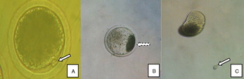
Some of the mature oocytes containing Pb I (mainly graded 1 and 2) were selected to be transferred into 0.5 ml TCM 199 medium supplemented with 10% NCS in a plastic tube, and centrifuged for 10 min at 15 000 g. The solution in the tube was gently inhaled and expelled using a Transferpettor pipette, then transferred into a watch-glass. The oocytes were transferred into a 50 μl microdrop of 20.0 g/l pronase in DPBS, digested for several minutes. The oocytes were transferred into 10% NCS in TCM 199 medium while the zona pellucida of the oocytes was thinned and expanded, and repeatedly inhaled and expelled until the Pb I was isolated from the oocyte (Fig. 1).
Isolation and collection of Pb I using the enzyme digestion method. (A) A normal matured oocyte, with the solid arrow indicating Pb I. (B) Morphological characteristics of an oocyte after centrifugation, with the dashed arrow indicating a part of the cytoplasm containing a concentration of lipids at one pole of the cytoplasm after centrifugation. (C) Free Pb I, indicated by the solid arrow, following enzyme digestion. (See online for a colour version of this figure.)

Micromanipulation
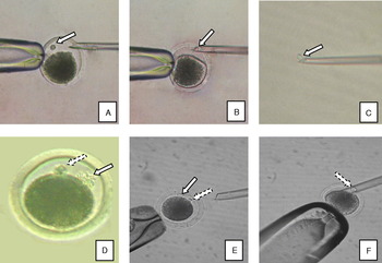
A one-step method was used to isolate Pb I and Pb II (Wang et al., Reference Wang, Tan and Zhou2008). Oocytes (or zygotes) were placed in 5 μg/ml cytochalasin B, 0.1 mol/l sucrose and 10% NCS in TCM 199 medium. The oocyte or zygote was fixed oriented towards 9 or 3 o'clock using a micropipette with the isolated polar body oriented towards 12 or 6 o'clock. A needle containing a small amount of solution pierced the zona pellucida of the oocyte or zygote, injecting the solution, then removed the polar body (see Fig. 2A–C for isolation of the Pb I and Fig. 2D–F for isolation of the Pb II). During isolation of the Pb II, the Pb I and Pb II in the zygote could be easily distinguished by their morphological characteristics, because the Pb I has disaggregated into a collection of scattered material, and the Pb II retains a complete and vigorous cell structure 20 h after in vitro fertilization.
(A)–(F) Principal steps for the isolation of Pb I and Pb II by micromanipulation. Pb I is indicated by a solid arrow and Pb II by a dashed arrow in each picture. The perivitelline space of each oocyte or zygote is expanded following treatment with 0.1 mol/l sucrose. There were obvious differences in the morphological characteristics of Pb I and Pb II in (D), (E) and (F); Pb II is clearly a complete cell, whereas Pb I is a collection of scattered material. (See online for a colour version of this figure.)

Preservation of polar bodies
Preservation at temperatures above zero
Some of the oocytes or fertilized eggs (zygotes) graded 1 and 2 by morphology were selected for culture in microdrops of essential TCM 199 medium at 4–7°C in a freezer or at 39°C in an incubator (5% CO2 and saturated humidity) for different times, respectively. The bioactivity and morphology of the oocytes were identified and graded at regular intervals.
Preservation under freezing conditions and cryopreservation
A glass capillary (diameter, 1.2 mm; length, 12 cm) was heated over an alcohol burner and drawn to form a 0.3-mm-diameter microtubule in the middle section of the capillary. The selected oocytes were transferred into EG20 de-icing fluid (ethylene glycol, EG:TCM 199 medium supplemented with 10% NCS at a ratio of 1:4) for 7 min, then into EG40 (TCM 199 medium supplemented with 40% EG and 0.5 mol/l sucrose) for about 30's. All of the oocytes treated with de-icing fluid were packed in the middle of the microtubules in batches; some of the microtubules were placed in a freezer (−20°C), and the remaining microtubules stored in a nitrogen canister (−196°C). After an interval of a day or month, the microtubules with oocytes or zygotes were removed from the freezer or the nitrogen canister, kept at room temperature in air for 10 s, and then immersed in a water bath at 37°C for 20 s. The de-icing fluid containing oocytes or zygotes was subsequently transferred into TCM 199 medium supplemented with 0.5 mol/l or 0.25 mol/l sucrose for 5 min, respectively, and finally washed three times with TCM 199 medium supplemented with 10% NCS before identification of their bioactivity and morphology.
Statistical analysis of data
The data from the experiments were analysed using the χ 2-squared text with SPSS 11.0 software, and expressed as average values.
Results
Effects of different culture times on the extrusion of polar bodies
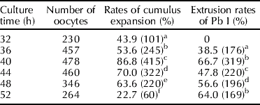
Cumulus cells were gradually expanded from oocytes during the initial process of in vitro culture of COCs, the expansion rates continuously increasing up to 40 h of culture, as did the extrusion rates of Pb I (see Table 1). However, following longer culture times, cumulus cells surrounded with oocytes were endlessly ablated from the oocytes, gathered together in disorder, and the extrusion rates of Pb I decreased. Although the extrusion rates showed little elevation from 48 h (56.6%) to 52 h of culture (64.0%), the proportion of the extruded Pb I with inferior morphological characteristics rose.
Extrusion rates of Pb I for different in vitro maturation times
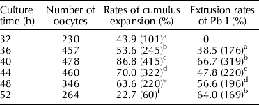
Different letters in the same column indicate significantly different values (P⩽0.05). The data in parentheses are numbers of oocytes in which cumulus cells were expanded or Pb I was extruded.
The data obtained from five experimental groups indicated that extrusion rates (1.8% to 5.9%) of Pb II did not change significantly from 0 h to 12 h of culture following in vitro fertilization, but significantly increased 16 h later, and the extrusion rates reached their the highest level (average 49.7%) at 20 h, thereafter clearly decreasing. So, a culture time of 20 h could be regarded as a suitable value in terms of the extrusion rate of porcine Pb II (see Table 2).
Extrusion rates of Pb II for different times after in vitro fertilization

Different letters in the same column indicate significantly different values (P⩽0.05). The data in parentheses are the number of fertilized eggs that extruded Pb II.
Optimal period for collection of Pb I and Pb II
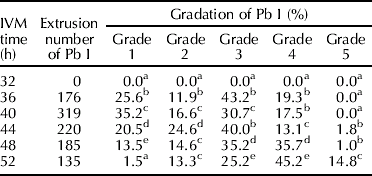
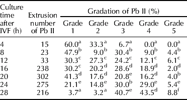
The data in Table 3 show that the total extrusion rates of Pb I and proportion of extruded Pb I changed significantly for different in vitro maturation (IVM) periods. Suitable culture times lay between 36 and 40 h, with 40 h being the optimal culture time, in which the extrusion number rose to 263 and there was a high proportion of active Pb I (grades 1, 2 and 3) of 51.8%. Following further culture, the total number and proportion of active Pb I decreased significantly, although the proportion of Pb I with low activity (that is, grade 4 and 5) clearly increased. Therefore, the optimal collection period for Pb I was close to 40 h of IVM.
Morphological gradation of Pb I at different in vitro maturation (IVM) periods
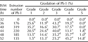
Different letters in the same column indicate significantly different values (P⩽0.05).
Four hours after in vitro fertilization (IVF), a few extrusions of Pb II could be observed. The number of Pb II extrusions increased significantly from 4 to 20 h. After 24 h culture, the extrusion number of active Pb II gradually decreased, whereas Pb II with low activity (grade 4 and 5) and dead morphological characteristics rose significantly. The optimal time for collection of Pb II was therefore between 16 h and 20 h, when most of the Pb II vigour (grade 1 and 2) and more than 50% of total Pb II was retained (see Table 4). Note that degenerated Pb I still existed in the perivitelline space after 16 h of culture, but its morphological characteristics allowed it to be distinguished easily from Pb II.
Morphological gradation of Pb II following different culture periods after in vitro fertilization (IVF)
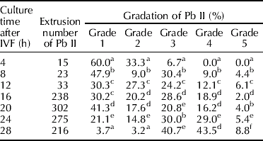
Different letters in the same column indicate significantly different values (P⩽0.05).
Comparison of collection effects using enzyme digestion and micromanipulation
When comparing the two methods of Pb I collection, the success and survival rates achieved using the enzyme digestion method for Pb I were significantly lower than those for the micromanipulation method. The recovery rate with enzyme digestion was only ~70.9% because of the limits of the operating field of vision under the microscope and the professional skills required to make observations. Also, the processes of centrifugation, enzyme digestion and mechanical aspiration might have caused the bioactivity of the Pb I to be affected slightly. Manipulation was found to be a suitable method for the collection of both Pb I and Pb II. Its collection rate could be higher than 95.5%, and the survival rate as high as 95.3%. However, enzyme digestion is still an option for Pb I collection, because of its ease of operation and the fact that its operation time is significantly less than that of the micromanipulation method.
Bioactivity of polar bodies under preservation at different temperatures
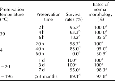
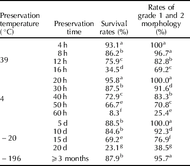
The results showed that the preservation of polar bodies was increasingly effective with lower preservation temperatures (see Table 5). At preservation conditions of 39°C, the survival time of Pb I was very short. Indeed, after 4 h, the survival rate was less than 63.3%. At 4°C conditions, 85.0% of Pb I could survive for ~40 h, whereas at −20°C, 95.0% of Pb I could survive for seven days. In comparison with the other preservation conditions, cryopreservation (−196°C) was the best, as 89.1% of Pb I could survive for more than three months. The preservation results for Pb II showed the same trends as those for Pb I (see Table 6) for different temperatures. At 39°C, 75.9% of Pb II could survive for 12 h of preservation, at 4°C 72.9% of Pb II survived for 40 h, at −20°C 84.6% of Pb II survived for 10 days, and at −196°C Pb II vigour did not change significantly over more than three months. Regardless of the preservation conditions used, the morphological characteristics of Pb I and Pb II were not significantly changed, except when the preservation time was unduly prolonged.
Differences in the activity and morphology of Pb I following preservation at different temperatures
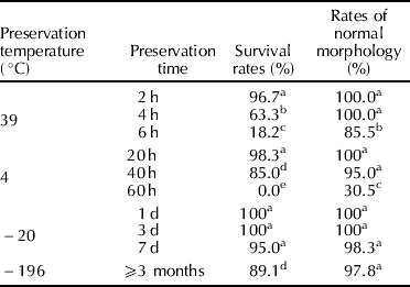
Different letters in the same column indicate significantly different values (P⩽0.05).
Differences in the activity and morphology of Pb II after preservation at different temperatures
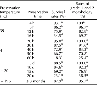
Different letters in the same column indicate significantly different values (P⩽0.05).
Discussion
There are some differences in the pattern of extrusion of polar bodies in different animals. It has been found that the highest proportion of mouse Pb I is extruded 12 h after hCG injection (Liu et al., Reference Liu, Sun and Wang2006). Evsikov and Evsikov (Reference Evsikov and Evsikov1995) considered that more than half of mouse Pb I would degenerate after ovulation, then disintegrate 12 h later, whereas the bioactivity of human Pb I could be maintained more than 20 h after ovulation. The extrusion peak of bovine Pb I appeared at 16 h of IVM, and the extrusion rate was as high as 73.5% after 24 h of IVM (Park et al., Reference Park, Kim, Kim, Park and Byun2005). In this study, extrusion of porcine Pb I began after 32 h of IVM, and at 40 h, the extrusion rate of Pb I was at its highest. During the process of extrusion of polar bodies, some will gradually degenerate and even disappear, so after 44 h of IVM, the extrusion rates for Pb I appear to have some irregular fluctuations. It might be thought that degeneration of Pb I would take place ~10–14 h after extrusion. Sun and colleagues (Reference Sun, Qin and Liu1996) reported that extrusion of bovine Pb II began after fertilization, and did not show apparent degeneration in its vigour for 18–24 h. From the beginning of its extrusion and reduction in extrusion number for porcine Pb II in this study, it was found that its vigour could be maintained for ~20 h. Gardner and colleagues (Reference Gardner1997) reported that mouse Pb II retained its complete morphological characteristics until the two-cell stage. It might therefore be inferred that the life of Pb II is longer than that of Pb I in mammals.
Porcine polar bodies could be successfully obtained using both methods described above, and these methods could be selected for use according to the different conditions. Micromanipulation is an effective and direct collection method of polar bodies; however, it requires professional skill from the operator, and the efficiency of collection depends on the availability of such skills. The enzyme digestion method is a more simple method suitable to the treatment of large samples, and it could become a highly effective method if it could be improved further. Because viewing of the Pb I was affected by the particular colour of porcine oocyte cytoplasm, Pb I could be easily missed and lost during isolation of the oocyte and during blowing and inhalation of the Pb I. The processes of centrifugation and enzyme digestion also caused physical and chemical damage to the polar bodies, so these problems would need to be resolved in order to improve the efficiency of this method. In addition, enzyme digestion was not suitable for the collection of Pb II, because Pb II interferes with the degenerate Pb I.
Choi and colleagues (Reference Choi, Fukasawa and Zhou1996) demonstrated that the degeneration and size of Pb I was controlled by the Mos/Mark gene, which regulates the processes of morphological degradation and loss in vigour of Pb I, but after fertilization, Mos/MARK lost its biological activity. This might imply that the vigour of Pb II is not influenced by Mos/MARK, and perhaps this is the reason why the survival time of Pb II was significantly longer than that of Pb I under preservation conditions between room temperature and 39°C. In our results we also found that the bioactivity of Pb I lasted longer than that of Pb II following a reduction in preservation temperature. This might be related to the structure of the polar bodies. There are complete caryotheca, cytolemma and a few organelles such as reticula and ribosomes in Pb II, whereas there are no such organelles other than a nucleus and a mutilated cytolemma in Pb I (Yanagimachi, Reference Yanagimachi1970; Watson, Reference Watson1993). So the more complex the structure of Pb II than that of Pb I, the more damaged it was. It was also found that lowering the temperature was good for the polar bodies in this study, suggesting some directions for further research about their preservation and utilization in the future.
Acknowledgements
This study was supported by the Natural Foundation of Jiangsu Province (no. BK2006160) and the Foundation of Jiangsu Academy of Agricultural Sciences (no. 6110817).