Introduction
Nowadays, clean hydrogen is one of the most hopeful energy carriers to substitute traditional fossil fuels. Moreover, for sustainable hydrogen production, solar photocatalytic water splitting has been deliberated as a promising approach [Reference Huang and Zhang1, Reference Chen, Shen, Guo and Mao2, Reference Wang, Jing and Ni3, Reference Shi, Cao, Bao, Zhao, Waterhouse, Fang, Wu, Tung, Yin and Zhang4]. Following the first research of hydrogen evolution via TiO2 photoelectrochemical water disintegration under UV-light irradiation was reported by Fujishima [Reference Fujishima and Honda5], semiconductor materials have prompted extensive attention as outstanding candidates for photocatalytic hydrogen evolution from water. However, in comparison to ultraviolet light which accounts for only about 4% of solar radiation, visible light accounts for about 45% [Reference Schultz and Yoon6]; hence many studies have been focused on the devise of photocatalysts with visible-light response for improving solar energy utilization, such as oxides [Reference Song, Li and Wang7, Reference Han, Chen, Zhang, Colmenares and Xu8, Reference Zhou, Shi, Shang, Zhao, Waterhouse, Wu, Tung and Zhang9], sulfides [Reference Zhu, Liu, Zhou, Fu, Guo, Li, Zhao, Huang, Liu and Kang10, Reference Li, Li, Wageh, Al-Ghamdi and Yu11, Reference Shang, Tong, Yu, Waterhouse, Zhou, Zhao, Tahir, Wu, Tung and Zhang12], graphite carbon nitride [Reference Liu, Chen, Xu, Zhang and Shen13, Reference Giannakoudakis, Travlou, Secor and Bandosz14], and oxynitride semiconductors [Reference Pan, Takata, Nakabayashi, Matsumoto, Shibata, Ikuhara and Domen15, Reference Chen, Takata and Domen16], etc.
For the photocatalysts, chalcogenide nanomaterials are drawing attention because of their suitable tunable band structures, which generally boost the separation and transport of photogenerated electron and hole, thus enhancing photocatalytic activity [Reference Zhang, Liu, Liu, Luo, Wang, Cai, Zeng, Yuan, Dong, Pei and Liu17, Reference Li and Lian18, Reference Coughlan, Ibanez, Dobrozhan, Singh, Cabot and Ryan19, Reference Zhao, Ding, Zhang, Li and Wang20]. Particularly, as a conventional semiconductor photocatalyst, zinc sulfide (ZnS) shows good activity for H2 evolution under noble-metal-free and UV-light conditions. However, owing to its wide band gap (3.6 eV), ZnS is not a visible-light-responsive photocatalyst. To improve its performance, many modifications have been put forward on ZnS, such as doping with different metal cations (i.e., Cu2+, Pb2+, Ni2+, In3+, Bi3+) [Reference Gaikwad, Tyagi, Betty and Sasikala21, Reference Sasikala, Shirole, Sudarsan, G. Jagannath, Sudakar, Naik, Rao and Bharadwaj22, Reference Gong, Lu, Wu, Huang, Zhu, Dang, Zhu, Lu and Huang23, Reference Kyne, Maguire, Obroin, Mcging, Mccann and Wright24, Reference Fang, Liu, Wang and Zhong25, Reference Fang, Zhai, Gautam, Li, Wu, Bando and Golberg26], combining with numerous narrow band gap semiconductors, and preparing solid solutions. Among those doped ZnS solid solution photocatalysts, CdxZn1−xS has been extensively studied for its efficient photocatalytic activity of generating hydrogen under visible light irradiation. Although many CdxZn1−xS catalysts have been synthesized and studied, such as CdxZn1−xS nanocomposites [Reference Jiang, Sun, Jia, Lu and Du27, Reference Shu, Wang, Wang, Li, Liu, Chen, Peng, Wang, Ruterana and Wang28, Reference Levchuk, Würth, Krause, Osvet, Batentschuk, Resch-Genger, Kolbeck, Herre, Steinrück, Peukert and Brabec29], nanotubes [Reference An, Feng, Liu, Wei, Du, Wang, Jin and Zhang30, Reference Han, Chen, Li, Yu and Zhou31], hexagonal [Reference Mei, Zhang, Schneider, Wang, Zhang, Su, Chen, Wang, Rogach and Pan32], microspheres [Reference Su, Zhang, Liu and Wang33, Reference Xing, Zhang, Yan and Guo34], core–shell [Reference Fang, Liu, Wang and Zhong25], and hollow dodecahedral structures [Reference Chen, Chen and Li35], the hydrogen production rate detected for most CdxZn1−xS is still inadequate and sometimes noble metals are also needed to facilitate hydrogen generation. Therefore, development of novel CdxZn1−xS structure with enhanced photocatalytic activity is highly required but still a challenge.
Metal–organic frameworks (MOFs) are a new kind of crystal porous materials, self-assembled with metal ions and organic linkers. Owing to their structural and functional tunability in addition to their ever-expanding scope of application, MOFs have turned into one of the most exciting classes of materials [Reference Zhu and Xu36, Reference Tian, He, Li, Zhao, Fang, Chen, Zhang, Zhang and Wang37]. Among them, homogenous bimetallic MOFs, formed by introducing different metal ions into a single framework, offer the ability to combine multiple active sites [Reference Liu, Zheng, Barpaga, Sabale, Arey, Derewinski, McGrail and Motkuri38]. Moreover, attributed to their advanced structure, bimetallic MOFs are a viable self-template or sacrificial template for fabricating porous or hollow homobimetallic materials, such as bimetallic oxides [Reference Fang, Zhou, Cai, Liu, Tan, Pan and Liang39, Reference Yu, Bai, Liu, Zhang, Chen, Zhang and Sun40], bimetallic sulfides [Reference Zhang, Guan, Yu and Lou41, Reference Huang, Song, Li, Tahir, Wang, Pan, Wang, Zhang and Zou42], and porous carbon materials [Reference Qian, Li, Hu and Huang43, Reference Huang, Mi, Zhang, Yu, Yu, Yuan, Kong and Xiong44]. The obtained derivatives preserve the original structural characteristics of the parent MOFs and generally have large surface areas, diffuse nano-sized subunits, and abundant active sites, giving them excellent performances in many applications [Reference Song, Zhu, Yang, Zhan, Pachfule, Tsumori and Xu45]. Furthermore, the synthesis of bimetallic MOFs is easy because of the good coordination ability of various metal ions with the organic skeleton. However, mesoporous CdxZn1−xS polyhedrons using bimetallic MOFs as precursors have not been reported.
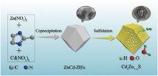
Herein, a facile method for fabricating bimetallic sulfide polyhedrons (CdxZn1−xS) with a mesoporous structure is reported (Scheme 1). In this method, a subclass of MOFs, homogeneous bimetallic zeolitic imidazolate frameworks (ZnCd-ZIFs), were used as self-templates and precursors to fabricate mesoporous CdxZn1−xS polyhedrons via in situ sulfidation reaction. The photocatalytic performance of the mesoporous CdxZn1−xS polyhedrons was explored via photocatalytic hydrogen production under visible-light irradiation and without any cocatalyst. In addition, the formation mechanism of the mesoporous CdxZn1−xS polyhedrons was studied in detail, and a possible photocatalytic enhancement mechanism on the basis of various characterization studies and theoretical analysis was also proposed. This facile bimetallic MOF-templated strategy may be extended to the preparation of other mesoporous bimetal sulfides for efficient visible-light-driven photocatalyst.
Description of the synthesis of mesoporous CdxZn1−xS polyhedrons.

Results and discussion
Formation of bimetallic zeolitic imidazolate frameworks
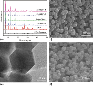
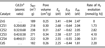
Bimetallic zeolitic imidazolate frameworks (ZnCd-ZIFs) were synthesized by coprecipitating Zn(NO3)2 and Cd(NO3)2 with 2-methylimidazole at room temperature in methanol solution. Figure 1(a) shows the powder X-ray diffraction (XRD) patterns of different samples. The ZnCd-ZIFs samples exhibit strong diffraction peaks similar to Zn-ZIFs(ZIF-8) [Reference Liu, Li, Wang, Wang, Zhang and Fan46], confirming the high crystallinity of ZnCd-ZIF crystals prepared successfully. Theoretically, the electronegativity of Cd2+(1.69) and of Zn2+(1.65) are similar, but the ionic radius of Cd2+(97 pm) is greater than that of Zn2+(74 pm) [Reference Yan, Fan, Chen, Pan, Zhang, Jiang and Su47]. Accordingly, as compared to the ZIF-8, it should be noted that all the XRD peak positions are slightly shifted to lower degrees with more Cd2+ introduced into ZIF-8 to form a series of ZnCd-ZIF samples, such as the highest characteristic peak position shifts from 7.48 to 7.38, 7.32, 7.30, and 7.28°, respectively. However, the feed atomic ratios of Cd/Zn are higher than the actual atomic ratios of Cd/Zn in ZnCd-ZIFs due to the different coordination abilities of Zn2+ and Cd2+ with 2-methylimidazole. The actual atomic ratios of Cd/Zn in ZnCd-ZIFs were detected by ICP (Table I). The SEM images [Figs. 1(b), 1(d), S1, and S2] and the TEM image [Fig. 1(c)] show that, when the Cd content was increased, the morphology of the series of ZnCd-ZIF samples is well preserved and that the as-synthesized ZnCd-ZIFs are of uniform polyhedral shape with a size of about 150 nm.
(a) XRD patterns of samples, (b) SEM image of ZnCd-ZIFs-1, (c) TEM image of ZnCd-ZIFs-1, (d) SEM image of ZnCd-ZIFs-3.

The structural properties and photocatalytic activities of the samples.

a The actual molar ratio detected by ICP.
Formation of mesoporous CdxZn1−xS polyhedrons
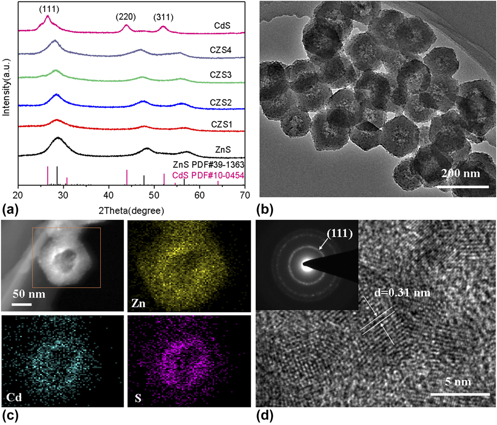
The CdxZn1−xS was prepared by in situ topotactic conversion of ZnCd-ZIFs. The XRD patterns [Fig. 2(a)] show that ZnS and CdS could be indexed as cubic ZnS (JCPDS no. 39-1363) and hawleyite CdS (JCPDS no. 10-0454), respectively. Compared with ZnS, the positions of XRD peaks on the (111) plane for each CdxZn1−xS sample shift slightly to lower degrees, from 28.59 to 28.52, 28.50, 28.32, and 28.14°, respectively, with the increase of Cd content. This investigation indicates that a solid solution is formed, rather than a physical mixture of CdS and ZnS [Reference Chen, Chen and Li35], and the change in the position of the diffraction peak of the CdxZn1−xS might be explained by the different cation radii between Zn2+ and Cd2+. The obtained CdxZn1−xS samples mostly maintain the polyhedral morphology of the ZnCd-ZIFs precursor (Fig. S3). The TEM image gives a further understanding of the polyhedron’s detailed and internal structure [Fig. 2(b)]. The sharp contrast between the edge and the center demonstrates the well-defined polyhedron structure. The uniform distribution of S and the metal is corroborated by the high-angle annular dark-field scanning transmission electron microscopy (HAADF-STEM) and elemental mapping images [Fig. 2(c)]. Besides, the d-spacing of the lattice fringes of the CZS1 is 0.31 nm [Fig. 2(d)], which conforms to the (111) plane.
(a) XRD patterns of samples, (b) TEM image of CZS1, (c) HAADF-STEM image and elemental mappings of CZS1, (d) HRTEM image of CZS1.

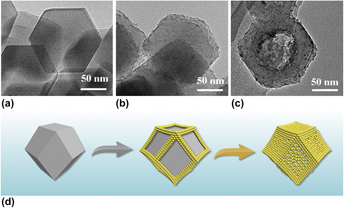
To further investigate the formation process of the mesoporous CdxZn1−xS polyhedrons, we conducted a time-dependent experiment (Fig. 3). Compared with the polyhedral shape of the ZnCd-ZIF precursor in Fig. 3(a), after sulfidation treatment for 0.5 h, the edges of these polyhedrons become rather rough in Fig. 3(b), indicating the initial sulfidation reaction occurred on the edges. When the reaction time was extended to 2 h, mesoporous polyhedrons were formed and a large amount of mesopores appeared in the center of these polyhedrons [Fig. 3(c)]. According to the time-dependent experiments described above, we could infer the formation of the mesoporous CdxZn1−xS polyhedrons via an anisotropic chemical etching/anion exchange and some mechanism similar to the Kirkendall effect [Fig. 3(d)] [Reference Han, Yu and Lou48, Reference Avci, Arinez-Soriano, Carne-Sanchez, Guillerm, Carbonell, Imaz and Maspoch49, Reference Indra, Song and Paik50, Reference Yu, Yu, Wu and Lou51]. At the beginning of the sulfidation procedure, TAA is hydrolyzed, which releases S2− ions slowly, which partially etches the edges of the polyhedrons via an exchange reaction of metal ions (Zn2+ and Cd2+ ions). The edges of the polyhedrons become rougher, and their planar surfaces remain the same. This finding reveals that the polyhedral edges, with possibly more defects and high curvature, provide more reaction sites for chemical etching/anion exchange than plane surfaces [Reference Yu, Yu, Wu and Lou51]. Therefore, the edges of the ZnCd-ZIF polyhedrons first form a thin layer of sulfides (CdxZn1−xS) in the first 0.5 h of reaction. Furthermore, as the reaction proceeded, the side surfaces with less etching are exposed and easy to be chemically etched. Thus, chemical etching/anion exchange occurs on the side of the intermediate planar surface. Based on the Kirkendall effect, the inward transport of S2− ions and the outward transport of the metal ions occur through the formed sulfides layer. The ionic radius of S2− ions is greater than the ionic radius of metal ions; therefore, the inward diffusion of S2− ions is more difficult than the outward diffusion of the metal ions. The unbalanced diffusion of reactants (metal ion and S2− ions) produces voids [Reference Yu, Zhang, Wu and Lou52, Reference Su, Ao, Liu and Wang53]. As the reaction continued, the freshly formed sulfide nuclei will provide prefabricated edge growth, while the central region of the polyhedrons would produce fewer sulfides. As a result, the mesoporous polyhedron structure, small mesopores formed at the edges, and large mesopores formed in the center is finally obtained.
(a) TEM image of ZnCd-ZIFs-1. Time-dependent formation of mesoporous CZS1: (b) 0.5 h, (c) 2 h. (d) Schematic diagram of the formation of mesoporous CdxZn1−xS polyhedron.

Characterization of samples
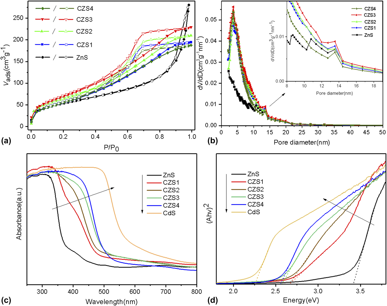
The specific surface areas of as-prepared samples have been characterized by nitrogen adsorption isotherm analysis [Fig. 4(a)]. ZnS can be classified as a type IV isotherm with a narrow hysteresis loop in the P/P 0 range of about 0.8–1.0, implying the existence of mesopores probably formed by the loose packing of nanoparticles. For ZnS, CSZ1, CSZ2, CSZ3, and CSZ4, the surface areas by Brunauer–Emmett–Teller (BET) are 189, 218, 238, 271, and 222 m2/g, respectively. Compared with ZnS, the surface area and pore volume (Table I) of CdxZn1−xS are significantly higher, indicating that the materials possessed more mesopores after replacing Zn2+ with Cd2+, which could have resulted from the particle expansion process as the ionic radius of Cd2+ is greater than the ionic radius of Zn2+. Figure 4(b) gives the pore size distribution of the CdxZn1−xS calculated by the Barrett–Joyer–Halenda (BJH) method using the adsorption branch of the isotherm curves, showing a main peak centered at 3.7 nm and large mesopores at approximately 13–14 nm. However, the higher Cd2+ content leads to more single CdS nanoparticle formation, which can be embedded in the pores or deposited on the surface of the sample, thereby reducing the pore volumes and specific surface areas. Since heterogeneous photocatalysis is mainly based on the surface of catalysts, a higher surface area would shorten the transmission distance of the charge carrier and supply more active sites to adsorb reactants, resulting in more effective photocatalysis [Reference Zhang, Yu, Jaroniec and Gong54]. In addition, the mesoporous structure is considered to promote the harvesting of light excitation and the diffusion of reactants and products. Therefore, CdxZn1−xS polyhedrons with mesoporous structure and high surface area have high potential in the catalytic activity in heterogeneous photocatalysis.
(a) Nitrogen isotherms, (b) pore size distribution, (c) UV-vis absorption spectrum, (d) the Kubelka–Munk plots to measure the direct band gap.

The photoelectrochemical properties of the as-prepared samples were further analyzed by UV-vis diffuse reflectance spectroscopy (UV-vis DRS) [Fig. 4(c)]. And Fig. 4(d) reveals the samples-derived E g based on the Kubelka–Munk function. Compared to ZnS, the photoabsorption edge of CdxZn1−xS shows a red shift, corresponding to the E g decreasing from 3.41 to 2.68, 2.67, 2.58, and 2.55 eV, respectively, with the increasing Cd content. These results demonstrate that incorporating Cd atoms into the ZnS can considerably reduce the E g values and thus lead to more efficient visible light harvesting, indicating that these mesoporous CdxZn1−xS polyhedrons may have excellent photocatalytic properties.
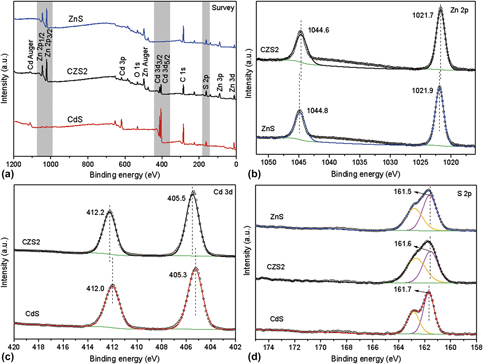
In order to further understand the changes in the surface chemical state of CdxZn1−xS after the incorporation of Cd, XPS analysis was performed. As observed in Fig. 5(a), Zn, Cd, and S elements are found in the survey scan. For ZnS sample, the observed binding energy of Zn 2p 3/2 is 1021.9 eV and that of Zn 2p 1/2 is 1044.8 eV [Fig. 5(b)], which are in agreement with the reported values for Zn2+ [Reference Fan, Wang, Sang and Wang55]. The Zn 2p 3/2 and Zn 2p 1/2 peaks are observed at 1021.7 eV and 1044.6 eV for the CSZ2, respectively, which are shifted by 0.2 eV slightly to lower values compared with ZnS. The positions of the Cd 3d 5/2 signal and Cd 3d 3/2 signal for CdS in the Cd 3d spectra [Fig. 5(c)] are at around 405.3 eV and 412.0 eV, respectively, corresponding to the values in the literature [Reference Jiang, Sun, Jia, Lu and Du56]. However, for CSZ2, the peak positions of Cd 3d shift 0.2 eV to higher values when compared to CdS, and other studies also show this [Reference Li, Meng, Zhou, Zheng, Wang, Yu and Gong57]. Meanwhile, in the S 2p spectra [Fig. 5(d)], the observed binding energy of S 2p 3/2 is 161.5 eV, 161.6 eV, and 161.7 eV for ZnS, CSZ2, and CdS, respectively, which demonstrates the existence of S2−. From these results, we could conclude that the presence of electronic coupling between ZnS and CdS and that the Cd atoms lose more electrons while the Zn atoms lose fewer electrons in CSZ2 [Reference Jiang, Sun, Jia, Lu and Du56]. The XPS analyses further demonstrate the formation of CdxZn1−xS solid solutions and exclude the presence of nitrogen and carbon residues in CdxZn1−xS.
XPS spectra of (a) survey, (b) Zn 2p, (c) Cd 3d, and (d) S 2p.

Photocatalytic H2 evolution activity
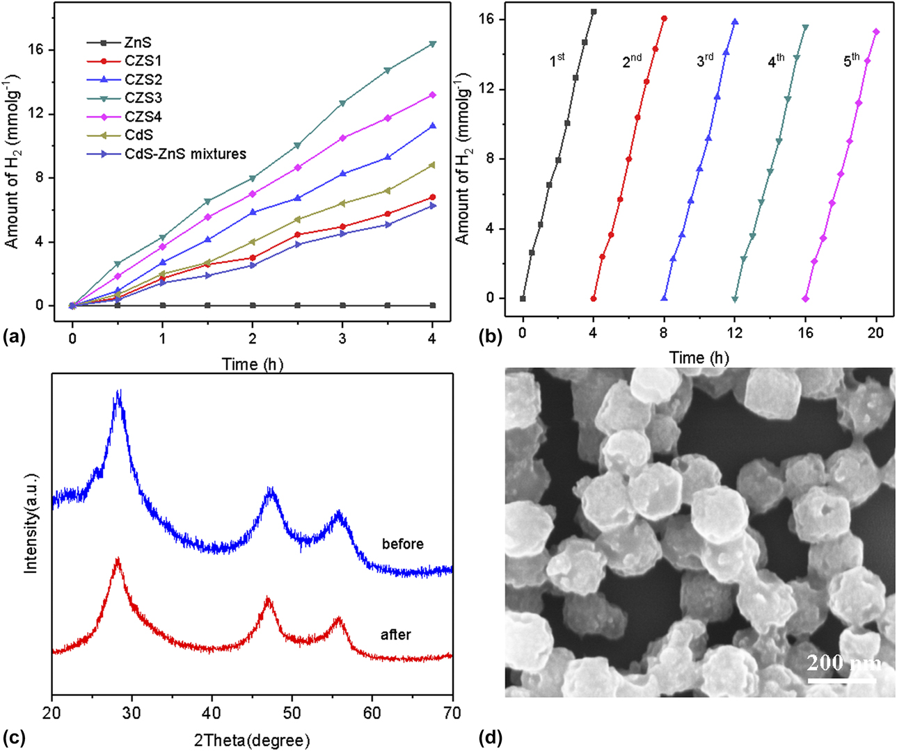
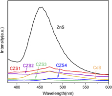
To evaluate the photocatalytic performances of the prepared samples, hydrogen evolution experiments were accomplished in water under cocatalyst-free and visible-light irradiation. The relation between hydrogen generation rate and the Cd/Zn ratio in the mesoporous CdxZn1−xS polyhedrons is investigated, as shown in Fig. 6(a), where mesoporous CdxZn1−xS polyhedrons exhibit an improved hydrogen production rate compared to ZnS, CdS, and CdS–ZnS mixture except for the CZS1 sample. The hydrogen production rates of ZnS, CdS, and CZS1 are 0, 2.20, and 1.73 mmol/(h g), respectively. CZS1 with 20% of Cd shows a higher BET result but a lower photocatalytic performance compared with pure CdS. As shown by the photoluminescence (PL) spectrum (Fig. 7), the CZS1 restrains the electron–hole pair recombination efficiency better than pure ZnS but not as good as pure CdS. In addition, as shown in Fig. 4(d), the E g of the CZS1 sample is larger than pure CdS, which results in its visible light utilization being less than that of pure CdS. These result in a lower photocatalytic performance of the CZS1 sample compared to pure CdS.
(a) Photocatalytic hydrogen evolution curves. (b) Recycling stability experiments in five consecutive cycles, (c) XRD patterns before and after recycling tests, (d) SEM image after recycling tests of CSZ3.

PL spectra of all as-prepared samples.

The hydrogen production rate of the CdS–ZnS mixture is 1.56 mmol/(g h), which is much lower than that of CZS3 [4.10 mmol/(g h)]. Meanwhile, the molar ratio of CdS to ZnS in the mixture is set to 0.42:0.58, which is exactly the same as the ratio of Cd to Zn in CZS3. Therefore, the photocatalytic performance of CdxZn1−xS can be improved by forming a solid solution mesoporous structure compared to physical mixing. Since ZnS is substantially incapable of producing hydrogen under visible light, the performance enhancement demonstrates that Cd plays a significant role in restraining the photoinduced electron–hole pair recombination. When the Cd content is 20% and 32%, the hydrogen evolution rate reaches 1.73 and 2.82 mmol/(h g), respectively. Upon increasing the Cd content to 42%, the sample (CZS3) displays the highest hydrogen evolution rate [4.10 mmol/(h g)]. With a further increase of Cd content to 49%, the rate of hydrogen evolution is reduced to 3.31 mmol/(h g). The enhancement in photocatalytic performance is due to the increased surface area and the reduction of band gap. However, single CdS nanoparticles formed by excess Cd may be deposited on the surface of the sample, resulting in a reduction in surface area [Reference Chen, Chen and Li35]. A reduction in surface area means less photogenerated carriers emerged, which ultimately decreases the photocatalytic activity. Furthermore, excess Cd might form clusters and become the recombination center of photogenerated electron–holes, which is also harmful to photocatalytic activity. Besides, the as-prepared photocatalyst (CSZ3) exhibits better photocatalytic hydrogen evolution activity than most of the reported metal sulfide photocatalysts (Table SI). In these reported metal sulfides systems, CoPt3 [Reference Wang, Li, Shu, Chen, Liu, Wang, Zhang and Wang58], Fe0.3Pt0.7 [Reference Shu, Wang, Wang, Li, Liu, Chen, Peng, Wang, Ruterana and Wang28], CoP and Pt [Reference Dai, Xu, Ge, Han, Gao, Li and Lu59] are employed as co-catalysts to promote photoactivity, respectively. However, their photoactivity is still unsatisfactory for the hydrogen evolution reaction. Pan research group, by modifying the absolute band edges of the photocatalysts, has showed better photoactivity [3.62 mmol/(g h)] than the aforementioned reported metal sulfide systems even under the cocatalyst-free condition. In this study, CdxZn1−xS without a co-catalyst is used as the photocatalyst, in which the absolute band edges are modified, and the mesoporous structure is endowed additionally, leading to good light absorption and fast charge carrier separation. Therefore, the prepared photocatalysts, mesoporous CdxZn1−xS polyhedrons, make the improvement of the photocatalytic performance.
Long-term stability is another important indicator for evaluating photocatalysts. Long-circulating photocatalytic performances of CSZ3 and the CdS–ZnS mixture were carried out, as shown in Figs. 6(b) and S4, respectively. After four hours of continuous hydrogen production, hydrogen was removed from the reaction system by evacuating several times and then the photocatalyst was recycled. After 5 cycles, the hydrogen production rates of CSZ3 and the CdS–ZnS mixture are reduced by about 7% and 25%, respectively, indicating that CSZ3 has excellent stability compared to the CdS–ZnS mixture. Moreover, the XRD and SEM results of CSZ3 after the recycles were studied to calculate the stability of CSZ3. No obvious change could be found from XRD [Fig. 6(c)], and mesoporous polyhedrons structure is retained [Fig. 6(d)], which may be the reason for the slight decrease in the hydrogen evolution rate of CSZ3 in the five-run test.
Photocatalytic mechanism investigation
Based on the above experiments and characterization results, the photocatalytic mechanism for the hydrogen evolution of mesoporous CdxZn1−xS polyhedrons from water has been proposed. As illustrated in Fig. S5, the conduction band potential (E CB) and the valence band potential (E VB) of CZ3 at the point of zero charge were −0.57 eV and 2.01 eV, respectively, calculated by the following formula: E CB = X − E c − 0.5 E g [Reference Guo, Chen, Song, Wang, Di and Qin60]. This formula has been widely used to estimate the band edge positions of sulfides and oxide semiconductors [Reference Zhu, Liu, Zhou, Fu, Guo, Li, Zhao, Huang, Liu and Kang10, Reference Pany and Parida61]. The photocatalytic hydrogen evolution activity of CdxZn1−xS was enhanced as follows: Firstly, a narrowed E g and matched E CB of the CdxZn1−xS would favor the light absorption and enhance photocatalytic performance. By tuning the composition of the components in CdxZn1−xS, fine-controlled bandgap engineering is achieved by adjusting the amount of Cd2+ in the bimetallic MOF precursor. Secondly, mesoporous structures, derived from bimetallic MOFs, could significantly enlarge the surface area (271 m2/g) of CdxZn1−xS. A larger specific surface could not only endow the CdxZn1−xS with a more effective light-harvesting process and produce more photogenerated carriers, but also accelerate electron separation and transmission along the surface and inside the pores. These two advantages ultimately improve the photocatalytic activity.
To prove this hypothesis, PL spectra were used to investigate the recombination/separation of photoinduced electron–hole pairs (Fig. 7). The emission spectra of all samples were tested at the excitation wavelength of 325 nm. The photocatalytic abilities of ZnS, CdxZn1−xS, and CdS correspond to the trends of the curve. For pure ZnS, it has a wide emission peak centered at around 460 nm. And a significant decrease in the fluorescence intensity of CdxZn1−xS is observed compared to ZnS, which is attributable to the efficient charge carrier transfer in CdxZn1−xS. Moreover, the relative intensity of CdxZn1−xS follows the order of CSZ3 < CSZ4 < CSZ2 < CSZ1, which is consistent with the observation that the lower the recombination rate, the higher the photocatalytic activity.
Conclusions
In summary, a facile bimetallic MOF (ZnCd-ZIF) sulfidation method has been developed to construct mesoporous CdxZn1−xS polyhedrons for photocatalytic hydrogen production. The Cd atoms are homogeneously dispersed in CdxZn1−xS due to the homogeneous composition of ZnCd-ZIFs precursor. Moreover, incorporating Cd atoms and the formation of mesoporous structure endow the CdxZn1−xS with a high surface area, effective light harvesting, and fast charge carrier separation. The mesoporous CSZ3 polyhedrons without any cocatalyst exhibit a significantly enhanced hydrogen evolution activity [4.10 mmol/(h g)] under visible-light irradiation. It is speculated that the aforementioned high photocatalytic activity is due to the balance between the CB potential and band gap width, fast charge carrier separation and migration ability, and a high surface area of the as-prepared photocatalysts. In addition, the mesoporous CdxZn1−xS polyhedrons show excellent photocatalytic durability and stability, indicating that they are promising for practical applications. The method developed here for constructing mesoporous polyhedrons may provide a way to prepare other metal sulfides for photochemical applications.
Experimental section
Materials
Cadmium nitrate tetrahydrate (Cd(NO3)2·4H2O), zinc nitrate hexahydrate (Zn(NO3)2·6H2O), 2-methylimidazole (C4H6N2) and thioacetamide (C2H5NS) were purchased from Aladdin (Shanghai, China). Methanol (CH3OH) and ethanol (C2H5OH) were supplied by Sinopharm Chemical Reagent Co., Ltd. (Shanghai, China). The chemical reagents were of analytical grade and were not further purified.
Synthesis of photocatalysts
ZnCd-ZIFs were synthesized according to a previous procedure with modifications [Reference Huang, Song, Li, Tahir, Wang, Pan, Wang, Zhang and Zou42]. Typically, Zn(NO3)2·6H2O and Cd(NO3)2·4H2O with different molar ratios of Zn2+/Cd2+ were dissolved in 150 mL of methanol to form a transparent solution, which was subsequently poured into 100 mL of methanol containing 2-methylimidazole (120 mmol). After stirring for 2 h, the solution was incubated at room temperature for 24 h without interruption. The total molar amount of Zn2+ and Cd2+ was fixed to be 15 mmol. The as-obtained white precipitates were centrifuged and repeatedly washed with methanol, resulting in the so-called ZnCd-ZIFs. Then, 64 mg of ZnCd-ZIFs and 128 mg of thioacetamide were added to 30 mL of ethanol with continuous stirring. After stirring for 15 min, the mixture was transferred into a Teflon-lined stainless steel autoclave and heated at 120 °C for 2 h. The resulting mesoporous CdxZn1−xS was collected by centrifugation, washed several times with ethanol, and dried at 70 °C for 12 h. A series of mesoporous CdxZn1−xS were prepared with different molar ratios of Zn2+/Cd2+ (15/0, 10.5/4.5, 7.5/7.5, 4.5/10.5, 1.5/13.5), denoted as ZnS, CSZ1, CSZ2, CSZ3, and CSZ4, respectively. For comparison, CdS was prepared as explained in Ref. Reference Chen, Chen and Li35.
Characterization
The crystalline structures were characterized by powder X-ray diffraction (XRD; Smartlab 9 kW, Rigaku, Tokyo, Japan) patterns, using Cu Kα radiation (λ = 0.154168 nm) at 40 kV and 40 mA. The surface morphology was obtained by a field-emission scanning electron microscope (FE-SEM; Hitachi S4800, Hitachi, Tokyo, Japan) and a transmission electron microscope (TEM; FEI Tecnai G2 F20S-TWIN, FEI Company, Hillsboro, Oregon) at an accelerating voltage of 200 kV. The content of Zn and Cd in each sample was determined by a PE Optima 2000DV (PerkinElmer, Waltham, Massachusetts) inductively coupled plasma (ICP) emission spectrometer. The optical properties were recorded in the range of 250–800 nm by a Lambda 950 ultraviolet–visible diffuse reflectance spectrophotometer (UV-vis DRS) using BaSO4 as the reference. The chemical states of the elements were carried out by an X-ray photoelectron spectroscopy (XPS; Thermo Fisher Scientific ESCALAB 250Xi, Thermo Fisher Scientific, Waltham, Massachusetts), using Al Kα radiation, and all the bonding energies were calibrated to the C 1s peak at 284.8 eV. The specific area was calculated using the BET method by the N2 (77.4 K) adsorption–desorption data obtained from BELSORP-mini II in a relative pressure range P/P 0 of 0.01 to 0.99. The PL spectra were investigated on a Hitachi F-4600 spectrometer, Hitachi, Tokyo, Japan.
Photocatalytic water splitting
Photocatalytic hydrogen generation experiments were carried out in a LABSOLAR-III photoreactor (Beijing PerfectLight Technology Co., Ltd., Beijing, China). Typically, 50 mg of as-prepared photocatalysts were dispersed in 100 mL of aqueous solution containing 0.70 M Na2S and 0.50 M Na2SO3 (sacrificial electron donor). The mixture was evacuated to completely remove air before irradiation. The light source used was a 300 W Xe lamp (PLS-SXE 300, CUV, Beijing PerfectLight Technology Co., Ltd., Beijing, China) with a UV-light cutoff filter (400 nm). A circulating water pump was used to maintain the temperature of the reaction system at room temperature. An online gas chromatograph (GC 9890A) equipped with a TCD detector was utilized to analyze the released hydrogen.
Supplementary material
To view supplementary material for this article, please visit https://doi.org/10.1557/jmr.2019.72.
Acknowledgments
This work was supported by the National Natural Science Foundation of China (Nos. 21476108, 21276125, 21776129, and 21706121), Natural Science Foundation of Jiangsu Province (No. BK20170995), General Program for University Natural Science Research of Jiangsu Province (No. 16KJB530003) and the Project of Priority Academic Program Development of Jiangsu Higher Education Institutions (PAPD).