Introduction
Attention-deficit hyperactivity disorder (ADHD) is a common neurodevelopmental disorder with core symptoms of inattention, hyperactivity, and impulsivity. ADHD symptoms typically start before adolescence and persist in about half of the ADHD patients to adulthood (Faraone & Larsson, Reference Faraone and Larsson2019). The presentation of ADHD symptoms may change from childhood to adulthood, and the level of severity fluctuates at different time points (Karam et al., Reference Karam, Rovaris, Breda, Picon, Victor, Salgado and Bau2017). Even though the number of ADHD symptoms may decrease as they grow up, academic underachievement and social dysfunction remain (Bussing et al., Reference Bussing, Mason, Bell, Porter and Garvan2010).
High heritability (70–80%) of ADHD has been reported (Larsson, Chang, D'Onofrio, & Lichtenstein, Reference Larsson, Chang, D'Onofrio and Lichtenstein2014), and the familial vulnerability of ADHD points toward a potential genetic cause in its etiology (Faraone & Larsson, Reference Faraone and Larsson2019). There is not yet a consensus on the genetic risk factors responsible for ADHD because the heritability of ADHD may come from the small effects of many common genetic variants (Faraone & Larsson, Reference Faraone and Larsson2019). According to the concept of endophenotype, or intermediate phenotype, unaffected relatives of probands with ADHD (asymptomatic carriers of the risk genes) are vulnerable to manifest some neurobiological characteristics as probands. Some studies have demonstrated similar changes in brain activation patterns during neuropsychological tasks in probands with ADHD and their unaffected relatives when compared to controls (Glahn et al., Reference Glahn, Knowles, McKay, Sprooten, Raventos, Blangero and Almasy2014; Pironti et al., Reference Pironti, Lai, Muller, Dodds, Suckling, Bullmore and Sahakian2014). For example, reduced functional MRI (fMRI) activations to a Stroop task measuring interference control were reported in both probands with ADHD and their co-twins (Godinez et al., Reference Godinez, Willcutt, Burgess, Depue, Andrews-Hanna and Banich2015). In two other studies, decreased activations during a response inhibition task (van Rooij et al., Reference van Rooij, Hoekstra, Mennes, von Rhein, Thissen, Heslenfeld and Hartman2015) and increased fMRI activations for reward anticipation (von Rhein et al., Reference von Rhein, Cools, Zwiers, van der Schaaf, Franke, Luman and Buitelaar2015) were found in both adolescents and adults with ADHD as well as their unaffected siblings. Therefore, neuroimaging studies may be useful to explore a familial risk marker of ADHD and to investigate how genes potentially affect brain circuits that lead to ADHD (Glahn et al., Reference Glahn, Knowles, McKay, Sprooten, Raventos, Blangero and Almasy2014).
Intrinsic functional connectivity in resting-state fMRI (rsfMRI) has been shown to recapitulate task-related coactivation patterns in task fMRI (Yeo et al., Reference Yeo, Krienen, Sepulcre, Sabuncu, Lashkari, Hollinshead and Buckner2011). In the resting brain, positive correlations in spontaneous fluctuations of the blood oxygen level-dependent (BOLD) signal can be found in regions within the same network (Fox et al., Reference Fox, Snyder, Vincent, Corbetta, Van Essen and Raichle2005). In the absence of task performance, anti-correlations can be observed between the default-mode network (DMN) (regions deactivate when engaging task) and cognitive control networks (regions activate when engaging task) (Fox et al., Reference Fox, Snyder, Vincent, Corbetta, Van Essen and Raichle2005; Kelly, Uddin, Biswal, Castellanos, & Milham, Reference Kelly, Uddin, Biswal, Castellanos and Milham2008). Although disturbed intrinsic functional connectivity has been demonstrated in probands with ADHD (Rubia, Reference Rubia2018), there has been only one study using rsfMRI to investigate the heritability of functional connectivity within families enriched for ADHD (Sudre et al., Reference Sudre, Choudhuri, Szekely, Bonner, Goduni, Sharp and Shaw2017). Specifically, Sudre et al. (Reference Sudre, Choudhuri, Szekely, Bonner, Goduni, Sharp and Shaw2017) applied independent component analysis to decompose intrinsic networks, which closely resembled the well-known large-scale networks. Their analysis focused on the seven major networks described by Yeo et al. (Reference Yeo, Krienen, Sepulcre, Sabuncu, Lashkari, Hollinshead and Buckner2011), and they found the heritability patterns of functional connectivity within DMN, frontoparietal network, and ventral attention network (VAN) (Sudre et al., Reference Sudre, Choudhuri, Szekely, Bonner, Goduni, Sharp and Shaw2017). However, this study did not explore the heritability of interaction among regions not within the same networks, although disrupted interaction between regions in different networks has been repeatedly reported in individuals with ADHD (Zhao et al., Reference Zhao, Li, Yu, Huang, Wang, Liu and Wang2017).
The DMN, which is associated with internally directed thoughts, has been proposed to be pivotal in the pathophysiology of ADHD (Sonuga-Barke & Castellanos, Reference Sonuga-Barke and Castellanos2007). Data-driven, network-based analysis could provide useful information about cross-network connectivity involving the DMN and other large-scale task-positive networks; however, cross-network connectivity often shows a more complex pattern (Sripada et al., Reference Sripada, Kessler, Fang, Welsh, Prem Kumar and Angstadt2014). For example, the core hub of the DMN, the precuneus/posterior cingulate cortex (PCC) (Castellanos et al., Reference Zhao, Li, Yu, Huang, Wang, Liu and Wang2008), has shown less anti-correlations (or increased connectivity) with the insula in VAN in subjects with ADHD (Sripada et al., Reference Sripada, Kessler, Fang, Welsh, Prem Kumar and Angstadt2014). However, decreased functional connectivity between the dorsal medial prefrontal cortex in DMN and the supplementary motor area in VAN has been found in subjects with ADHD (Sripada et al., Reference Sripada, Kessler, Fang, Welsh, Prem Kumar and Angstadt2014). Therefore, we chose to conduct a seed-based functional connectivity analysis for the present study. Although aberrant functional connectivity is evident in rsfMRI studies of ADHD (Castellanos et al., Reference Castellanos, Margulies, Kelly, Uddin, Ghaffari, Kirsch and Milham2008; Sripada et al., Reference Sripada, Kessler, Fang, Welsh, Prem Kumar and Angstadt2014), no study has explored whether the connectivity among brain regions cross different functional networks is a familial risk marker of ADHD.
To investigate the connectivity among regions crosses different networks, we employed a seed-based voxel-wise connectivity analysis. This approach enables a direct comparison of the connectivity map among groups to examine the intrinsic connectivity comprehensively. Since there is limited knowledge in siblings of probands with ADHD, we believe the results from seed-based analysis would be easier to interpret and informative as well. Additionally, a whole-brain approach could also explore the neural compensation mechanisms of the unaffected siblings to provide novel information. Since the intrinsic functional connectivities between the precuneus/PCC and middle frontal gyrus, inferior frontal gyrus (IFG), and occipital regions have been reported to be associated with the performance in inhibitory control in ADHD (Barber et al., Reference Barber, Jacobson, Wexler, Nebel, Caffo, Pekar and Mostofsky2015), we also analyzed whether brain–behavior relationships could be found in our samples. The primary aim of the study is to compare the intrinsic functional connectivity between the precuneus/PCC and other brain regions in probands with ADHD, their unaffected siblings, and typically developing controls. Furthermore, we aim to examine the associations between functional connectivity and ADHD symptoms and neuropsychological functions. Based on previous findings reporting heritability of within-network connectivity within ADHD families (Sudre et al., Reference Sudre, Choudhuri, Szekely, Bonner, Goduni, Sharp and Shaw2017), we expected to find evidence of aberrant connectivity among regions cross different networks in both probands with ADHD and unaffected siblings when compared to controls.
Methods
Participants and procedures
This work was approved by the Research Ethics Committee of National Taiwan University Hospital prior to its implementation (approved number, 201204071RIC; ClinicalTrials.gov number, NCT01682915). All participants and their parents (if the participants are under the age of 18) provided written informed consent after the procedures and purpose of the study were explained to them. We recruited 60 sibling pairs of adolescents and adults with ADHD, aged 15–40 years old, consecutively, and their unaffected biological siblings if they met the inclusion and exclusion criteria of the current study. Probands with ADHD were clinically diagnosed according to the DSM-IV diagnostic criteria by board-certified child psychiatrists at National Taiwan University Hospital, Taipei, Taiwan. Exclusion criteria for probands with ADHD included a history of psychosis, bipolar disorders, autism spectrum disorder, neurological disorders, and a Full-scale IQ score of <80. Controls were enrolled if there was no history of medical or neuropsychiatric illness, nor a current or past history of using psychotropic agents. Sixty controls were selected to match individually in age (±1 year) and gender with probands with ADHD.
All the participants, and their parents if they are under the age of 18, received psychiatric interviews about the diagnosis of the participants with the Chinese version of the Schedule for Affective Disorders and Schizophrenia for School-Age Children – Epidemiologic Version (K-SADS-E) (Gau, Chong, Chen, & Cheng, Reference Gau, Chong, Chen and Cheng2005), the Wechsler Adult Intelligence Scale-Revised (WAIS-R) (Wechsler, Reference Wechsler1981) or Wechsler Intelligence Scale for Children-third edition (WISC-III) (Cockshott, Marsh, & Hine, Reference Cockshott, Marsh and Hine2006) according to their age, and MRI assessments. The unaffected siblings and controls were also assessed with the K-SADS-E interview to confirm that they did not have ADHD. In addition, those who were currently taking methylphenidate were asked to discontinue medication at least one week before the MRI.
Clinical measures
The Chinese version of the K-SADS-E
The K-SADS-E is a semi-structured interview scale for the systematic assessment of both lifetime and current (past 6 months) diagnosis of mental disorders (Gau et al., Reference Gau, Chong, Chen and Cheng2005). All participants were assessed by the Chinese K-SADS-E interview to obtain the information on lifetime and current psychiatric symptoms and diagnoses according to the DSM-IV diagnostic criteria. The Child Psychiatry Research Group in Taiwan developed the Chinese K-SADS-E via a two-stage translation (Gau et al., Reference Gau, Chong, Chen and Cheng2005). The ADHD supplement was further modified into adult version by the corresponding author (SSG) to include information about the age onset of ADHD and the medication history for treating ADHD (Tsai, Tseng, Yang, & Gau, Reference Tsai, Tseng, Yang and Gau2019). This semi-structured interview scale is a reliable and valid instrument to assess child (Chiang, Chen, Lo, Tseng, & Gau, Reference Chiang, Chen, Lo, Tseng and Gau2015) and adult (Chang, Chiu, Wu, & Gau, Reference Chang, Chiu, Wu and Gau2013; Lin, Yang, & Gau, Reference Lin, Yang and Gau2016) psychiatric disorders in Taiwan, and has been widely used in a variety of studies (e.g. Chiang et al., Reference Chiang, Chen, Lo, Tseng and Gau2015; Gau & Huang, Reference Gau and Huang2014; Shang, Lin, Tseng, & Gau, Reference Shang, Lin, Tseng and Gau2018). The details of the K-SADS-E interview training and the best estimate of each DSM-IV psychiatric diagnosis have been described elsewhere (Gau et al., Reference Gau, Lin, Cheng, Chiu, Tsai and Soong2010; Lin et al., Reference Lin, Yang and Gau2016) and are provided upon request.
Neuropsychological measurement
The Conners' Continuous Performance Test (CCPT)
The CCPT is a 14 min computerized task measuring processes related to attention, response inhibition, signal detection for individuals older than 6 years old (Conners and Staff., Reference Conners2000). There are 360 trials in total with letters that appear on the computer screen, one at a time. Respondents are required to press the spacebar when any letter except the letter ‘X’ is shown on the screen. The paradigm has six blocks and three sub-blocks, each containing 20 trials. Each sub-block has a different inter-stimulus interval of 1, 2, and 4 s with display time of 250 ms, and is presented randomly. Indices we used in this study included omission errors, commission errors, and detectability. Omission errors refer to the number of times not responding to a target, while commission errors refer to the number of incorrect responses to non-target, the letter ‘X’. Detectability is the ability to discriminate non-targets from targets, and a higher score indicates worse performance in discrimination.
Statistical analysis
To compare demographics and characteristics of the participants among the three groups, we used SAS 9.2 version (SAS Institute, Cary, NC, USA) for data analysis. The descriptive results are displayed as the mean and standard deviation for the continuous variables; and number and frequencies for categorical variables. The general linear model was used in three group comparison analyses with the Bonferroni correction method to adjust p values in the post-hoc pairwise analysis. The α value was preselected at the level of p < 0.05.
MRI data acquisition
All magnetic resonance imaging (MRI) scans were collected on a Siemens 3T MRI system (TIM Trio, Siemens, Erlangen, Germany) with a 32-channel phased-array head coil at the National Taiwan University Hospital, Taipei, Taiwan. A sagittal localizer was used to define axial planes orthogonal to the sagittal image, which makes imaging plane parallel to the anterior commissure–posterior commissure. High-resolution T1-weighted images covering the whole head were acquired using a 3D magnetization-prepared rapid gradient echo (MPRAGE) sequence: repetition time (TR) = 2000 ms, echo time (TE) = 2.98 ms, inversion time = 900 ms, field of view (FOV) = 256 × 192 × 208 mm3, matrix size = 256 × 192 × 208, yielding an isotropic resolution of 1 × 1 × 1 mm3. Participants also were instructed to relax and remain still with their eyes closed during a 7 min 39 s resting-state scan consisting of 180 functional volumes using an EPI sequence (TR = 2000 ms; TE = 24 ms; flip angle = 90°; FOV = 256 × 256 mm2; matrix size = 64 × 64; 34 axial slices acquired in an interleaved descending order; slice thickness = 3 mm; voxel size = 4 × 4 × 3 mm3). T1-weighted images were visually inspected for artifacts, such as blurriness and motion, to ensure that data quality of all final samples met the ratings of ‘fair’ or higher quality based on the procedures of quality control in the Human Connectome Project (Marcus et al., Reference Marcus, Harms, Snyder, Jenkinson, Wilson, Glasser and Van Essen2013).
Preprocessing of functional MRI images
Image preprocessing was performed using AFNI (version 16.1.10) and FSL (version 5.0.7). Preprocessing included skull stripping to remove non-brain tissue, motion correction, spatial smoothing using a Gaussian kernel of 6 mm full width at half-maximum, grand-mean scaling to normalize the global 4D data, and temporal filtering (0.005–0.1 Hz). The T1-weighted image was registered to Montreal Neurological Institute (MNI) space using linear [FLIRT (FMRIB's linear image registration tool)] and non-linear [FNIRT (FMRIB's nonlinear image registration tool)] algorithms implemented in FSL, and the registration matrix was applied to move the resting-state fMRI data image into MNI space. The CSF and the white matter masks were generated by segmenting individual subjects' T1-weighted images (FSL FAST). Lastly, motion parameters, CSF signal, and the white matter signal were regressed out as nuisance covariates. We did not perform global signal regression, because it is likely to enhance negative correlations and to bias group differences in fMRI analyses, and it has shown to cause anti-correlations in studies of negatively correlated networks (Murphy, Birn, Handwerker, Jones, & Bandettini, Reference Murphy, Birn, Handwerker, Jones and Bandettini2009).
We identified two probands with ADHD, three siblings, and two control subjects with >2 mm maximum displacement in either x, y, z directions, or >2 degrees of angular rotation. These subjects, with excess motion and their corresponding, matched subjects were excluded from further analysis.
Intrinsic functional connectivity of resting-state activity analyses
We used a seed-based approach to investigate intrinsic functional connectivity based on bilateral precuneus/PCC, which is known to play a core role in DMN and engages a variety of cognitive process (Utevsky, Smith, & Huettel, Reference Utevsky, Smith and Huettel2014). The a priori precuneus seed (Talairach coordinates: ±7, −60, 21) was chosen based on the literature (Sheline, Price, Yan, & Mintun, Reference Sheline, Price, Yan and Mintun2010), and converted to MNI coordinates (7, −63, 20 and −8, −63, 20) using an online tool: MNI<->Talairach with Brodmann Areas, v1.09 (http://sprout022.sprout.yale.edu/mni2tal/mni2tal.html). We created a 5 mm radius sphere around these two coordinates as the seed regions of interest. We extracted the time-series data from individual subjects and used the averaged time course from the seed region as a regressor. The general linear model analysis was conducted by correlating the regional time series against all other voxels within the brain to determine temporally coherent networks.
A whole-brain approach was used to explore group differences in intrinsic functional connectivity. We first tested whether there is a difference among the three groups (probands with ADHD, siblings, controls) with F test in one-way ANOVA using a non-parametric permutation test (FSL randomize with 10 000 permutations) with threshold-free cluster enhancement approach and with family-wise error rate (FWE) to correct for multiple comparisons (Smith & Nichols, Reference Smith and Nichols2009; Winkler, Ridgway, Webster, Smith, & Nichols, Reference Winkler, Ridgway, Webster, Smith and Nichols2014). A cluster-level FWE-corrected p < 0.05 was used. After detecting significant differences by one-way ANOVA, we then examined which specific pair of group means showed difference and the direction of the difference (positive or negative) by two-sample unpaired t tests in functional connectivity between probands with ADHD v. controls as well as siblings v. controls. A two-sample paired t test was used to compare probands with ADHD and their siblings. To avoid an increased α error level by implementing multiple two-group comparisons by t tests, only the clusters found in three-group comparison, rather than the regions detected by t tests, were considered significant group differences in our study.
Correlation analysis was performed to analyze whether the strength of intrinsic functional connectivity between the precuneus/PCC and the clusters showing a difference in three-group comparisons was correlated with the CCPT performance. To control for the inflation of the type I error in multiple comparisons, we performed multiple linear regression with the backward elimination method to determine the significance of the correlations. Here the ADHD, unaffected sibling, and control groups were computed separately.
To investigate the functional implications of differences in intrinsic functional connectivity with the precuneus/PCC, we used a general linear model to test whether ADHD symptoms and the strength of intrinsic functional connectivity are correlated in probands with ADHD. Both the lifetime symptoms and current symptom counts were used in the voxel-wise analysis. The ADHD symptom count values were demeaned and used as a regressor of interests. Randomized permutation (FSL's randomize) test was also used to obtain inferences for the relationship between the behavioral measure and dependent variable with the FWE-corrected significance threshold of p < 0.05. Here we analyzed with lifetime and current ADHD symptoms separately.
Results
Sample characteristics
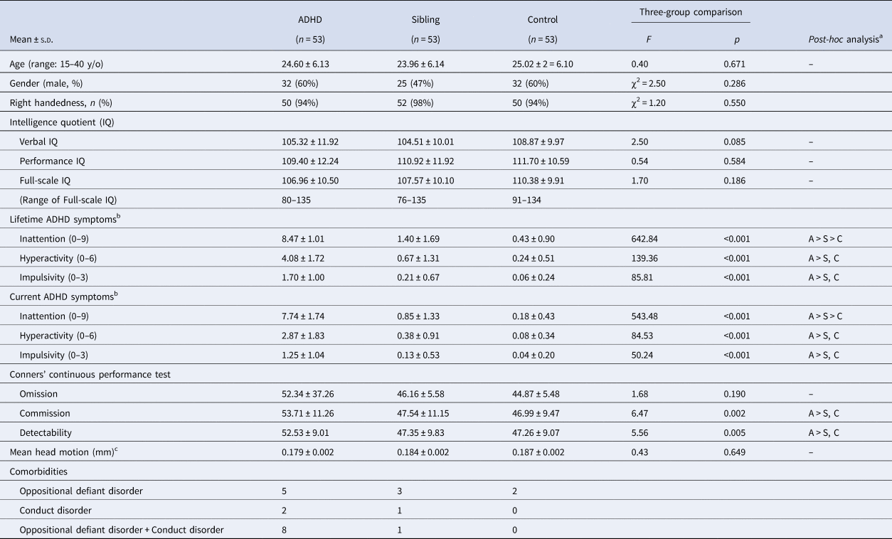
After excluding subjects with excess motion and their corresponding matched subjects, 53 subjects in each group were included for analysis (159 in total). Twenty-two of them were inattentive type, 30 were combined type, and one was hyperactive-impulsive type in childhood; 36 of them were inattentive type, and 17 were combined type currently. We found no significant group differences among the proband group, the unaffected sibling group, and the control group in age (24.60 ± 6.13, 23.96 ± 6.14, and 25.02 ± 6.10 years old, respectively), gender (male: 32, 25, and 32, respectively), handedness, full-scale IQ, and measurements of head motion during scanning (Table 1). Probands with ADHD had significantly higher ADHD symptoms when compared to the other two groups (p < 0.05). Unaffected siblings showed a subclinical and intermediate level of inattention symptoms, but low hyperactivity and impulsivity symptoms similar to the controls. Eight probands with ADHD had comorbid oppositional defiant disorder (ODD) and conduct disorder (CD), five of them had ODD, and two of them had CD in their lifetime (Table 1). Eighteen patients with ADHD have been treated with methylphenidate medication, with an average treatment duration of 26 months (range 1–162 months, s.d. = 47).
Demographics and characteristics of the participants

A, patients with ADHD; S, unaffected siblings; C, typically developing controls, CCPT, Conners' Continuous Performance Test.
a Pairwise comparison by post-hoc analysis with Bonferroni test.
b The sum of the symptom counts according to the Schedule for Affective Disorders and Schizophrenia for School-Age Children – Epidemiologic Version.
c Averaged maximal head movement for each volume.
Group differences in intrinsic functional connectivity
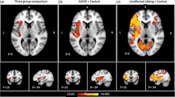
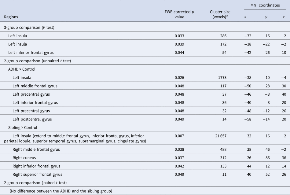
Comparing intrinsic functional connectivity with the precuneus/PCC among the three groups, we identified three significant clusters, including the left insula and left IFG (p FWE < 0.05) (Table 2 and Fig. 1a). When compared to controls, both probands with ADHD and unaffected siblings showed increased functional connectivity with the precuneus/PCC in regions overlapping with the clusters found in three-group comparison (Table 2, Fig. 1b and c). In addition, no brain region showed a reduction in connectivity with the precuneus/PCC in probands with ADHD and unaffected siblings compared to controls. We did not detect any significant differences in any brain regions between probands with ADHD and unaffected siblings using independent sample t test or paired two-sample t test with randomized permutation (Table 2). After we repeated the same analyses (t test) while controlling for gender effect or controlling for age, gender, and FIQ at the same time, the results are the same that we cannot detect any significant differences in any brain regions between probands with ADHD and unaffected siblings.
Comparisons in intrinsic functional connectivity with the DMN among the ADHD, unaffected siblings, and control groups. (a) Regions showing a significant difference in the three-group comparison. (b) Regions showing increased functional connectivity in probands with ADHD when compared to controls. (c) Regions showing increased functional connectivity in unaffected siblings when compared to controls. Images are shown in MNI space. Red–yellow represents regions where intrinsic functional connectivity significantly differ between groups [family-wise error rate (FWE) corrected for multiple comparisons, p FWE < 0.05; non-parametric permutation test with 10 000 permutations].

Differences in resting-state functional connectivity among adults with ADHD, their unaffected siblings, and controls
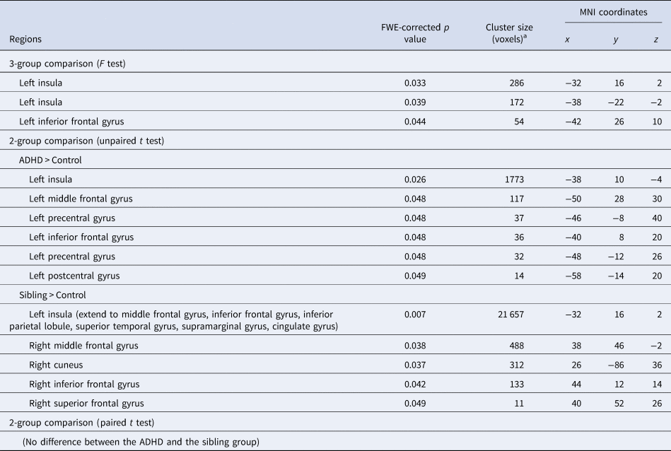
ADHD, attention-deficit hyperactivity disorder; FWE, family-wise error; MNI, Montreal Neurological Institute.
a The normalized voxel was resampled to the size of isotropic 2 mm.
Post-hoc correlation analysis revealed that commission error (r = −0.329, p = 0.017, online Supplementary Fig. S1A in supplement) and detectability (r = −0.289, p = 0.038, online Supplementary Fig. S1B in supplement) of the CCPT were negatively correlated with the intrinsic functional connectivity in the control group. The significant effects remained only for commission error in the final model (regression coefficient estimate β = –0.33, p = 0.017, R 2 = 0.11). These findings suggest that higher intrinsic functional connectivity was associated with less commission error in the control group. On the other side, such correlations were absent in probands with ADHD (commission error: r = −0.059, p = 0.674; detectability: r = 0.004, p = 0.979) and unaffected siblings (commission error: r = −0.223, p = 0.109; detectability: r = −0.221, p = 0.111). Besides, we found that the intrinsic functional connectivity in these clusters had no correlation with ADHD symptoms within probands with ADHD and within a combined group of probands with ADHD and siblings.
Connectivity–ADHD symptoms correlations in probands with ADHD
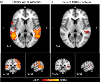
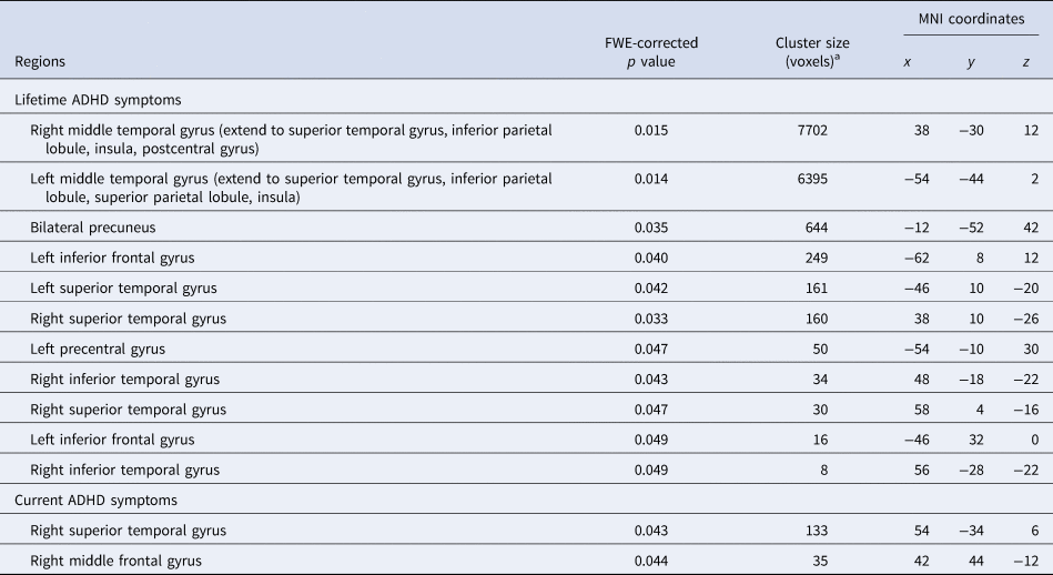
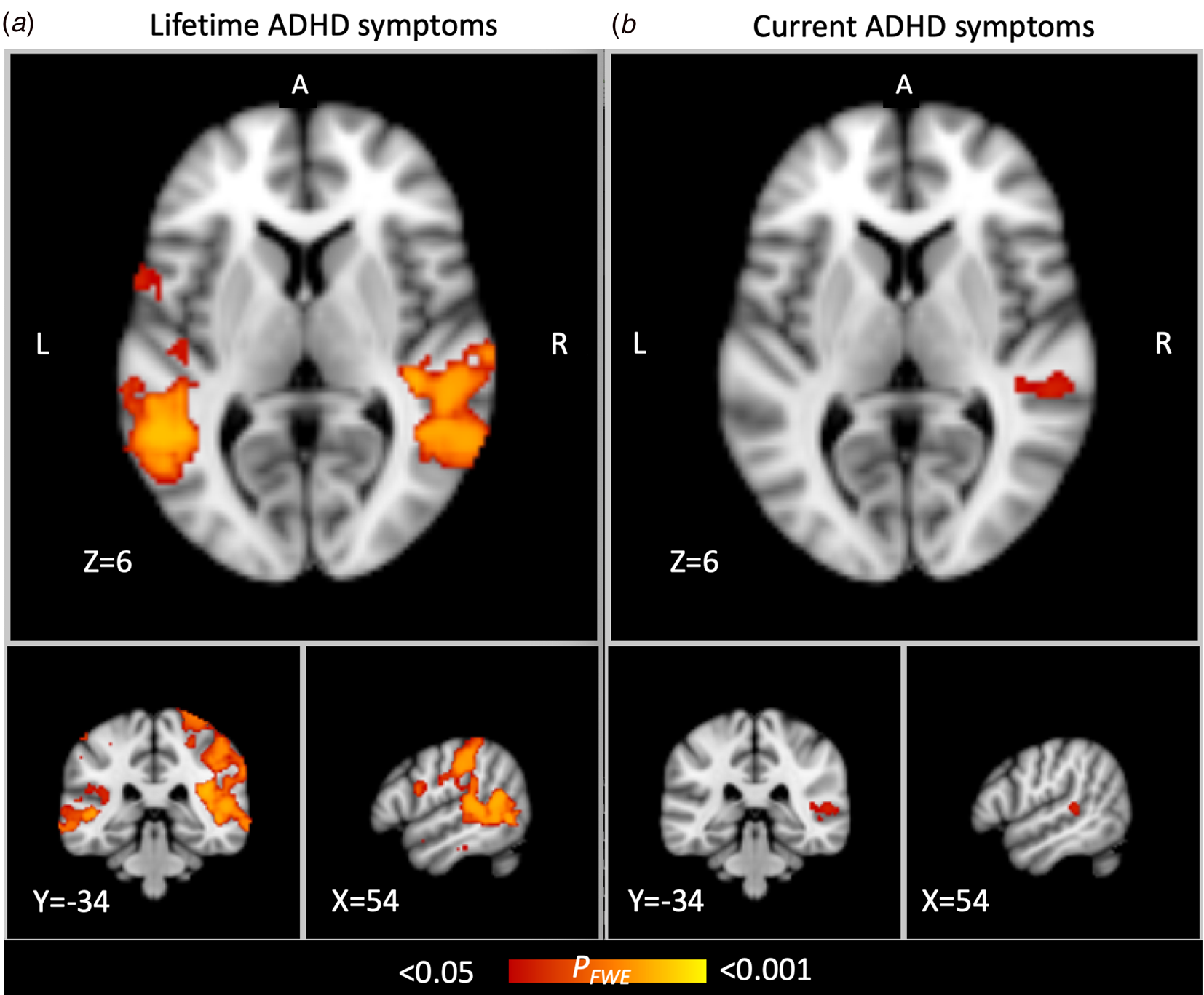
We found that lifetime ADHD symptoms positively correlated with intrinsic functional connectivity with peak voxels centered at the bilateral middle temporal gyrus in probands with ADHD and extended to the superior temporal gyrus, inferior parietal lobule, superior parietal lobule and insula, and also right postcentral gyrus (right-side cluster size = 61 616 mm3, p FWE-corrected = 0.015; left-side cluster size = 51 160 mm3, p FWE-corrected = 0.014), as well as other clusters in the left IFG, left precentral gyrus, and right inferior temporal gyrus (Table 3 and Fig. 2a). Current ADHD symptoms positively correlated with intrinsic functional connectivity in the right superior temporal gyrus and the right middle frontal gyrus (Table 3 and Fig. 2b). On the other hand, although unaffected siblings may have subclinical ADHD symptoms, no relationships between brain connectivity and ADHD symptoms were found.
Significant associations between ADHD symptoms and intrinsic functional connectivity with the DMN in the ADHD group. (a) Regions showing positive correlations between lifetime ADHD symptoms with intrinsic functional connectivity. (b) Regions showing positive correlations between current ADHD symptoms with intrinsic functional connectivity. Red–yellow represents regions where intrinsic functional connectivity significantly differ between groups [family-wise error rate (FWE) corrected for multiple comparisons, p FWE < 0.05; non-parametric permutation test with 10 000 permutations].
Regions with significant correlations between functional connectivity and clinical symptoms in adults with ADHD
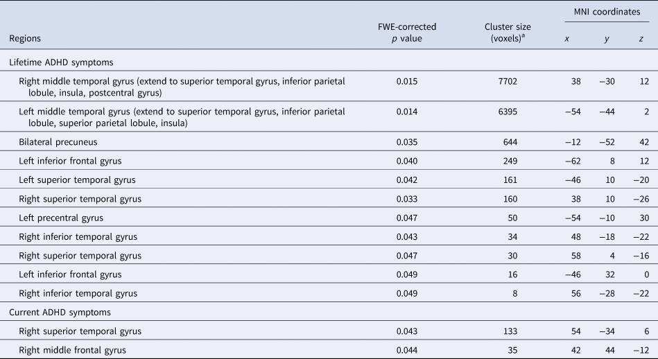
ADHD, attention-deficit hyperactivity disorder; FWE, family-wise error; MNI, Montreal Neurological Institute.
a The normalized voxel was resampled to the size of isotropic 2 mm.
Discussion
Using a sample of probands with ADHD, their unaffected siblings, and matched control participants, we compared the intrinsic functional connectivity between the precuneus/PCC and other brain regions to identify familial risk markers of ADHD. One of the major findings is that both probands with ADHD and unaffected siblings showed increased functional connectivity between the precuneus/PCC and key brain regions of salience and frontoparietal network (i.e. the left insula and left inferior frontal gyrus) when compared to controls. Increased intrinsic functional connectivity in these clusters was correlated with better performance in response inhibition (less commission error) in the CCPT only in the control group. This finding confirmed our hypothesis of aberrant functional connectivity among regions cross different networks in both probands with ADHD and unaffected siblings. Another major finding is that the severity of ADHD symptoms positively correlated with functional connectivity with the precuneus/PCC in bilateral fronto-parietal-temporal regions. The correlation involved larger brain regions with lifetime ADHD symptoms than with current symptoms in probands with ADHD.
Consistent with the previous study reporting increased intrinsic connectivity between the precuneus/PCC and the anterior insula in subjects with ADHD (Sripada et al., Reference Sripada, Kessler, Fang, Welsh, Prem Kumar and Angstadt2014; Zhao et al., Reference Zhao, Li, Yu, Huang, Wang, Liu and Wang2017), we further demonstrated increased connectivity in unaffected siblings of probands with ADHD. The insula cluster where both probands with ADHD and unaffected siblings showed increased functional connectivity than controls in three-group comparison is a major hub of the salience network (Uddin, Reference Uddin2015). The anterior insula is important for the behavioral inhibition and attentional control toward an unexpected stimulus (Bonnelle et al., Reference Bonnelle, Ham, Leech, Kinnunen, Mehta, Greenwood and Sharp2012), and the function of the salience network is to detect a salient event and to initiate appropriate responses to stimuli by engaging cognitive control networks while suppressing DMN (Sridharan, Levitin, & Menon, Reference Sridharan, Levitin and Menon2008). The only published study using rsfMRI in families with ADHD has reported the within-network functional connectivity in DMN, frontoparietal network, and VAN was heritable (Sudre et al., Reference Sudre, Choudhuri, Szekely, Bonner, Goduni, Sharp and Shaw2017). Although the salience network was not defined and analyzed in this study, the anterior insula has been included as part of VAN for analysis (Yeo et al., Reference Yeo, Krienen, Sepulcre, Sabuncu, Lashkari, Hollinshead and Buckner2011). Several studies have conceptualized VAN and salience networks as distinct entities that adults showed increased segregation of these two networks than children (Farrant & Uddin, Reference Farrant and Uddin2015). Task-based fMRI studies provide further evidence supporting familial influence on the salience network (Godinez et al., Reference Godinez, Willcutt, Burgess, Depue, Andrews-Hanna and Banich2015). For example, probands with ADHD and their co-twins, when compared to matched controls, showed decreased activation during a Stroop test in the anterior insula and the anterior cingulate cortex, both of them are major hubs of the salience network (Godinez et al., Reference Godinez, Willcutt, Burgess, Depue, Andrews-Hanna and Banich2015). In accordance with task-based fMRI results, our findings suggest that aberrant functional connectivity between the precuneus and insula during rest is a familial risk marker of ADHD.
The posterior insula is connected with motor and somatosensory areas (Yeo et al., Reference Yeo, Krienen, Sepulcre, Sabuncu, Lashkari, Hollinshead and Buckner2011), at its descending pathways terminates in the brainstem to regulate physiological reactivity to salient stimuli (Craig, Reference Craig2002). Another function related to the posterior insula is the subjective experience of time (Wittmann, Simmons, Aron, & Paulus, Reference Wittmann, Simmons, Aron and Paulus2010), and time perception and reproduction have been reported to be less precise in children with ADHD and their unaffected siblings than controls (Rommelse, Oosterlaan, Buitelaar, Faraone, & Sergeant, Reference Rommelse, Oosterlaan, Buitelaar, Faraone and Sergeant2007). Although the posterior insula has been less investigated in ADHD, our finding implies the potential role of the posterior insula in ADHD.
The left IFG cluster we found in three-group comparison is located within the frontoparietal network (Yeo et al., Reference Yeo, Krienen, Sepulcre, Sabuncu, Lashkari, Hollinshead and Buckner2011), in which the heritability of intrinsic functional connectivity has been reported in families with ADHD (Sudre et al., Reference Sudre, Choudhuri, Szekely, Bonner, Goduni, Sharp and Shaw2017). The IFG and the anterior insula tend to coactivate in many neuroimaging studies during inhibitory control and error-related activity (Tops & Boksem, Reference Tops and Boksem2011). The IFG has been reported to have decreased activation during the stop-signal task, a kind of response inhibition task, in probands with ADHD and unaffected siblings, compared to controls (van Rooij et al., Reference van Rooij, Hoekstra, Mennes, von Rhein, Thissen, Heslenfeld and Hartman2015). Increased activation of the IFG showed increased activation during the counting Stroop task (the number of words was consistent or inconsistent with the meaning of the word in Chinese), which involved inhibition and inference control (Fan, Shang, Tseng, Gau, & Chou, Reference Fan, Shang, Tseng, Gau and Chou2018). Our finding adds to the literature that intrinsic functional connectivity between DMN and frontoparietal network during rest may be a familial risk marker of ADHD.
No difference between probands with ADHD and unaffected siblings supports our hypothesis that aberrant connectivity with the precuneus is a familial risk marker. However, we did not detect any brain region showing neural compensation, which might have a protective effect from ADHD in unaffected siblings. When we repeated the t test while controlling for gender effect or controlling for age, gender, and Full-scale IQ at the same time, the results are the same that we cannot detect any significant differences.
As to the clinical implication of the brain regions identified in the three-group comparison (the clusters in the left insula and left IFG), we found that increased intrinsic functional connectivity between the precuneus/PCC and these regions was associated with less commission error and higher detectability in the control group. Lack of correlation in probands with ADHD and unaffected siblings is consistent with a previous study reporting positive or negative correlations between intrinsic functional connectivity and working memory only in the control group, not in ADHD (Zhao et al., Reference Zhao, Li, Yu, Huang, Wang, Liu and Wang2017). Our finding supports the notion that disrupted correlation might influence the cognitive process and therefore suggests neuropsychological impairment (Zhao et al., Reference Zhao, Li, Yu, Huang, Wang, Liu and Wang2017).
Previous studies reported decreased functional connectivity within DMN, VAN, and executive control network to be associated with more ADHD symptoms (Francx et al., Reference Francx, Oldehinkel, Oosterlaan, Heslenfeld, Hartman, Hoekstra and Mennes2015; McCarthy et al., Reference McCarthy, Skokauskas, Mulligan, Donohoe, Mullins, Kelly and Frodl2013; Sudre et al., Reference Sudre, Choudhuri, Szekely, Bonner, Goduni, Sharp and Shaw2017). Our finding that lifetime ADHD symptoms positively correlated with intrinsic functional connectivity between the precuneus/PCC and bilateral fronto-parietal-temporal regions in probands with ADHD demonstrated the correlation between higher cross-network connectivity and more ADHD symptoms. Combining with the findings in the literature, increased ADHD symptoms may be related to decreased functional integration within networks (decreased within network connectivity) and decreased functional segregation across networks (increased connectivity among regions cross different networks).
Methodological limitations
There are some limitations to our study. First, 18 probands with ADHD have been treated with methylphenidate. Although they were asked to discontinue methylphenidate at least one week before the MRI and neuropsychological assessment to minimize the influence of medication, long-term effects of medication on functional connectivity should still be considered. Second, psychiatric comorbidities such as ODD, CD, and depression are increased in subjects with ADHD (Lin et al., Reference Lin, Yang and Gau2016). Although no previous study has reported a significant effect of these comorbidities on the findings of functional connectivity in ADHD studies, we still provide information about these comorbidities that allow readers to evaluate the potential influence of comorbidities on our findings. Lastly, this study is a cross-sectional design which limits our interpretation of the meaning of the difference in ADHD sibling pairs and controls. It is not sure that the aberrant intrinsic functional connectivity is heritable neuropathology within ADHD families or the consequences of a compensatory neurodevelopment process.
In conclusion, this study demonstrates increased functional connectivity between major hubs in DMN and salience network and in DMN and frontoparietal networks that account for the underlying familial risks of ADHD. These networks correspond to the triple network model, which suggests altered connectivity among DMN, salience network, and central executive network, and play a prominent role in ADHD and several other psychiatric disorders (Menon, Reference Menon2011). Hence, the network-based analysis would be an interesting next step according to our current findings.
Supplementary material
The supplementary material for this article can be found at https://doi.org/10.1017/S0033291720003529
Financial support
All the authors report no financial relationships with commercial interests. This work was supported by the Ministry of Science and Technology (NSC101-2321-B-002-079; MOST103-2314-B-002-021-MY3), National Taiwan University Hospital (NTUH101-S1910), Chen-Yung Foundation, and National Health Research Institute (NHRI-EX107-10404PI), Taiwan.
ClinicalTrials.gov number, NCT01682915
Abbreviated title: Functional connectivity as a familial risk marker for ADHD