Introduction
Head and neck cancers account for over 8000 new cancer cases and 2700 deaths per year in the United Kingdom. The most commonly encountered distant metastases are pulmonary.Reference Ferlito, Shaha, Silver, Rinaldo and Mondin1 The presence of distant metastases is a significant factor in prognosis, and is often the ultimate event that leads to patient mortality. The presence of distant metastases usually means a poor prognosis and may be a deterrent to radical treatment of the primary head and neck tumour. It is self-evident that an effective screening programme for distant metastases would improve our ability to counsel these patients regarding important therapeutic decisions and end-of-life issues.
While it may seem logical to offer screening to all head and neck cancer patients, our enthusiasm for evaluating distant metastases must be balanced against the reality of medical economics in an environment of increasing cost-consciousness. A well conceived algorithm for screening holds the potential for cost-savings, in comparison with the current practice of apparently random and ill-defined screening. However, there is currently no evidence-based consensus on radiological screening. The value of chest computed tomography (CT) and chest X-ray has yet to be determined. Furthermore, the benefits of early detection as regards to patient outcome have not been thoroughly evaluated. Some authors have recommended chest X-ray in all cases, with chest CT reserved for advanced disease.Reference de Bree, Deurloo, Snow and Leemane2, Reference Brouwer, Bree, Hoekstra, Langendijk, Casterlijns and Leemans3 Other departments have adopted a local policy of performing chest CT on all patients.Reference Houghton, Hughes, Garvey, Beasley, Hamilton and Gerlinger4
In the United Kingdom, the National Institute for Health and Clinical Excellence (NICE) guidelines on head and neck cancer management have not recommended routine screening of pulmonary metastases in newly diagnosed cases of head and neck squamous cell carcinoma (SCC).5 These guidelines conclude that the low level of evidence, due to a paucity of good quality studies, precludes the effective use of limited resources. However, the three studies evaluated by NICE were observational surveys of small patient cohorts.Reference Warner and Cox6–Reference Tan, Greener, Seikaly, Rassekh and Calhoun8 We believe there is potential gain in the identification of metastases and second primary cancers in this group of patients, who are clearly at risk.
The aims of this study were: to evaluate the benefit of chest X-ray and chest CT as screening investigations in patients with newly diagnosed head and neck SCC; to determine the incidence of pulmonary metastases or synchronous pulmonary lesions; and to evaluate the factors associated with positive radiological findings. The results of this study were also compared with those of the studies evaluated by NICE.
Patients and methods
A retrospective review of consecutive patients diagnosed over a five-year study period, between January 2000 and December 2005, was performed. Patients were identified from the hospital cancer registry and were limited to those with a diagnosis of primary head and neck SCC. Patients with SCC in other regions were excluded, as were those with recurrent primary disease. Patients who had not undergone a radiological procedure performed for screening at the time of diagnosis were also excluded.
Two consultant radiologists routinely reported cases referred by the local multidisciplinary team (MDT) for head and neck cancer. The reference standard was clinical observation. Postero-anterior chest X-ray views were obtained. Chest X-ray findings were reported as either normal with no evidence of metastases or abnormal with evidence of metastases. In patients with indeterminate chest X-ray findings, the chest CT was referred to as the ‘gold standard’.
Chest CT was performed using a General Electric (Chalfont St Giles, United Kingdom) multislice scanner. The following radiological criteria were used: for pulmonary metastases, smoothly defined and subpleurally located lesions; for bronchogenic carcinoma, solitary, spiculated and centrally located lesions; and for mediastinal lymph node metastases, a short axial diameter greater than 10 mm. Chest CT findings were reported as follows: normal with no evidence of metastasis; abnormal with evidence of metastasis or synchronous tumour; or suspicious. Suspicious nodules were followed up with another CT scan at three months. This was an arbitrary period agreed upon by the local MDT, as there was no national consensus on a suitable interval for repeat CT scanning. In order to compare the efficacies of chest X-ray and chest CT, we analysed all patients in whom both investigations had been performed at the time of diagnosis and within three months of each other. It was also locally agreed that an interval exceeding three months may invalidate comparison of the chest X-ray and chest CT findings. The repeat scan was regarded as normal if there was no progression of the suspicious nodules. Patients with metastatic or synchronous disease on CT were regarded as having ‘positive CT findings’. All patients with a normal chest CT were categorised as having ‘negative CT findings’.
Demographic information included age at diagnosis and gender. Tumour (T) and node (N) classifications were according to the American Joint Committee on Cancer and Union Internationale Contre le Cancer staging system. Statistical data were analysed using the chi-square test where appropriate.
Results
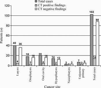
Between January 2000 and December 2005, 102 patients (63 men and 39 women) fulfilling the study criteria were identified from the hospital head and neck cancer registry. Of these, 83 patients (52 men and 31 women) had a chest X-ray and a chest CT within three months of each other. Fourteen patients had a chest X-ray–CT interval longer than three months, and five other patients had only chest CT performed. The mean age at diagnosis was 67 years (range 33–91 years). Head and neck SCC occurred most commonly in the larynx (39 per cent), followed by the oral cavity (20 per cent) and oropharynx (18 per cent) (Figure 1). Seven patients had primary carcinoma of unknown origin. These patients had initially presented with cervical lymphadenopathy, which subsequently revealed SCC on lymph node biopsy.
Distribution of cancer by site, showing numbers of positive and negative computed tomography (CT) findings (n = 102).

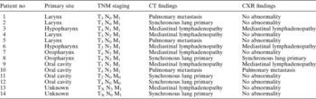
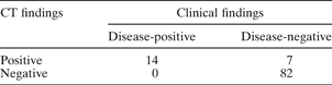
In total, 14 patients (14 per cent) had positive chest CT scan findings (Table I). Of these, three patients had pulmonary metastases, six had mediastinal lymphadenopathy and five had synchronous lung tumour (one larynx, two oral cavity, one oropharynx and one unknown primary). The most common primary site was the oral cavity and oropharynx, followed by the larynx. Nine patients had clinically advanced (T3 or T4) tumours at presentation. The chest CT was reported as suspicious in seven patients, but a repeat chest CT showed no radiological alteration. These patients were therefore managed as having no metastatic disease. The sensitivity and specificity of chest CT were 100 and 92.1 per cent, respectively (Table II). Twenty-nine per cent of all patients with an unknown primary cancer were found to have positive chest CT findings; of these, one patient had a synchronous bronchogenic carcinoma.
Details of patients* with positive CT findings

* n = 14. No = number; TNM = tumour–node–metastasis; CT = computed tomography; CXR = chest X-ray
Sensitivity and specificity of chest CT: total patients*

* n = 102. Sensitivity = 100%; specificity = 92.1%. CT = computed tomography
All 14 patients with positive CT findings had a chest X-ray performed within three months of the chest CT. Of these, nine patients' chest X-rays were reported as normal. Of the remaining five patients, three chest X-rays were reported as showing mediastinal lymphadenopathy, one as pulmonary metastasis and one as bronchogenic carcinoma. The findings in all five positive chest X-rays were verified by the corresponding chest CT, without discrepancy. In total, there were 10 positive chest X-ray reports. Besides the five chest X-ray results discussed above, five other chest X-rays were reported as having pulmonary metastasis. However, these lesions were found to be reported as benign on chest CT. Subsequent review of these five patients' case notes showed no development of pulmonary involvement within the first 12 months of diagnosis.
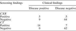
The sensitivity for the detection of pulmonary involvement by chest X-ray was only 35.7 per cent, if CT was used as the gold standard (Table III). The specificity of chest X-ray was marginally higher than that of chest CT, at 92.7 per cent. If statistical analysis of CT screening was limited to the 83 patients with a CT–chest X-ray interval of less than three months, then the sensitivity of CT remained at 100 per cent but the specificity dropped marginally to 89.8 per cent (Table III).
Sensitivity and specificity of CXR VS chest CT: patients with cxr–CT interval <3 months*

* n = 83. For chest X-ray (CXR), sensitivity = 35.7% and specificity = 92.7%. For chest computed tomography (CT), sensitivity = 100% and specificity = 89.8%.
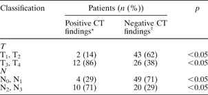
In our study, higher-staged SCC tumours were significantly associated with positive CT findings (Table IV). In patients with a positive chest CT scan, 86 per cent had T3 or T4 tumours, compared with 38 per cent of those with a negative chest CT (p < 0.05). In addition, 71 per cent of patients with positive CT findings had N2 or N3 nodal disease, compared with 29 per cent of those with negative findings (p < 0.05). Sixty patients in our cohort had either T1 or T2 tumours, of which only three had positive chest CT findings (Table I). Two of these patients had primary cancers of the oral cavity and one had a hypopharyngeal tumour.
Correlation of T and N classifications with chest CT findings

* n = 14; †n = 88. T = tumour; N = node; CT = computed tomography
Discussion
Synchronous tumours and pulmonary metastases are not uncommon in patients with head and neck cancers. The lung remains the most common site for distant metastasis in cases of head and neck SCC. The published incidence of pulmonary metastases varies widely, ranging between 4 and 25 per cent.Reference de Bree, Deurloo, Snow and Leemane2, Reference Houghton, Hughes, Garvey, Beasley, Hamilton and Gerlinger4, Reference Houghton, McGarry, Stewart, Wilson and McKenzie9 In our study, the incidences of synchronous bronchogenic tumour and metastasis were 5 and 9 per cent, respectively. The most common primary site of cancer was the larynx; this contrasts with previous studies, which found that metastasis from the oral cavity and oropharynx were more common.Reference Nillsen, Murthy, McClymont and Denholm10 This difference may be due to sampling bias within our study population. Patients who had not undergone a screening chest CT were excluded from the study. There was no local protocol on screening of newly diagnosed head and neck SCC. The four consultant surgeons (two ENT and two maxillo-facial) within the Multidisciplinary (MDT) adopted a different approach to management and radiological investigation. This meant that a greater proportion of some cancer sites may not have been screened and, hence, some metastases may have been missed.
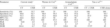
In our unit, chest CT has been shown to be an effective screening tool, offering high accuracy (91.5 per cent) and high negative predictive value (100 per cent). Chest CT is more sensitive than chest X-ray, although chest X-ray appears to be slightly more specific than CT. The results of our study also compare well with those of the three studies evaluated by NICE (Table V).
Comparison of results with studies evaluated by NICE5

* n = 14, †n = 4, ‡n = 3, **n = 1, where n represents number of positive chest findings for ‘gold standard’ investigations. NICE = National Institute for Health and Clinical Excellence; CT = computed tomography; CXR = chest X-ray; PPV = positive predictive value; NPV = negative predictive value; PLR = positive likelihood ratio; NLR = negative likelihood ratio; DOR = diagnostic odds ratio
Of these studies, those by NICE, and Warner and Cox, compared the use of chest X-ray with CT in screening for pulmonary malignancy in head and neck cancer patients; they found that CT was more accurate than chest X-ray, with accuracies of 95 and 92 per cent for CT versus 93 and 85 per cent for chest X-ray, respectively.5, Reference Warner and Cox6 The sensitivity of CT was 100 per cent in both studies and the sensitivities of chest X-ray were 33 and 25 per cent, respectively. The specificities of CT were 95 and 91 per cent and those of chest X-ray were 98 and 95 per cent, respectively.
The third study, which evaluated CT with chest X-ray versus chest X-ray alone in patients with advanced head and neck cancer, found that chest X-ray alone was more accurate than CT with chest X-ray, with accuracies of 96 and 88 per cent, respectively.Reference Arunachalam, Putnam, Jennings, Messersmith and Robson7 However, this study reported only one patient with metastatic chest disease; given the methodological limitations of the study, the results should be interpreted with caution. In their conclusion, the authors did not recommend routine use of CT. The low sensitivity of chest X-ray reported in this study may be due in part to the fact that the majority of lung metastases are peripherally situated, where chest X-ray tends to be less reliable.Reference Troell and Terris11
In the current study, it can be argued that the favourable results of CT were due to bias in methodology.
Firstly, the study cohort was small, although significantly larger than that of the NICE study. This was due to the differing practices of each local consultant surgeon; hence, not all new patients within the study period underwent CT scanning. It was beyond the scope of the current study to investigate patients who had not undergone a CT but who may have had distant metastases at the time of diagnosis.
Secondly, the two radiologists on the MDT were not blinded to patients' clinical details or to the findings of the other imaging modality. If the same radiologist read all the films, their awareness of the result of one imaging modality could easily bias their interpretation of the second modality.
Thirdly, the reference standard was clinical observation, as histological confirmation was not obtained.
While it is undeniable that more robust studies are required, many serious ethical issues are raised. It would be difficult to imagine how randomisation of patients could be achieved, given that up to one-quarter of patients may have had distant metastases. It is unlikely that patients would give consent to ‘opt out’, were they aware of such a high incidence. Furthermore, the need to maintain clinical excellence within a specialty-specific MDT would preclude the involvement of radiologists who did not routinely report chest CTs. Randomisation and blinding of the radiologists to clinical details would not be easy to achieve. These limitations were also recognised in the studies evaluated by NICE. Nevertheless, we believe that the benefits of screening such patients compare favourably in the current climate of medical cost-savings, which mitigates against blanket CT scanning.
There are obvious discrepancies of opinion regarding CT screening. Kesti-Santti et al. concluded that there was no indication for routine chest CT scanning, as only two of their patients (of 100) had pulmonary metastasis at presentation.Reference Kesti-Santti, Markkola, Makitie, Back and Atula12 Conversely, other authors have already recommended routine CT scanning of all patients with advanced tumours.Reference Loh, Brown, Baker, Gilbert, Gullane and Irish13, Reference Mercader, Gatenby, Mohr, Fisher and Caroline14 For example, Loh et al. reported that 64 per cent of patients with CT-positive findings had T4 tumours at diagnosis, with 73 per cent having N2 or N3 nodal disease.Reference Loh, Brown, Baker, Gilbert, Gullane and Irish13 In our study, 86 per cent of patients had T3 or T4 tumours, and 71 per cent had N2 or N3 lymphadenopathy. However, we feel that there is a strong case for routine CT scanning for all newly diagnosed cases of head and neck SCC. Routine screening with chest CT is appropriate, given the need for patients and their families to make very difficult therapeutic decisions regarding the appropriateness of radical surgery, participation in experimental trials and the option of palliative care only. Knowledge regarding metastatic disease may be an essential determinant in this decision for many patients and their families. As such, chest CT scanning has become our unit's policy following the current study, while chest X-ray is no longer performed for screening.
While identifying pulmonary involvement does not always influence management, it can change opinion, from treating the primary cancer surgically, with post-operative chemo-radiotherapy, to treating it with chemo-radiotherapy alone. The chest CT also acts as a useful baseline when there is a need to perform later scanning for comparison.
There is now increasing interest in the use of positron emission tomography (PET) scanning in the treatment planning process of patients with head and neck cancer.Reference Gregoire, Bol, Geets and Lee15 In head and neck carcinoma cases, the outcome of PET scanning varies widely, which may be due to the same biases and limitations faced by most studies evaluating the value of CT scanning. Many authors have claimed that combining PET with CT increases the accuracy of metastasis detection. For example, Pauleit et al. reported a sensitivity of 93 per cent, a specificity of 79 per cent and an accuracy of 83 per cent in their cohort of 18 patients who underwent a combination of PET and CT.Reference Pauleit, Zimmerman, Stoffels, Bauer, Risse and Fluss16 In a larger study of 547 cancer patients who underwent PET and CT scanning for initial staging purposes, the incidence of a second primary cancer was 4.8 per cent.Reference Choi, Lee, Kwon, Shim, Baek and Park17 Out of the 45 lesions reported as malignant, only 24 were subsequently proven to be a second primary. The sensitivity and positive predictive value were 91 and 69 per cent, respectively. However, this study was not limited to head and neck SCC. Jeong et al. claimed to achieve a high sensitivity, specificity and accuracy (91.8, 98.9 and 97.1 per cent, respectively) with a combination of PET and CT, in their study cohort of 12 patients with advanced head and neck cancer.Reference Jeong, Baek, Son, Ki Chung, Kyung Lee and Young Choi18 While such published data may show an improvement, compared with CT scanning alone, the results must be interpreted with caution, as many of these studies are non-randomised, non-blinded analyses of small patient cohorts.
• The most commonly encountered distant metastases are pulmonary, which is a significant factor in the prognosis of cancer patients
• There is no consensus on the radiological screening of patients with newly diagnosed head and neck cancer
• An effective screening programme for distant metastases would improve our ability to counsel patients regarding important therapeutic decisions and end-of-life issues
• In this study, the incidence of pulmonary involvement (metastases and primary bronchogenic tumours) was 14 per cent
• Chest CT was found to be an effective screening tool for head and neck cancer patients
• National guidelines (e.g. those from the National Institute for Health and Clinical Excellence) should be re-evaluated in the light of CT scans becoming more cost-effective and readily available
Positron emission tomography is not without drawbacks, not least its cost and availability and the skills required. During the current study, it was not available locally and therefore was not a practical solution for scanning all new cases managed by the MDT. To our knowledge, PET scanning is not currently used as a screening tool in the United Kingdom. Furthermore, PET scans are only able to detect metabolically active lesions 5 mm and larger. In the current study, some of the lesions reported on CT were less than 5 mm and may not have been metabolically active. The lack of anatomical detail is also a major drawback. Despite claims of high accuracy, use of PET may not abrogate the need for conventional imaging. Until PET becomes more widely available, its use as a screening tool for head and neck SCC may be limited to the research arena.
Conclusion
This study adds additional data to the debate regarding pre-treatment chest CT screening in newly diagnosed cases of head and neck SCC. While the superiority of chest CT, compared with chest X-ray, in screening for metastasis or synchronous lung tumour is already known, the cost burden of screening all new head and neck SCC patients is significant. In this era of increased scrutiny of healthcare costs, it may be impossible to offer CT screening to all head and neck SCC patients.
However, the incidence of distant metastasis in clinically advanced tumours is compelling. Therefore, we recommend chest CT screening in newly diagnosed cases of head and neck SCC, in particular patients with highly staged disease and those with primary tumours of the oral cavity.
The current NICE guideline needs to be reappraised in the light of more recent studies on the role of CT screening. While the use of a combination of PET and CT studies in head and neck cancer screening may make a difference to the treatment and outcome of the disease, further studies are needed to evaluate the efficacy of this approach.