I. INTRODUCTION
The healing of large bone defects resulting from trauma, resection for tumors, infection, and congenital diseases represents a difficult challenge in orthopedic surgery. Reference Yang, Wang, Hou, Guo and Liu1 Periprosthetic infection rates are in the range 1–2.5% for primary implants Reference Pauksch, Hartmann, Rohnke, Szalay, Alt, Schnettler and Lips2 and are even higher for revision surgeries. Reference Renaud, Lavigne and Vendittoli3 Infected implants can represent a severe threat to the patient's health and provide a significant economic burden to the healthcare system. Clinically, bone defects can be reconstructed through the use of various bone grafts. However, current treatments based on the use of autologous bone grafts and allografts suffer from several limitations. Consequently, there is a growing interest in the development of synthetic bone grafts. Reference Henslee, Spicer, Yoon, Nair, Meretoja, Witherel, Jansen, Mikos and Kasper4,Reference Ye, Xu, Gao, Yan, Zhao, Tu, Zhang, Duan, Sommer, Mostoslavsky, Kaplan, Wu, Zhang, Wang and Chen5
Bioactive glasses undergo specific reactions in vivo, resulting in the formation of a carbonated hydroxyapatite (HA) layer on the surface of the glass that leads to strong bonding with bone and soft tissues. Reference Cao and Hench6 Ions released from the glass during its conversion to HA have been shown to enhance the proliferation and osteogenic differentiation of osteoprogenitor cells. Reference Tsigkou, Jones, Polak and Stevens7,Reference Sun, Wei, Liu, Li, Li, Wang and Meng8 While silicate-based bioactive glasses have been studied and applied for decades, borate-based bioactive glasses have been developed and evaluated more recently for biomedical applications. Reference Liang, Rahaman, Day, Marion, Riley and Mao9–Reference Han and Day11 Borate-based bioactive glasses have more favorable processing characteristics and more controllable conversion rates to HA than the widely-studied silicate 45S5 bioactive glass. Reference Huang, Day, Kittiratanapiboon and Rahaman12 Boron has also been shown to be beneficial for processes, such as embryogenesis, healthy bone growth, immune function, and psychomotor skills. Reference Nielsen13 It has been reported that boron released from borate-based bioactive glasses can enhance the proliferation and migration of human umbilical vein endothelial cells, the formation of tubules in vitro and the secretion of proangiogenic cytokines, such as interleukin-6 and basic fibroblast growth factor. Reference Durand, Gongora, Lopez, Boccaccini, Zago, Baldi and Gorustovich14 Trace quantities of boron (B) have also been found to be necessary for the optimal health of rats. Reference Forrer, Wenker, Gautschi and Lutz15,Reference Forrer, Gautschi and Lutz16 Previous studies showed that borosilicate bioactive glasses can stimulate more rapid bone formation in a rat tibial defect model in vivo when compared to silicate bioactive glasses. Reference Zhang, Jia, Gu, Xiao, Liu, Wang, Zhang, Huang, Rahaman and Day10,Reference Bi, Jung, Day, Neidig, Dusevich, Eick and Bonewald17
The oligodynamic effect of Ag has been used for thousands of years. Furthermore, silver (Ag) ions have been considered to have a broad spectrum of antibacterial activity which is significant for the inhibition of bacterial colonization associated with biomaterial infections. The ability to inhibit biofilm formation has been confirmed in several studies. Reference Pauksch, Hartmann, Rohnke, Szalay, Alt, Schnettler and Lips2,Reference Gristina, Naylor and Myrvik18–Reference Kalishwaralal, BarathManiKanth, Pandian, Deepak and Gurunathan20 Staphylococcus aureus (S. aureus) and Staphylococcus epidermidis are the most common organisms responsible for infections. Reference Moojen, Spijkers, Schot, Nijhof, Vogely, Fleer, Verbout, Castelein, Dhert and Schouls21,Reference Harris, Tosatti, Wieland, Textor and Richards22 It has been found that bacteria have a lower tendency to develop resistance to Ag-containing biomaterials and that Ag is able to exert a bactericidal effect when used in small concentrations. Reference Clement and Jarrett23 Despite the established role of Ag as an antibacterial agent, the feasibility of using Ag-containing biomaterials in clinical applications still depends on several factors, including safety issues associated with the amount of Ag released from the biomaterials. Consequently, it is necessary to consider any adverse effects of Ag-containing biomaterials on bone cells. Another issue is that effects of the Ag doping on the degradation and conversion of bioactive glasses are largely unknown.
The objective of this study was to evaluate the effects of doping borosilicate bioactive glass scaffolds with varying amounts of Ag2O (0.05–1.0 wt%) on the bioactivity, compressive strength, antibacterial activity, and cytocompatibility of the scaffolds in vitro. The degradation and conversion of the scaffolds to HA and the release of Ag ions from the scaffolds were studied as a function of immersion time in simulated body fluid (SBF). The cytocompatibility of the scaffolds was investigated using assays of cell viability and alkaline phosphatase (ALP) activity of murine preosteoblastic MC3T3-E1 cells while the cell morphology was examined using confocal laser scanning microscopy (CLSM). The antibacterial activity of the scaffolds was evaluated from their ability to inhibit the growth of S. aureus, a Gram-positive bacterium that is commonly associated with infected implants, and Escherichia coli (E. coli), a Gram-negative bacterium. Reference Percival, Bowler and Russell24
II. EXPERIMENTAL PROCEDURE
A. Preparation of bioactive glass scaffolds
The parent borosilicate bioactive glass (designated BG) with the composition 6Na2O, 8K2O, 8MgO, 22CaO, 36B2O3, 18SiO2, 2P2O5 (mol%) and the bioactive glass doped with 0.05, 0.5, and 1.0 wt% Ag2O (designated by BG-0.05Ag, BG-0.5Ag, and BG-1.0Ag, respectively) were prepared by conventional melting and casting. A mixture composed of the requisite amounts of Na2CO3, K2CO3, MgCO3, CaCO3, SiO2, H3BO3, NaH2PO4, AgNO3 (analytical grade; Sinopharm Chemical Reagent Co., Ltd. China) was melted in a platinum crucible for 4 h at 1250 °C and quenched between cold steel plates.
The glass was crushed, ground, and sieved to give particles of size < 50 μm (Fig. S1), which were used to prepare porous scaffolds with a trabecular microstructure by a foam replication technique. Reference Liu, Huang, Fu, Yao, Wang, Pan and Lu25 Briefly, a slurry composed of the glass particles (53 wt%), anhydrous ethanol (44 wt%), and ethyl cellulose (3 wt%) used as a binder (analytical grade, Sinopharm Chemical Reagent Co. Ltd.) was prepared by stirring the mixture for 5 h at room temperature. Then a polyurethane foam with open porosity of ∼50 pores per inch (Shanghai No. 6 Plastic Co., Ltd, China) was immersed in the slurry to coat the surface of the foam with the slurry. The as-coated foams were dried, heated for 2 h at 400 °C to decompose the foam and then for 2 h at 550 °C to densify the glass particles into a strong network. The porous cylindrical constructs (5 mm in diameter) were sectioned into the appropriate thickness for subsequent experiments.
B. Degradation and conversion of bioactive glass scaffolds in SBF
As-fabricated scaffolds of the four glasses in the shape of cylinders (5 mm in diameter × 10 mm) were tested in compression to measure their strength using a Zwick/Roell Z010 servo-hydraulic testing instrument(Ulm, Germany). The constructs were tested at a cross-head speed of 0.5 mm min−1 using a 1 kN load cell. Five specimens of each group were tested and the strength was expressed as an average ± standard deviation (SD).
The degradation of the scaffolds in SBF was studied by measuring the weight loss of the scaffolds and the pH of the medium. Reference Kokubo and Takadama26 Briefly, samples of the scaffolds (1.0 g) were immersed in 100 mL SBF at 37 °C, with the medium completely replaced with fresh medium at predetermined time points. The pH of the SBF was measured using a pH meter. After removal from the SBF, the samples were rinsed three times with anhydrous ethanol, dried at 80 °C for 10 h and weighed. The weight loss of the reacted scaffolds was defined as (W o–W t)/W o, where W o was the initial (unreacted) mass and W t was the mass at an immersion time t. The concentration of Ag ions released from the scaffolds into the SBF was measured using inductively-coupled plasma optical emission spectrometry (Perkin Elmer Model Optima 2100 ICP, USA).
The scaffolds were examined in a field-emission scanning electron microscopy (FESEM, Quanta 200 FEG, Netherlands) equipped with an energy dispersive x-ray spectrometer (EDS) (Quanta 200 FEG). The scaffolds were ground into a powder and analyzed using x-ray diffraction (XRD) (D/max 2550 V, Rigaku, Cu Kα, Tokyo, Japan) and Fourier transform infrared (FTIR) spectroscopy (Perkin Elmer Multiscope) to determine phase and compositional changes resulting from the reaction in SBF. The oxidation state of the Ag and other elements at the surface of the scaffolds was determined using x-ray photoelectron spectroscopy (XPS) (Physical Electronics PHI 5802 equipped with a monochromatic Al Kα source).
C. In vitro cell culture
The degradation of the scaffolds in an aqueous medium resulted in the release of ions from the glass which can affect the biocompatibility of the scaffolds. Consequently, the effects of the ions dissolved from the glass (referred to as the ionic dissolution product of the scaffolds) on the proliferation and alkaline phosphate activity of MC3T3-E1 cells were studied in vitro. The MC3T3-E1 cells (subclone 14), a mouse preosteoblastic cell line, were obtained from the Cell Bank of the Chinese Science Academy (Shanghai, China).
The scaffolds were immersed in high glucose Dulbecco's modified Eagle's medium (DMEM) without serum Reference King, King and Hughes27 (100 mg scaffold per mL medium) for 24 h at 37 °C. After centrifugation and filtration through a 0.2 μm bacterial filter, the medium was collected and used as a stock solution. The stock solutions obtained from the four groups of scaffolds and fresh DMEM (control medium) were supplemented with 10% fetal bovine serum and 1% penicillin/streptomycin (P/S) and used in the cell culture.
MC3T3-E1 cells were seeded in 96-well plates at an initial density of 3 × 10³ cells per well and cultured in extracts of the ionic dissolution product of the scaffolds (100 μL) for 1, 3, and 7 days, with the media replaced every 2 days. Cells cultured in media without the ionic dissolution product of the bioactive glass were used as the control group. Then 10 μL of the Cell Counting Kit-8 solution (CCK-8, Dojindo Molecular Technologies, Inc. Japan) was added to each well and the system was incubated for 1.5 h. The absorbance was measured at 450 nm on a plate reader.
For the osteogenic differentiation assay, complete medium supplemented with 50 mg/mL ascorbic acid and 10 mM β-glycerol phosphate was used. MC3T3-E1 cells were seeded in 24-well plates (1 × 10⁴ cells per well), cultured in osteogenic differentiation medium containing the ionic dissolution product of the scaffolds, and assayed on days 7 and 14 to determine their ALP activity. Briefly, the cells were washed with phosphate-buffered saline (PBS) to remove the medium and lysed in 200 μL of 0.2% Triton X-100 (Sigma Aldrich, Australia). Lysates were centrifuged at 14,000 rpm for 5 min at 4 °C. Then, 50 μL of the supernatant was mixed with 150 μL of the ALP assay working solution according to manufacturer's protocol (QuantiChrom™ Alkaline Phosphatase Assay Kit, Bioassay Systems). The total protein content was measured using a Bicinchoninic acid protein assay kit (Thermo Scientific, Australia). The optical density (OD) was measured at 405 nm on a plate reader.
MC3T3-E1 cells were also seeded directly on the scaffolds to examine their adhesion and spreading. The cells were seeded on individual scaffolds (5 mm in diameter × 3 mm) placed in each well of a 48-well plate at an initial concentration of 1 × 10⁴ cells per well. After 48 h, the scaffolds were rinsed with PBS (pH = 7.4) and fixed with 2.5% glutaraldehyde in PBS for 1 h at 4 °C. Then the scaffolds were dehydrated in a graded ethanol series (30, 50, 60, 70, 80, 90, and 100%), coated with gold and examined in an FESEM (Quanta 200 FEG).
The morphology of the MC3T3-E1 cells cultured in the ionic dissolution product of the four groups of scaffolds was examined. The cells were seeded in the ionic dissolution product (500 µL) in 24-well plates (104 cells per well). After 24 h, the cells were fixed in 4% paraformaldehyde for 30 min at 4 °C. Then 0.1% Triton X-100 was added for 5 min before staining the F-actin cytoskeletons. The samples were incubated with FITC-Phalloidin (Enzo Life Sciences, UK) at room temperature in the dark for 30 min. Finally, the nuclei were stained with 4′,6-diamidino-2-phenylindole dihydrochloride (DAPI) (Invitrogen). The cell morphology was observed using CLSM (Leica, Germany).
D. In vitro antibacterial activity of the scaffolds
S. aureus (ATCC 25923) and E. coli (ATCC 25922) were used as two model bacteria to evaluate the antibacterial activity of the scaffolds. Both qualitative analyses on Luria–Bertani (LB) solid medium and quantitative analyses in LB liquid medium were used. All samples were sterilized under ultraviolet light for 2 h before use.
Qualitative analysis was performed by determining the inhibition zones around the scaffolds for E. coli and S. aureus cultured on LB agar plates. Briefly, frozen E. coli and S. aureus were activated with fresh LB. The bacterial suspension (100 μL, 108 cfu mL−1) was spread onto the agar plates and scaffold discs (diameter = 10 mm; mass ∼ 100 mg) were placed on the agar plate. After incubation for 24 h at 37 °C, the inhibition zones were observed. To qualitatively evaluate their long-term antibacterial efficacy, scaffolds previously immersed for 3 days in SBF at 37 °C were also studied under the same conditions.
In the quantitative analysis of the antibacterial activity of the scaffolds, E. coli or S. aureus bacterial suspension (100 μL; 108 cfu mL−1) was added to 5 mL LB liquid medium with an OD value of 0.1–0.2 at 600 nm in individual test tubes. Then, one scaffold of mass = 0.45 g was added to each test tube. Reference Zheng, Wang, Wen, Shen, Zhu and Shi28 After incubation for 24 h at 37 °C with gentle shaking (100 rpm), the absorbance of the bacterial suspension was measured at 600 nm using a Lambda 25 UV–Vis spectrophotometer. The percent bacterial inhibition was determined as
 $${\rm{Percent}}\,{\rm{bacterial}}\,{\rm{inhibition}} = {{100\left( {{I_{\rm{c}}} - {I_{\rm{s}}}} \right)} \over {{I_{\rm{c}}}}}\quad ,$$
where I c and I s are the average OD of the control group and the experimental group, respectively. The morphology of the bacteria adhering to the scaffolds was examined using FESEM (Quanta 200 FEG).
E. Statistical analysis
The data (n = 3) were presented as a mean ± SD. Analysis for differences between groups was performed using one-way analysis of variance followed by Tukey's test. Differences were considered to be statistically significant for P < 0.05 (*) or P < 0.01 (**).
III. RESULTS
A. Degradation and conversion of scaffolds in vitro
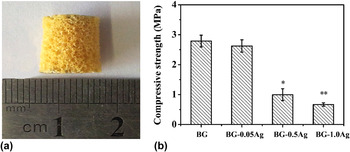
The as-fabricated scaffolds had a trabecular microstructure [Fig. 1(a)], composed of a nearly fully dense glass phase, interconnected pores of size 200–400 μm and a porosity of 85 ± 3%. There was no observable difference in the microstructure of the four groups of scaffolds. The compressive strength of the scaffolds was dependent on the amount of Ag in the glass [Fig. 1(b)]. The strength of the BG-0.05Ag scaffolds (2.7 ± 0.2 MPa) was not significantly different from the BG scaffolds (2.8 ± 0.2 MPa) but the strength of the BG-0.5Ag scaffolds (1.0 ± 0.2 MPa) and the BG-1.0Ag scaffolds (0.8 ± 0.1 MPa) was significantly lower. The strength of all four groups of scaffolds is comparable to the lower range of values reported for human trabecular bone (2–12 MPa), Reference Erol, Mouriňo, Newby, Chatzistavrou, Roether, Hupa and Boccaccini29 which indicates that they can be used in the repair of nonloaded bone.
(a) Optical image of as-fabricated BG-1.0Ag scaffold and (b) compressive strengths of BG and BG-Ag scaffolds. Mean ± SD; n = 5. Difference when compared to BG (*P < 0.05, **P < 0.01).

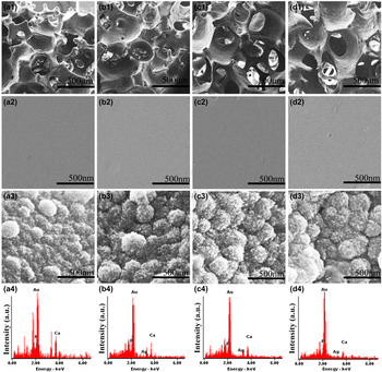
The surface of the glass phase of the as-fabricated BG and BG-Ag scaffolds had a relatively smooth surface [Fig. 2(a1)–2(d2)]. After immersion in SBF, the glass surface became rougher with granular features, as shown in Fig. 2(a3)–2(d3) for the scaffolds immersed in SBF for 90 days. There were no observable differences in morphology among the four scaffolds before and after immersion in SBF. EDS analysis [Fig. 2(a4)–2(d4)] showed that the surface of the reacted scaffolds had a high concentration of calcium and phosphorus, with the Ca/P atomic ratio in all four groups of scaffolds equal to approximately 1.67, the value for stoichiometric HA.
FESEM images for the (a1, a2) BG, (b1, b2) BG-0.05Ag, (c1, c2) BG-0.5Ag, and (d1, d2) BG-1.0Ag scaffold before soaking in SBF; FESEM images for the (a3) BG, (b3) BG-0.05Ag, (c3) BG-0.5Ag, and (d3) BG-1.0Ag scaffolds after soaking in SBF for 90 days; EDS analysis for the (a4) BG, (b4) BG-0.05Ag, (c4) BG-0.5Ag, and (d4) BG-1.0Ag.

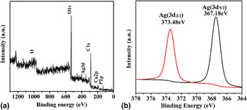
XPS survey scans of the BG-1.0Ag scaffolds that were immersed in SBF for 90 days [Fig. 3(a)] showed the presence of peaks corresponding to Ag(3d), O(1s), Ca(2p), and P(2p), as well as the C(1s) peak that often results from carbon contamination. The oxidation state of the Ag was determined from XPS high-resolution scans of the Ag(3d) peak [Fig. 3(b)]. Fitting of the Ag(3d) doublet indicated Ag(3d 5/2) and Ag(3d 3/2) binding energies of 367.18 and 373.48 eV, respectively, indicating that the Ag existed in the monovalent oxidation state. Reference Boronin, Koscheev and Zhidomirov30
(a) XPS spectrum and (b) Ag(3d) line spectrum of BG-1.0Ag scaffold after immersion for 90 days in SBF.

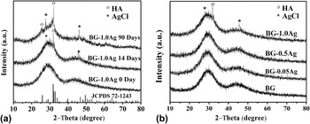
XRD of the BG-1.0Ag scaffolds that were immersed in SBF for 90 days [Fig. 4(a)] showed peaks that corresponded to a reference HA, Ca10(PO4)6OH2 (JCPDS 72-1243), indicating the conversion of the glass to HA. The pattern also showed small peaks at 28° and 46° which corresponded to AgCl, presumably formed by reaction of the Ag ions released from the glass with Cl ions from the SBF. For an immersion time of 14 days, there was little difference in the XRD patterns of these groups of scaffolds, except that the pattern for the BG-1.0Ag scaffolds showed small peaks corresponding to HA and AgCl [Fig. 4(b)].
XRD patterns for (a) BG-1.0Ag after immersion in SBF for 0, 14, 90 days; (b) BG and BG-Ag scaffolds after immersion for 14 days. For comparison, the pattern of a reference HA (JCPDS 72-1243) is shown.

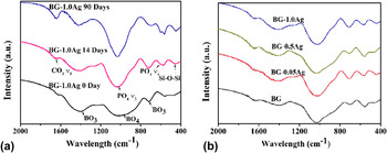
FTIR spectra [Fig. 5(a)] show compositional changes of the BG-1.0Ag scaffold as-fabricated and after immersion for 14 and 90 days in SBF. The spectrum of the as-fabricated scaffold showed resonances at 1350–1500 cm−1 and 650–750 cm−1 that are attributed to the B–O stretching mode and the bending mode of BO3 groups, respectively, while the resonance at 900–1100 cm−1 is attributed to the B–O stretching mode of BO4 group. Reference Liu, Rahaman and Day31 Immersion of the scaffold for 14 days in SBF resulted in a strong reduction in the intensity of the resonances associated with the borate group while resonances attributed to vibrations of the phosphate group dominated the spectrum, such as a strong resonance at ∼1050 cm−1 attributed to the (PO4)3− ν3 resonance and the weaker resonance at ∼600 cm−1 attributed to the (PO4)3− ν4 resonance. With an increase in the immersion time to 90 days, the (PO4)3− ν3 resonance increased in intensity while the (PO4)3− ν3 at ∼600 cm−1 was split into two peaks which is characteristic of the presence of HA. The spectrum also showed a resonance at 460 cm−1 corresponding to the bending vibration of the Si–O–Si bond. The resonance at 1670 cm−1, attributed to C–O in CO3 2−, indicated the formation of a carbonated-substituted HA, presumably due to the presence of dissolved CO2 in the SBF. There was no clear difference among the FTIR spectra for the four groups of scaffolds immersed in SBF for 14 days [Fig. 5(b)]. In general, the FTIR results are consistent with the XRD analysis described above.
FTIR spectra for (a) BG-1.0Ag scaffold after immersion in for 0, 14, 90 days; (b) BG and BG-Ag scaffolds after immersion in SBF for 14 days.

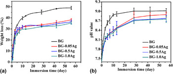
The weight loss of the four groups of scaffolds is shown in Fig. 6(a) as a function of immersion time in SBF. As the conversion of the glass to HA is accompanied by a weight loss, the data can be used as a measure of the conversion kinetics. The weight loss for all four groups of scaffolds showed the same trend; it increased rapidly during the first 7–10 days and slowed considerably thereafter, reaching a nearly constant value within ∼30 days. At any immersion time, the weight loss of the Ag-doped scaffolds was lower than the BG scaffolds. When the experiments were stopped (at 56 days), the weight loss of the BG scaffolds was 49%, compared to 38, 36 and 35%, respectively, for the BG-0.05Ag, BG-0.5Ag and BG-1.0Ag scaffolds. The increase in pH of the SBF showed a similar trend [Fig. 6(b)]; it increased from the starting value of 7.4–9.02, 8.90, 8.78, and 8.75, respectively, for the BG, BG-0.05Ag, BG-0.5Ag and BG-1.0Ag scaffolds at an immersion time of 56 days. The lower weight loss of the Ag-doped scaffolds and the lower pH of the SBF indicate that the Ag reduced the degradation and conversion of the glass to HA.
(a) Weight loss of the four group of scaffolds and (b) the pH of the medium as a function of immersion time of the scaffolds in SBF (starting pH = 7.4).

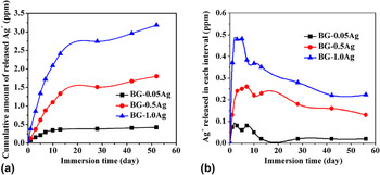
The cumulative amount of Ag ions released from the BG-Ag scaffolds into the SBF showed the same trend for all four groups of scaffolds but, at any immersion time, it increased with increasing amount of Ag in the glass [Fig. 7(a)]. The amount of Ag released into the SBF within specific time intervals showed a rapid increase during the first 7–10 days [Fig. 7(b)]. Thereafter, the amount of Ag released from the BG-0.05Ag scaffold almost ceased. In comparison, the BG-0.5Ag and BG-1.0Ag scaffolds showed a sustained release for up to 56 days when the experiments were terminated. At 56 days, the cumulative amount of Ag ions released into the SBF was 1.8 and 3.2 ppm, respectively, for the BG-0.5Ag and BG-1.0Ag. These cumulative amounts of Ag released into the SBF at 56 days are equal to 31 and 36%, respectively, of the Ag initially present in the as-fabricated BG-0.5Ag and BG-1.0Ag scaffolds, indicating the potential for sustained release of Ag from those two scaffolds.
(a) Cumulative amount of Ag ions released from Ag-doped bioactive glass scaffolds as a function of immersion time in SBF; (b) amount of Ag released from the scaffolds at specific times into SBF.

B. Proliferation and ALP activity of MC3T3-E1 cells cultured on scaffolds
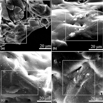
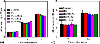
FESEM images of MC3T3-E1 cells cultured on the surface of the four groups of scaffolds for 48 h appeared to indicate that the cells attached and spread well on the surface of the scaffolds (Fig. 8). There was no marked difference in the morphology of the cells cultivated in the ionic dissolution product of the four groups of glass scaffolds. Figure 9(a) shows the results of CCK-8 assays for the proliferation of MC3T3-E1 cells cultured in the ionic dissolution product of the four groups of scaffolds and in the control group (media without the ionic dissolution product). For each group, cell growth increased with incubation time from 1 day to 7 days but there was no significant difference among the groups at each incubation time (P > 0.05), indicating that the Ag doping had no effect on cell proliferation. ALP activity, an early indicator of osteoblastic differentiation, was used to evaluate the effect of the ionic dissolution product of the scaffolds on the differentiation of MC3T3-E1 cells. The ALP activity of the cells increased as the culture time increased from 7 to 14 days [Fig. 9(b)], but there was no significant difference among the groups at each incubation time (P > 0.05).
FESEM micrographs of MC3T3-E1 cells cultured for 48 h on the surface of the four groups of scaffolds: (a) BG, (b) BG-0.05Ag, (c) BG-0.5Ag, (d) BG-1.0Ag.

Quantitative measurement of (a) proliferation and (b) ALP activity of MC3T3-E1 cells cultured in extracts of the ionic dissolution product of four groups of scaffolds for the times shown. Control: under identical culture conditions without the ionic dissolution products of the glass. Mean ± SD; n = 3, P > 0.05 as control.

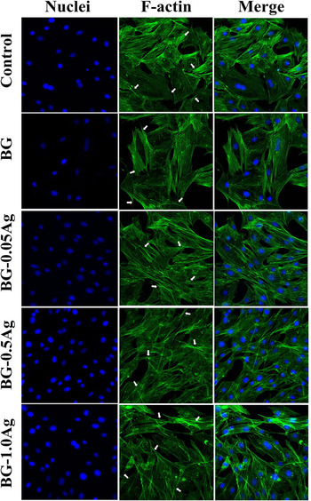
CLSM images of MC3T3-E1 cells cultured in the ionic dissolution product of the scaffolds and in the control media are shown in Fig. 10. The cells showed morphologies with large elongation (white arrow) and there was no observable difference in the cell morphology among the groups. In general, these CLSM images provide support for the results of the CCK-8 assays which showed that the ionic dissolution products of all four scaffold groups were favorable for the proliferation of MC3T3-E1 cells with no observable cytotoxicity.
CLSM images of MC3T3-E1 cells cultured for 24 h in extracts of the ionic dissolution product of four groups of scaffolds (BG, BG-0.05Ag, Bg-0.5Ag, BG-1.0Ag) showing nucleus stained with DAPI (blue) and F-actin stained with FITC-phalloidin (green). (White arrow: prominent filopodia and unidirectional lamellipodia extensions.)

C. Antibacterial activity of scaffolds in vitro
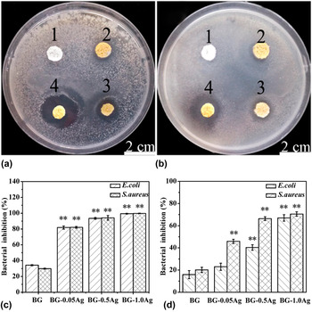
Images of the bacterial inhibition zones around the four groups of scaffolds [Figs. 11(a) and 11(b)] showed that the BG-Ag scaffolds inhibited the proliferation of E. coli and S. aureus. When compared to the BG scaffolds (scaffold 1), there were clear inhibition zones around the BG-0.05Ag, BG-0.5Ag, and BG-1.0Ag scaffolds and the diameter of the inhibition zone increased with the Ag concentration in the scaffold (scaffolds 2–4). These observations were supported by the quantitative analysis of antibacterial activity by the scaffolds in LB liquid medium which showed an increase in the percent bacterial inhibition with an increase in the amount of Ag in the scaffold [Fig. 11(c)]. The BG scaffolds showed a bacterial inhibition of ∼35% which was presumably due to the release of B and other ions from the glass. While the bacterial inhibition increased to ∼80% for the BG-0.05Ag scaffolds, the effect was presumably limited by the low concentration of Ag released from the glass. In comparison, the BG-0.5Ag and BG-1.0Ag scaffolds showed a bacterial inhibition of >90%. Together, the results in Figs. 11(a)–11(c) showed that the antibacterial activity of the scaffolds was dependent on the concentration of Ag released from the scaffolds and that the BG-0.5Ag and BG-1.0Ag scaffolds were most effective in inhibiting the bacterial growth.
Growth inhibition zones of (a) E. coli and (b) S. aureus around the four groups of scaffolds after incubation for 24 h on LB agar plates (1: BG; 2: BG-0.05Ag; 3: BG-0.5Ag; 4: BG-1.0Ag); (c) percent bacterial inhibition of E. coli and S. aureus by the four groups of scaffolds after incubation for 24 h; (d) percent bacterial inhibition of E. coli and S. aureus by the four groups of scaffolds after the scaffolds were immersed for 3 days in SBF and then incubated for 24 h. Mean ± SD; n = 3. Difference between groups (*P < 0.05, **P < 0.01).

The long-term antibacterial efficacy of the scaffolds was also evaluated. The scaffolds were immersed in SBF for 3 days; then they were incubated in the LB liquid medium and analyzed for their antibacterial activity. The immersion in SBF reduced the antibacterial activity of the scaffolds [Fig. 11(d)], which was presumably due to the lower amount of Ag released from the scaffolds after the three-day treatment in SBF. However, the percent bacterial inhibition of E. coli and S. aureus by the BG-1.0Ag scaffolds was still 66 and 71%, respectively. In comparison, the percent bacterial inhibition of E. coli and S. aureus by the BG-0.5Ag scaffolds was 40 and 66%, respectively. The results of the antibacterial assays indicate that the BG-0.5Ag scaffolds could provide long-term bacterial inhibition to S. aureus while the BG-1.0Ag could provide long-term bacterial inhibition to both E. coli and S. aureus.
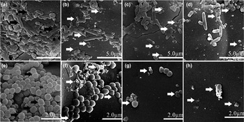
The effects of the BG-Ag scaffolds on the morphology and membrane integrity of E. coli and S. aureus colonies are shown in Fig. 12. The E. coli colonies changed from rod-shape when cultured on the BG scaffolds [Fig. 12(a); black arrow] to a corrugated morphology and even merged into the surface [Figs. 12(b)–12(d); white arrow] of the BG-Ag scaffolds. The S. aureus cells showed smooth, intact surfaces, and undamaged cells going through binary fission on the surface of the BG scaffolds [Fig. 12(e); black arrow]. Cell debris and completely lysed cells were observed on the BG-Ag scaffolds [Figs. 12(f)–12(h); white arrow]. These results indicated that the BG-Ag scaffolds had the ability to prevent the bacterial colonization and biofilm formation of E. coli and S. aureus with increasing amount of Ag in the scaffold.
FESEM images of (a–d) E. coli and (e–f) S. aureus seeded on the surface of the four groups of scaffolds and incubated for 24 h, (a, e) BG, (b, f) BG-0.05Ag, (c, g) BG-0.5Ag, (d, h) BG-1.0Ag. The black arrows indicate the round, smooth or cells going through binary fission while the white arrows indicate the corrugated, damaged, and lysed cells.

IV. DISCUSSION
Prevention of bacterial colonization is an important consideration in reducing the risk of implant infection. One approach to reduce the incidence of bacterial colonization is to impart antibacterial properties to the biomaterial implants. The scaffolds of bioactive ceramics or bioactive glass with improved biological properties are promising implants for bone regeneration. In the present study, scaffolds of a borosilicate bioactive glass doped with 0.05–1.0 wt% Ag2O showed a desirable combination of cytocompatibility, conversion to HA, and antibacterial activity which make them potential candidate implants for bone repair which could also provide a lower risk of infection.
In addition to their composition, the microstructure (or architecture) of the scaffolds, such as pore size, porosity, and pore interconnectivity, plays a key role in supporting bone ingrowth. Interconnected pores of size (diameter or width) ∼100 µm are generally considered to be the minimum requirements to permit tissue ingrowth and function in porous scaffolds, but pores of size > 300 µm may be required for enhanced bone ingrowth and formation of capillaries. Reference Jones and Hench32 The BG and BG-Ag scaffolds created in the present study had a “trabecular” microstructure similar to human trabecular bone, with pores in the range 200–400 µm and a porosity of ∼85% that are favorable for bone infiltration.
Doping the borosilicate glass with 0.05–1.0 wt% Ag2O reduced its rate of degradation and conversion to HA (or a calcium phosphate material) but resulted in the sustained release of Ag ions from the glass into SBF which increased with increasing concentration of Ag in the glass. The HA formed on the surface of the glass, due to the reaction of Ca2+ ions released from the glass with PO4 3− ions present in the medium, had a porous granular structure [Fig. 2(a3)–2(d3)]. The porous granular HA layer formed on the surface of the scaffolds during the in vitro cell culture could contribute to enhancing the differentiation of the MC3T3-E1 cells. Reference Ma, Kotaki, Inai and Ramakrishna33
The reaction between the Ag ions released from the scaffolds and anions present in the SBF can lead to insoluble precipitates on the surface of the glass, to a reduction in the concentration of anions in the solution (such as phosphate ions) and, presumably, to a reduction in the rate of conversion to HA. The formation of insoluble Ag-containing precipitates is supported by the EDS and XPS spectra that showed the presence of Ag on the surface of the converted BG-Ag scaffolds. The XPS spectra of the BG-1.0Ag scaffold (Fig. 3) showed that the Ag in the glass existed as Ag ions in the monovalent oxidation state and not as Ag nanoparticles (or clusters). As dissolved Ag ions play a major role in the biological action against microorganisms, Reference Baer, Foldbjerg, Hayashi, Sutherlans and Autrup34,Reference Kittler, Greulich, Diendorf, Koller and Epple35 the presence of the Ag ions in the glass could provide for more controllable release. As the glass degrades and converts to HA, the Ag ions are released into the medium. The XRD patterns of the BG-Ag scaffolds showed small peaks corresponding to AgCl which increased in intensity with increasing amount of Ag in the glass. This is consistent with the release profile Ag ions from the glass which showed a larger amount of Ag ions released into the SBF with increasing amount of Ag in the glass scaffolds (Fig. 7). All the BG-Ag scaffolds showed a rapid release of Ag ions in the first 7–10 days of immersion in SBF. Thereafter, the release of Ag ions from the BG-0.05Ag scaffolds almost ceased, whereas the BG-0.5Ag and BG-1.0Ag scaffolds showed a slower but sustained release until the experiments were terminated after 56 days (8 weeks). Thus, the BG-0.5Ag and BG-1.0Ag scaffolds might have the ability to provide long-term antibacterial activity.
The proliferation and ALP activity of MC3T3-E1 cells cultured in the ionic dissolution product of the BG, BG-0.05Ag, BG-0.5Ag, and BG-1.0Ag scaffolds showed no dependence on the Ag concentration in the glass (Fig. 9). It is expected that the Ag in the ionic dissolution product of the scaffolds existed freely as Ag ions and not as bound Ag that is reported to lose its functionality. Reference Harris, Tosatti, Wieland, Textor and Richards22 Furthermore, Ag-containing implants appear to have a positive effect in improving wound healing. Reference Percival, Bowler and Russell24 These data indicate that for the Ag2O concentrations used (up to 1.0 wt%), the Ag-doped scaffolds were not toxic to MC3T3-E1 cells in vitro.
The cytotoxicity of Ag has received considerable attention. Reference Park, Neigh, Vermeulen, de la Fonteyne, Verharen, Briedé, van Loveren and de Jong36–Reference Rai, Yadav and Gade38 A previous study showed that below a certain threshold concentration, Ag ions had little effect on cell proliferation whereas above the threshold level, cell proliferation decreased markedly with increasing Ag concentration. Reference Albers, Hofstetter, Siebenrock, Landmann and Klenke39 Humans typically ingest small amounts of Ag daily, some of which is resorbed by the intestine while the major portion is excreted by the liver. Reference Clement and Jarrett23 It has been reported that an Ag ionic concentration of <200 ppb in the blood is considered to be normal in vivo. Reference Ewald, Hösel, Patel, Grover, Barralet and Gbureck40 In the present study, the highest amount of Ag ions released from the BG-1.0Ag scaffold was 0.48 ppm (Ag concentration = 0.004 mM) [Fig. 7(b)]. This concentration is still lower than the minimal inhibitory concentration of AgNO3 solution for the osteoblast viability and differentiation (Ag concentration = 0.048 mM or 5.18 ppm) reported by Albers et al. Reference Albers, Hofstetter, Siebenrock, Landmann and Klenke39 After the peak in the release profile, the release of Ag ions decreased markedly. The results showed that the release profile of Ag from the scaffolds was not cytotoxic. The normal concentration of B in human plasma is 13 ± 3 μM (140 ± 32 ppm). Reference Hunt41,Reference Park, Li, Shcheynikov, Zeng and Muallem42 In the present study, the highest amount of B released from the four groups of scaffolds (98 ppm for BG scaffolds) was below the normal concentration in human plasma [Fig. S2]. Furthermore, concerns about the toxicity of B in vivo have been alleviated by results showing that borosilicate and borate glasses are nontoxic in small animals. Reference Gorustovich, López, Guglielmotti and Cabrini43
Both the quantitative and qualitative antibacterial assays showed that the BG-Ag scaffolds could provide both short-term and long-term antibacterial activity, which is a desirable property for biomedical applications requiring therapeutic activity. Whereas the BG scaffolds showed little ability to inhibit the growth of E. coli and S. aureus (Fig. 11), there were clear inhibition zones around the BG-0.05Ag, BG-0.5Ag, and BG-1.0Ag scaffolds and the diameter (or width) of the inhibition zone increased with the Ag concentration in the scaffold. The ability of the BG-Ag scaffolds to inhibit the growth of the bacterial strains was also supported by the quantitative analysis of antibacterial activity by the scaffolds in LB liquid medium [Fig. 11(c)]. After they were pretreated for 3 days in SBF, the BG-Ag scaffolds showed a lower antibacterial activity, particularly for E. coli [Fig. 11(d)]. This was presumably due to a lower release of Ag ions from the scaffolds as a result of the SBF pretreatment. The bacterial inhibition by the BG-0.5Ag scaffolds pretreated in SBF was more effective against S. aureus (66%) than E. coli (40%) but the pretreated BG-1.0Ag scaffolds still showed a bacterial inhibition of ∼70% for both bacterial strains. FESEM images of the bacterial strains cultured on scaffolds indicated that the BG-Ag scaffolds had the ability to prevent the bacterial colonization and biofilm formation of E. coli and S. aureus which increased with increasing concentration of Ag in the scaffold (Fig. 12).
A mechanism that has been suggested for the antibacterial activity of Ag is that Ag ions penetrate the cell wall, incorporate within the cell and subsequently convert the DNA into a condensed form, leading to damage or even death of the microorganisms. Reference Jones and Hench32 The greater susceptibility of Gram-positive bacteria has also been reported by Reddy et al. Reference Reddy, Feris, Bell, Wingett, Hanley and Punnoose44 The different response between Gram-positive and Gram-negative bacteria has been suggested to be closely related to the differences in cell wall structure, cell physiology, metabolism or degree of contact. Reference Kohanski, Dwyer and Collins45,Reference Brogden46 Based on their cytocompatibility and ability to inhibit bacteria, the BG-0.5Ag and BG-1.0Ag appear to be promising bioactive glasses for bone repair with reduced risk of bacterial infection.
V. CONCLUSIONS
Scaffolds of a parent borosilicate glass doped with 0.05–1.0 wt% Ag2O were created and evaluated in vitro to determine the effect of the Ag concentration on the bioactivity, cytocompatibility, and antibacterial activity of the scaffolds. Doping the glass with Ag2O resulted in a reduction in the degradation and conversion of the scaffolds to HA in SBF at 37 °C. When immersed in SBF, the scaffolds doped with 0.5 and 1.0 wt% Ag2O showed a sustained release of Ag ions into the medium for over 56 days. The ions released from the parent or Ag-doped bioactive glass were not toxic to mouse preosteoblastic MC3T3-E1 cells. The scaffolds doped with 0.5 and 1.0 wt% Ag2O showed a bacterial inhibition of >90% against S. aureus and E. coli in vitro. After pretreatment for 3 days in SBF at 37 °C, the scaffolds doped with 1.0 wt% Ag2O showed a bacterial inhibition of 66 and 71%, respectively, against S. aureus and E. coli, indicating their potential for longer-term bacterial inhibition. These borosilicate bioactive glass scaffolds doped with 0.5 or 1.0 wt% Ag2O are promising for bone repair coupled with providing a lower risk of bacterial infection.
Supplementary Material
To view supplementary material for this article, please visit http://dx.doi.org/jmr.2015.243.
ACKNOWLEDGMENTS
This work was supported by the Science and Technology Commission of Shanghai Municipality (Grant No. 12JC1408500) and the National Natural Science Foundation, China (Grant Nos. 51072133, 81201377 and 51372170).