




No CrossRef data available.
Article contents
Unravelling the phylogenetic relationships of African trypanosomes of suids
Published online by Cambridge University Press: 12 July 2001
Abstract
African trypanosomes of the subgenera Nannomonas and Pycnomonas have been recorded from both wild and domestic suids. However, complete descriptions of some of these trypanosomes with regard to host range, pathogenicity, transmission and distribution are still lacking. Neither the recently described Trypanosoma (Nannomonas) godfreyi nor Trypanosoma (Nannomonas) congolense Tsavo have been isolated from mammalian hosts, while Trypanosoma (Pycnomonas) suis remains the rarest of the Salivarian trypanosomes. The only isolate presumed to be of the latter species is maintained at the Kenya Trypanosomiasis Research Institute, Nairobi. We present here the results of characterization of this isolate by morphology, tsetse transmission, the use of species-specific DNA probes and DNA sequence analysis. Morphology in stained blood smears revealed a small trypanosome with a free flagellum. Experimental transmission through Glossina morsitans morsitans showed a developmental cycle typical of subgenus Nannomonas. A positive identification was obtained with species-specific PCR primers for T. congolense Tsavo; moreover, the sequence of the SSU rRNA gene was almost identical to that of T. congolense Tsavo on database. In phylogenetic analysis of the SSU rRNA genes of Salivarian trypanosomes, T. congolense Tsavo grouped with T. simiae rather than T. congolense, suggesting that the name T. simiae Tsavo is more appropriate.
Information
- Type
- Research Article
- Information
- Copyright
- © 2001 Cambridge University Press
INTRODUCTION
In tropical Africa, trypanosomiasis is a disease of such importance to animal health that it has shaped agricultural development and continues to constrain livestock production across much of the continent. The epidemiology of trypanosomiasis in tropical Africa is complex. A wide range of domestic and wild mammals can serve as hosts and there are large numbers of different tsetse and trypanosome species and subspecies involved. The application of molecular methods to the identification of pathogenic trypanosomes has greatly improved our understanding of the epidemiology of trypanosomiasis, but has also raised new questions. For example, there is evidently a significant proportion of trypanosome infections, which cannot be identified by the available DNA probes (McNamara, Laveissière & Masiga, 1995; Masiga et al. 1996a; Solano et al. 1996; Woolhouse et al. 1996; Lehane et al. 2000). The importance of such omissions is highlighted by the discovery that one such unidentified trypanosome represented a new species, Trypanosoma (Nannomonas) godfreyi, which was subsequently shown to be widespread in tropical Africa and probably pathogenic to pigs (McNamara et al. 1989; McNamara & Snow, 1991; McNamara, Mohammed & Gibson, 1994; Woolhouse, Hargrove & McNamara, 1993; Lehane et al. 2000). A further new genotype of subgenus Nannomonas was discovered by Majiwa et al. (1993) in a tsetse fly (Glossina pallidipes) caught in the Tsavo National Park in Kenya, and was named Trypanosoma (N.) congolense Tsavo type.
On the other hand, no DNA probes at all are available for the tsetse-transmitted trypanosomes of subgenus Pycnomonas. This subgenus contains the single species T. suis, which is known from only a handful of reports. Ochmann originally named T. suis in 1905, after finding it in a herd of sick pigs in Dar-es-Salaam, Tanzania (Hoare, 1972; Stephen, 1986). There is one further report of T. suis in a warthog from the Lake Rukwa area of Tanzania in 1912 (Stephen, 1986), before the thorough work of Peel and Chardome, working in Burundi in the 1950s, who gave a full account of the life-cycle and morphology of T. suis (Peel & Chardome, 1954a, b). They described T. suis as a short, stout monomorphic trypanosome with a free flagellum. Its cycle of development in the tsetse fly involved the gut and salivary glands like T. brucei; however, salivary gland forms were reputedly not infective, full maturation taking place in the hypopharynx of the fly. T. suis was placed in a new subgenus Pycnomonas on the basis of its life-cycle in the fly and was considered to be an intermediate evolutionary stage between development in the proboscis or the salivary glands (Hoare, 1972).
Since the rediscovery of T. suis by the Belgian workers, there have been 2 other reports of this trypanosome (a) from the salivary glands of G. vanhoofi caught in the Irangi forest in Congo, west of lake Kivu; stained metacyclics were identified by morphology (Van den Berghe & Zaghi, 1963) and (b) from G. pallidipes caught just south of Mombasa on the Kenya Coast (Janssen & Wijers, 1974). This trypanosome isolate produced a chronic infection in pigs, in contrast to the majority of trypanosomes isolated from tsetse in the same study, which were identified as T. simiae since they caused an acute infection in pigs (Janssen & Wijers, 1974). The trypanosome from the chronic infection showed many of the characteristics of T. suis, according to the brief account given (Janssen & Wijers, 1974), and was later identified as T. suis from the morphology of stained specimens by Stephen (1986). In an attempt to elucidate the phylogenetic affinities of T. suis, we characterized this same isolate from cryopreserved material held at the Kenya Trypanosomiasis Research Institute. However, as detailed below, our study has shed more light on the nature of T. congolense Tsavo than T. suis, and has highlighted the value of genotypic taxonomic descriptors.
MATERIALS AND METHODS
Trypanosomes
The cryopreserved T. suis isolate, KETRI 1864, consisted of blood from a pig, which had been infected via in vivo feeding of 54 G. pallidipes collected on 6 June 1970 from Muhaka forest, Ukunda, Kenya. KETRI 1864 was used to infect 3 pigs, and their infected blood was subsequently frozen as KETRI 3436. All pigs developed parasitaemia with varying degrees of pathogenicity (C. M. Mwendia, J. N. Makumi, J. M. Ngotho, J. M. Ndung'u, unpublished observations). The infected pig blood was cryopreserved and specimens stained for microscopy. DNA was subsequently prepared from frozen blood and transformed procyclics. The isolate was not cloned at any stage and thus could potentially consist of a mixture of trypanosome genotypes.
PCR analysis and DNA sequencing
Species-specific PCR tests for T. congolense savannah, forest, Kilifi and Tsavo types, T. godfreyi and T. brucei were carried out using DNA from KETRI 3436 as template. The primer sequences used were those described by Masiga et al. (1992), with the addition of primers specific for T. godfreyi (Masiga, McNamara & Gibson, 1996b) and T. congolense Tsavo (Majiwa et al. 1993).
The miniexon and small subunit ribosomal RNA (SSU rRNA) repeat units were also amplified from this template DNA using published PCR primer sequences (Murthy, Dibbern & Campbell, 1992, Maslov et al. 1996). The miniexon repeat unit was cloned using a plasmid vector designed for PCR products, and was sequenced using an ABI automated sequencer. The sequence of the SSU rRNA was obtained direct from the PCR fragment as described (Stevens et al. 1999a).
Sequence alignment
SSU rRNA sequences were aligned primarily on the basis of their secondary structure (Neefs et al. 1990). Subsections of the alignment, between regions of high homology were subaligned using the program Clustal W (Thompson, Higgins & Gibson, 1994), before final adjustments were made by eye. The KETRI 3436 sequence was aligned to the 64 trypanosome SSU rRNA sequences analysed previously (Stevens, Noyes & Gibson, 1998; Stevens et al. 1999a, b). The sequences of miniexon transcripts and upstream region (∼100 nt) were aligned using Clustal W.
Phylogenetic analysis
Analysis of 2 data sets was performed. The full data set of 65 SSU rRNA sequences (results not presented) was analysed to establish the phylogenetic position of KETRI 3436 relative to other trypanosome species. Trypanosoma sequences were compared with a range of outgroup taxa (Trypanoplasma borreli, Crithidia spp., Leishmania spp.) and the tree was rooted on Bodo caudatus. The suitability of free-living bodonid taxa as outgroups for phylogenetic studies of trypanosomatids has been established by a number of studies using a range of ribosomal and protein coding genes (e.g. Stevens et al. 1999a and references therein).
A second analysis on a subset of 22 taxa, including all available SSU rRNA sequences of T. (Nannomonas) spp., T. (Trypanozoon) spp. and T. (Duttonella) vivax was performed allowing a finer degree of phylogenetic resolution to be obtained. The alignment of Stevens et al. (1999a) was used as a starting point and reduced from 2509 to 2410 informative positions after the deletion of superfluous blanks in the reduced data set; at the same time the number of included characters increased from 1809 to 1852, as reliable alignment of previously excluded characters became possible. A diverse selection of Trypanosoma spp. was included as outgroups including the type species, T. lewisi, T. theileri and T. cruzi of the subgenera Herpetosoma, Megatrypanum and Schizotrypanum respectively, together with T. grayi and T. varani. Analysis of the full data set supported the use of these taxa as suitable outgroup species.
Bootstrapped maximum parsimony analyses with 100 replicates were performed for both the full data set (65 taxa) and the Salivarian clade subset (22 taxa). The number of included taxa necessitated the use of an heuristic search strategy to find the most parsimonious trees. Phylogenetic analyses were performed using PAUP* 4 (beta version 4.0b4a; Swofford, 1998); the default options of PAUP were used: TBR branch swapping, zero length branches collapsed and 10 random addition sequences (bootstrap analyses used simple addition).
Tsetse fly infections
Groups of 30–50 teneral male G. m. morsitans were fed on procyclic forms of KETRI 3436 in sterile horse blood; some feeds were supplemented with 0.06 M glucosamine to inhibit midgut lectin (Maudlin & Welburn, 1987). Flies were dissected 27–46 days later and the midgut, proboscis and salivary glands of each fly were examined for the presence of trypanosomes.
RESULTS
Morphology
Examination of the stained blood smears from pigs infected with KETRI 1864 showed short trypanosomes typical of subgenus Nannomonas, but with a pronounced undulating membrane and an obvious free flagellum (Fig. 1A). Comparison with original specimens of T. suis prepared by E. Peel and M. Chardome from infected pig blood shows that KETRI 1864 is not as stout or as granular as T. suis and the undulating membrane is not as pronounced (Fig. 1B). No specimens of T. congolense Tsavo were available for comparison, but Zweygarth et al. (1994) described the bloodstream forms as having a marginal kinetoplast close to the posterior end, sometimes with a free flagellum; the undulating membrane was not always visible.

Fig. 1. Giemsa-stained trypanosomes of (A) KETRI 1864 and (B) Trypanosoma suis in thin smears of pig blood. The T. suis specimen is an original preparation of E. Peel and M. Chardome from the collection of C. A. Hoare.
PCR identification
DNA from KETRI 3436 produced a fragment of the expected size with primers for T. congolense Tsavo (Fig. 2); no positive control reaction could be performed, since no DNA of T. congolense Tsavo was available and the original isolate has been lost (P. A. O. Majiwa and E. Zweygarth, personal communication). Primers specific for 6 other trypanosome species or subgroups (T. brucei; T. congolense savannah, forest, Kilifi; T. simiae, T. godfreyi) failed to produce an amplified fragment. These species-specific PCR tests can detect less than a single trypanosome; therefore, although KETRI 3436 was uncloned, it is not contaminated with a significant proportion of any of the 6 trypanosome species or subgroups above. However, from these results, we cannot rule out the possibility that KETRI 1864 or 3436 is a mixture of T. congolense Tsavo and T. suis, for which we have no species-specific PCR test. Inhibitory substances were not present in the KETRI 3436 DNA sample, since each specific PCR reaction worked well when the KETRI 3436 DNA was spiked with the DNA under test (not shown).

Fig. 2. PCR identification of KETRI 3436. Lanes 1–3 primers specific for Trypanosoma godfreyi; lanes 4–6 primers specific for T. congolense savannah; lanes 7–9 primers specific for T. congolense forest; lanes 10, 11 primers specific for T. congolense Tsavo. Reactions in lanes 1, 4, 7 and 10 were control reactions with no DNA added; reactions in lanes 2, 5 and 8 are positive controls with the appropriate DNA added; reactions in lanes 3, 6, 9 and 11 are test reactions with KETRI 3436 DNA added. Only lane 11 with the T. congolense Tsavo primers shows an amplified band of the expected size for KETRI 3436 DNA; no positive control DNA was available for this reaction. Sizes are shown in base pairs (bp). The broad low molecular weight band is unused primers.
DNA sequence comparison
In the SSU rRNA sequence alignment of 65 trypanosome sequences (Stevens et al. 1998, 1999a, b) the necessary elimination of hypervariable regions resulted in a difference of only a single nucleotide between KETRI 3436 and T. congolense Tsavo. Using a restricted data set of 22 taxa, and an alignment including 1852 of 2410 characters, KETRI 3436 and T. congolense Tsavo differed by 2 nucleotides. Absolute sequence differences among the 11 taxa of subgenus Nannomonas analysed are presented in Table 1.

The miniexon repeat unit of KETRI 3436 amplified by PCR was 508 bp in length. Comparison of the sequence of the miniexon transcript and upstream region of KETRI 3436 with those of other Salivarian trypanosomes revealed greatest similarity to T. simiae (Fig. 3). Indeed, these are the only 3 sequences that can be aligned with confidence, as large parts of the T. congolense, T. brucei and T. vivax sequences show little homology. The KETRI 3436 sequence differed by 24 of 249 nucleotides (∼10%) from T. simiae Ban7 and 29 of 249 nucleotides (∼12%) from T. simiae KETRI 2431. The 2 T. simiae sequences differed by 15 of 249 nucleotides or ∼6%. In contrast, the difference between KETRI 3436 and T. congolense savannah sequences was 109 of 249 nucleotides (44%), moreover with problematic alignment of the 3′ end of the miniexon transcript.

Fig. 3. Sequence alignment of spliced leader transcript and upstream region of salivarian trypanosomes. The 39 nucleotide spliced leader sequence is in shaded box. Arrows indicate the region covered by the primers used for PCR amplification; note that this sequence was not determined directly for KETRI 3436. Dots indicate homology; dashes indicate gaps introduced by the alignment; asterisks indicate complete identity of all 6 sequences. Note that the Trypanosoma congolense sequence extends further than any of the other sequences and ends in a pyrimidine tract composed of C rather than T residues. Accession numbers; KETRI 3436 AJ278533; T. simiae KETRI 2431 X90847; T. simiae Ban 7, X99907; T. congolense, M17786; T. brucei, X00633; T. vivax, X00631.
Phylogenetic analysis
Parsimony analysis of the restricted data set of 22 taxa produced 2 equally parsimonious trees (length 537). Subtree agreement analysis indicated that the 2 trees were identical for 21/22 taxa and that the trees differed only in the position of 2 outgroup taxa, namely the position of T. lewisi relative to that of T. cruzi. The resulting phylogram is shown in Fig. 4. Robust bootstrap support is provided for subgenus Trypanozoon (100%) and for distinct T. congolense (100%) and T. simiae (95%) clades within subgenus Nannomonas, which itself is only poorly supported (52%). T. congolense Tsavo and KETRI 3436 cluster together with 100% bootstrap support within the well-supported T. simiae clade.

Fig. 4. Phylogram constructed by bootstrapped (100 replicates) maximum parsimony analysis of 22 Trypanosoma spp. SSU rRNA sequences, and rooted on the non-Salivarian taxa: T. cruzi, T. lewisi, T. theileri, T. grayi and T. varani. The tree is derived from the 2 most parsimonious trees of length = 537 (RI = 0.8627, CI = 0.6577), based on an alignment of 1852 included characters (nucleotide sites). Bootstrap values for all major nodes are given and all branches receiving bootstrap support values >50% are shown; relationships failing to achieve this level of support are shown as polytomies (i.e. branch points at which 3 or more branches arise from the ancestral line). For details of taxa see Stevens et al. (1999a).
Tsetse infection
A total of 98 flies were dissected between 27 and 46 days after the infected feed. A total of 24 (∼25%) had infected midguts and 3 (∼3%) had trypanosomes in the proboscis. The proboscis infections were quite light, with only very few trypanosomes. No flies had trypanosome-infected salivary glands.
DISCUSSION
The KETRI isolate of T. suis (KETRI 3436) has been shown unequivocally to be T. congolense Tsavo by a specific PCR test based on satellite DNA and by comparison of the DNA sequence of the SSU rRNA gene with that of T. congolense Tsavo. Moreover, the morphology of bloodstream forms of KETRI 3436 is similar to the description given for T. congolense Tsavo by Zweygarth et al. (1994), and the host range restriction to pigs (C. M. Mwendia, J. N. Makumi, J. M. Ngotho, J. M. Ndung'u, unpublished) is also in accord with results for T. congolense Tsavo (Zweygarth et al. 1994). The possibility that KETRI 3436, which was uncloned, is a mixed infection is contradicted by the failure of PCR amplification with various other trypanosome species-specific primers, and the fact that the general primers for the trypanosome SSU rRNA amplified only the T. congolense Tsavo sequence.
Our phylogenetic analysis of the SSU rRNA sequences of Salivarian trypanosome species clearly shows T. congolense Tsavo to be more closely related to T. simiae and T. godfreyi than any of the 3 T. congolense subgroups, and this result is supported by the similarity of the miniexon repeat sequences of KETRI 3436 and T. simiae. The host range of both T. congolense Tsavo and T. godfreyi appears to be restricted to pigs (Zweygarth et al. 1994; McNamara et al. 1994; C. M. Mwendia, J. N. Makumi, J. M. Ngotho, J. M. Ndung'u, unpublished observations), while the pig is the recognized and most susceptible host for T. simiae (Stephen, 1986). The apparent association of the T. simiae clade with suid hosts suggests a possible reason for the divergence of this clade from the T. congolense clade, which is associated with a much wider range of ungulate hosts. Interestingly, in a recent large-scale survey of trypanosome infections in G. pallidipes on a cattle ranch near Tanga in Tanzania, mixed infections were frequently detected in the flies by PCR and were segregated into 2 groups by statistical analysis: T. simiae, T. godfreyi and T. congolense Tsavo on the one hand, and T. vivax, T. congolense savannah and Kenya Coast on the other (Lehane et al. 2000). This intriguing segregation of mixed infections is most readily explained if individual flies became infected by feeding primarily on suids harbouring trypanosomes of the T. simiae clade, or ruminants harbouring, T. vivax, T. congolense savannah and Kenya Coast (Lehane et al. 2000).
Comparison of the sequence of the miniexon transcript of KETRI 3436 with other trypanosomes of subgenus Nannomonas supports the proposal that T. congolense Tsavo is related to T. simiae: the KETRI 3436 miniexon transcript is very similar to that of T. simiae. In view of the molecular and host range evidence, we therefore propose that T. congolense Tsavo should more appropriately be named T. simiae Tsavo.
The next question is whether T. simiae Tsavo (T. congolense Tsavo) is in fact T. suis. These 2 trypanosomes are alike in having a free flagellum, which is not a typical characteristic of trypanosomes of subgenus Nannomonas. They also share a similar host range, being infective only to pigs (Peel & Chardome, 1954a, b; Zweygarth et al. 1994; C. M. Mwendia, J. N. Makumi, J. M. Ngotho, J. M. Ndung'u, unpublished observations). However, the crucial diagnostic feature of T. suis identified by Peel & Chardome (1954a, b) is its developmental cycle within the tsetse fly, which involves midgut, salivary glands and proboscis. The experimental fly transmissions of KETRI 3436 carried out here failed to demonstrate salivary gland infections, although few flies overall developed mature infections. However, G. m. morsitans may be an inappropriate host for T. simiae Tsavo. Both KETRI 3436 and the original T. congolense Tsavo isolate from Kenya (Majiwa et al. 1993; Zweygarth et al. 1994) came from infected G. pallidipes. In the Tanzanian study referred to above, G. pallidipes showed a high prevalence of T. congolense Tsavo (T. simiae Tsavo) by PCR analysis of infected proboscides: T. congolense Tsavo (T. simiae Tsavo) was present in 69 of 352 flies (approx. 20%) in which the trypanosomes could be identified by PCR (Lehane et al. 2000). Thus G. pallidipes may be a more efficient vector for T. simiae Tsavo than G. m. morsitans. No salivary gland infections were found by dissection in this survey of a total of 3741 flies (Lehane et al. 2000). The lack of salivary gland infections, despite a high prevalence of T. simiae Tsavo, suggests that T. suis and T. simiae Tsavo are unlikely to be one and the same trypanosome.
We thank Dan Masiga, Phelix Majiwa and Eric Zweygarth for help and advice on this project and The Wellcome Trust for funding. We thank Mrs Jenny Berry for assistance with experimental fly transmission, Drs Elizabeth Opiyo and Alan Robinson of the International Atomic Energy Agency, Vienna for provision of tsetse puparia, and Dr Mick Bailey for guidance on digital image capture.
References
HOARE, C. A. (1972). The Trypanosomes of Mammals. Blackwell, Oxford.
JANSSEN, J. A. H. A. & WIJERS, D. J. B. (1974). Trypanosoma simiae at the Kenya coast. A correlation between virulence and the transmitting species of Glossina. Annals of Tropical Medicine and Parasitology 68, 5–19.Google Scholar
LEHANE, M. J., MSANGI, A. R., WHITAKER, C. J. & LEHANE, S. M. (2000). Segregation of trypanosome species among mixed infections in Glossina pallidipes and evidence that dissection-based identification of infection types is not reliable. Parasitology 120, 583–592.CrossRefGoogle Scholar
MAJIWA, P. A. O., MAINA, M., WAITUMBI, J. N., MIHOK, S. & ZWEYGARTH, E. (1993). Trypanosoma (Nannomonas) congolense: molecular characterization of a new genotype from Tsavo, Kenya. Parasitology 106, 151–162.CrossRefGoogle Scholar
MASIGA, D. K., SMYTH, A. J., HAYES, P. J., BROMIDGE, T. J. & GIBSON, W. C. (1992). Sensitive detection of trypanosomes in tsetse flies by DNA amplification. International Journal for Parasitology 22, 909–918.CrossRefGoogle Scholar
MASIGA, D. K., MCNAMARA, J. J. & GIBSON, W. C. (1996b). A repetitive DNA sequence specific for Trypanosoma (Nannomonas) godfreyi. Veterinary Parasitology 62, 27–33.Google Scholar
MASIGA, D. K., MCNAMARA, J. J., LAVEISSIÈRE, C., TRUC, P. & GIBSON, W. C. (1996a). A high prevalence of mixed trypanosome infections in tsetse flies in Sinfra, Côte d'Ivoire, detected by DNA amplification. Parasitology 112, 75–80.Google Scholar
MASLOV, D. A., LUKES, J., JIRKU, M. & SIMPSON, L. (1996). Phylogeny of trypanosomes as inferred from the small and large subunit rRNAs: implications for the evolution of parasitism in the trypanosomatid protozoa. Molecular and Biochemical Parasitology 75, 197–205.CrossRefGoogle Scholar
MAUDLIN, I. & WELBURN, S. C. (1987). Lectin mediated establishment of midgut infections of Trypanosoma congolense and T. brucei in Glossina morsitans. Tropical Medicine and Parasitology 38, 167–170.Google Scholar
MCNAMARA, J. J., DUKES, P., SNOW, W. F. & GIBSON, W. C. (1989). Use of DNA probes to identify Trypanosoma congolense and T. simiae in tsetse from The Gambia. Acta Tropica 46, 55–61.Google Scholar
MCNAMARA, J. J. & SNOW, W. F. (1991). Improved identification of Nannomonas infection in tsetse flies from The Gambia. Acta Tropica 48, 127–136.Google Scholar
MCNAMARA, J. J., MOHAMMED, G. & GIBSON, W. C. (1994). Trypanosoma (Nannomonas) godfreyi sp. nov. from tsetse flies in the Gambia: biological and biochemical characterization. Parasitology 109, 497–509.Google Scholar
MCNAMARA, J. J., LAVEISSIERE, C. & MASIGA, D. K. (1995). Multiple trypanosome infections in wild tsetse in Côte D'Ivoire detected by PCR analysis and DNA probes. Acta Tropica 59, 85–92.CrossRefGoogle Scholar
MURTHY, V. K., DIBBERN, K. M. & CAMPBELL, D. A. (1992). PCR amplification of mini-exon genes differentiates Trypanosoma cruzi from T. rangeli. Molecular and Cellular Probes 6, 237–243.CrossRefGoogle Scholar
NEEFS, J.-M., VAN DE PEER, Y., HENDRIKS, L. & DEWACHTER, R. (1990). Compilation of small ribosomal subunit RNA sequences. Nucleic Acids Research 18, 2237–2243.CrossRefGoogle Scholar
PEEL, E. & CHARDOME, M. (1954a). Trypanosoma suis Ochmann, 1905, a porcine trypanosome from the Belgian Congo with metacyclic development in the salivary glands of Glossina. Transactions of the Royal Society of Tropical Medicine and Hygiene 48, 288.Google Scholar
PEEL, E. & CHARDOME, M. (1954b). Trypanosoma suis – Ochmann 1905 – trypanosome monomorphe pathogène de mammifères, évoluant dans les glandes salivaires de Glossina brevipalpis Newst., Mosso (Urundi). Annales de la Société belge de Médicine Tropicale 34, 277–296.Google Scholar
SOLANO, P., REIFENBERG, J. M., AMSLERDELAFOSSE, S., KABORE, I., CUISANCE, D. & DUVALLET, G. (1996). Trypanosome characterization by polymerase chain reaction in Glossina palpalis gambiensis and G. tachinoides from Burkina Faso. Medical and Veterinary Entomology 10, 354–358.CrossRefGoogle Scholar
STEPHEN, L. E. (1986). Trypanosomiasis. A Veterinary Perspective. Pergamon Press, Oxford.
STEVENS, J., NOYES, H. & GIBSON, W. (1998). The evolution of trypanosomes infecting humans and primates. Memórias do Instituto Oswaldo Cruz 93, 669–676.CrossRefGoogle Scholar
STEVENS, J. R., NOYES, H. A., DOVER, G. A. & GIBSON, W. C. (1999a). The ancient and divergent origins of the human pathogenic trypanosomes, Trypanosoma brucei and T. cruzi. Parasitology 118, 107–116.Google Scholar
STEVENS, J. R., TEIXEIRA, M. M. G., BINGLE, L. E. H. & GIBSON, W. C. (1999b). The taxonomic position and evolutionary relationships of Trypanosoma rangeli. International Journal for Parasitology 29, 749–757.Google Scholar
SWOFFORD, D. L. (1998). PAUP*. Phylogenetic Analysis Using Parsimony (*and Other Methods), Version 4. Sinauer Associates, Sunderland, Massachusetts.
THOMPSON, J. D., HIGGINS, D. G. & GIBSON, T. J. (1994). CLUSTAL W: improving the sensitivity of progressive multiple sequence alignment through sequence weighting, positions-specific gap penalties and weight matrix choice. Nucleic Acids Research 22, 4673–4680.CrossRefGoogle Scholar
VAN DEN BERGHE, L. & ZAGHI, A. J. (1963). Wild pigs as hosts of Glossina vanhoofi Henrard and Trypanosoma suis Ochmann in the Central African forest. Nature, London 197, 1126–1127.CrossRefGoogle Scholar
WOOLHOUSE, M. E. J., HARGROVE, J. W. & MCNAMARA, J. J. (1993). Epidemiology of trypanosome infections of the tsetse fly Glossina pallidipes in the Zambezi valley. Parasitology 106, 479–485.CrossRefGoogle Scholar
WOOLHOUSE, M. E. J., MCNAMARA, J. J., HARGROVE, J. W. & BEALBY, K. A. (1996). Distribution and abundance of trypanosome (subgenus Nannomonas) infections of the tsetse fly Glossina pallidipes in Southern Africa. Molecular Ecology 5, 11–18.CrossRefGoogle Scholar
ZWEYGARTH, E., MIHOK, S., MAJIWA, P. A. O. & KAMINSKY, R. (1994). A new Nannomonas-type trypanosome: isolation, in vitro cultivation and partial characterization. Proceedings of the First International Congress of Parasitology and Tropical Medicine on Trends in the Management and Control of Tropical Diseases (ed. B. Sinniah), pp. 188–190.
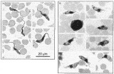
Fig. 1. Giemsa-stained trypanosomes of (A) KETRI 1864 and (B) Trypanosoma suis in thin smears of pig blood. The T. suis specimen is an original preparation of E. Peel and M. Chardome from the collection of C. A. Hoare.
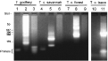
Fig. 2. PCR identification of KETRI 3436. Lanes 1–3 primers specific for Trypanosoma godfreyi; lanes 4–6 primers specific for T. congolense savannah; lanes 7–9 primers specific for T. congolense forest; lanes 10, 11 primers specific for T. congolense Tsavo. Reactions in lanes 1, 4, 7 and 10 were control reactions with no DNA added; reactions in lanes 2, 5 and 8 are positive controls with the appropriate DNA added; reactions in lanes 3, 6, 9 and 11 are test reactions with KETRI 3436 DNA added. Only lane 11 with the T. congolense Tsavo primers shows an amplified band of the expected size for KETRI 3436 DNA; no positive control DNA was available for this reaction. Sizes are shown in base pairs (bp). The broad low molecular weight band is unused primers.
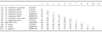
Table 1. Absolute nucleotide differences between Trypanosoma (Nannomonas) spp. SSU rRNA sequences
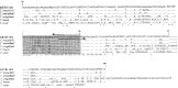
Fig. 3. Sequence alignment of spliced leader transcript and upstream region of salivarian trypanosomes. The 39 nucleotide spliced leader sequence is in shaded box. Arrows indicate the region covered by the primers used for PCR amplification; note that this sequence was not determined directly for KETRI 3436. Dots indicate homology; dashes indicate gaps introduced by the alignment; asterisks indicate complete identity of all 6 sequences. Note that the Trypanosoma congolense sequence extends further than any of the other sequences and ends in a pyrimidine tract composed of C rather than T residues. Accession numbers; KETRI 3436 AJ278533; T. simiae KETRI 2431 X90847; T. simiae Ban 7, X99907; T. congolense, M17786; T. brucei, X00633; T. vivax, X00631.

Fig. 4. Phylogram constructed by bootstrapped (100 replicates) maximum parsimony analysis of 22 Trypanosoma spp. SSU rRNA sequences, and rooted on the non-Salivarian taxa: T. cruzi, T. lewisi, T. theileri, T. grayi and T. varani. The tree is derived from the 2 most parsimonious trees of length = 537 (RI = 0.8627, CI = 0.6577), based on an alignment of 1852 included characters (nucleotide sites). Bootstrap values for all major nodes are given and all branches receiving bootstrap support values >50% are shown; relationships failing to achieve this level of support are shown as polytomies (i.e. branch points at which 3 or more branches arise from the ancestral line). For details of taxa see Stevens et al. (1999a).
 You have
Access
- 26
- Cited by