Introduction
Alveolar echinococcosis (AE), caused by the metacestode stage of the fox tapeworm Echinococcus multilocularis, represents one of the most severe parasitic zoonosis with endemic areas in the Northern hemisphere (Deplazes et al., Reference Deplazes, Rinaldi, Alvarez Rojas, Torgerson, Harandi, Romig, Antolova, Schurer, Lahmar, Cringoli, Magambo, Thompson, Jenkins, Thompson, Deplazes and Lymbery2017). The life cycle of E. multilocularis is perpetuated between canid definitive hosts such as foxes (Vulpes vulpes) and small rodents (mainly voles) as natural intermediate hosts. Humans can accidentally acquire the infection through ingestion of eggs shed in the feces of a definitive host. Metacestode tissue grows like a malignant tumour-like lesion into the surrounding liver tissue and can infiltrate adjacent organs and tissues producing distant metastases. If not appropriately treated, parasite expansion will eventually lead to organ failure and death (Kern et al., Reference Kern, Menezes da Silva, Akhan, Müllhaupt, Vizcaychipi, Budke, Vuitton, Thompson, Deplazes and Lymbery2017).
The current strategies for treating human AE are surgical resection of parasite lesions accompanied by chemotherapy with benzimidazoles (BMZ) carbamate derivatives, and for inoperable cases, chemotherapy alone is the only option. Albendazole (ABZ) is the most common and effective antiparasitic drug for AE treatment. Alternatively, mebendazole may be used if ABZ is not available or not tolerated (Siles-Lucas et al., Reference Siles-Lucas, Casulli, Cirilli and Carmena2018). However, after treatment interruption, the recurrence of the disease is commonly reported as the effect of BMZ is parasitostatic rather than parasitocidal (Kern et al., Reference Kern, Menezes da Silva, Akhan, Müllhaupt, Vizcaychipi, Budke, Vuitton, Thompson, Deplazes and Lymbery2017). Moreover, after its oral administration, ABZ shows erratic absorption from the gastrointestinal tract leading to low drug levels in plasma. Consequently, ABZ often has to be administered life-long at higher doses causing severe side-effects in some patients (Daniel-Mwambete et al., Reference Daniel-Mwambete, Torrado, Cuesta-Bandera, Ponce-Gordo and Torrado2004). To overcome such problems, it is necessary to develop new chemotherapeutic alternatives for the treatment of AE that provide greater efficacy and bioavailability, low toxicity and reduced side-effects.
Drug repurposing is a strategy that accelerates the drug development process as a result of reducing costs and risks and decreasing time to market (Baker et al., Reference Baker, Ekins, Williams and Tropsha2018). Important drug repurposing efforts have been recently directed against several helminth infections (Panic et al., Reference Panic, Duthaler, Speich and Keiser2014). Particularly in the experimental chemotherapy of AE, a variety of anticancer, antifungal and antiprotozoal drugs have demonstrated in vitro effects against the larval stage of E. multilocularis (Lundström-Stadelmann et al., Reference Lundström-Stadelmann, Rufener, Ritler, Zurbriggen and Hemphill2019). However, only a few of these compounds showed efficacy in infected mice and only mefloquine, and amphotericin B alone or combined with nitazoxanide were assayed in humans (Siles-Lucas et al., Reference Siles-Lucas, Casulli, Cirilli and Carmena2018).
Dichlorophene (DCP) is a halogenated phenolic compound used as bactericide and fungicide in cosmetic product formulations (Yamarik, Reference Yamarik2004). It was evaluated in vitro and in vivo as an anthelmintic drug. In vitro studies include the use of DCP on Hymenolepis nana (Sen and Hawking, Reference Sen and Hawking1960), E. multilocularis (Sakamoto, Reference Sakamoto1973), Taenia saginata (Grinenko, Reference Grinenko1964), and Trichostrongylus colubriformis (Rapson et al., Reference Rapson, Jenkins and Topley1985). Several in vivo studies have been conducted with DCP against Anoplocephala perfoliata (Fukui et al., Reference Fukui, Kaneko and Ogawa1960), Moniezia expansa, M. benedeni (Fukui, Reference Fukui1960), Ancylostoma caninum (Miller, Reference Miller1966) and E. granulosus (Gemmell, Reference Gemmell1958). DCP is used as a narrow-spectrum cestocide for treatment of dogs and cats against Taenia spp. (Maddison et al., Reference Maddison, Page and Church2008). Moreover, DCP has been assessed in humans to lower the parasite burden of the intestinal tapeworms T. saginata, H. nana and Ascaris lumbricoides (Jackson, Reference Jackson1956; Adams and Seaton, Reference Adams and Seaton1959; Biagi et al., Reference Biagi, Gómez Orozco and Robledo1959).
However, DCP has a very low aqueous solubility and is poorly absorbed after oral administration (Maddison et al., Reference Maddison, Page and Church2008). In this context, the development of nanodevices for drug delivery offers new treatment options that improve the solubility, the absorption and minimize secondary effects and toxicity (Irache et al., Reference Irache, Esparza, Gamazo, Agüeros and Espuelas2011). Nanoparticles are defined as particles with a size in the range of 1 to 100 nm at least in one of the three dimensions. They can be classified into organic and inorganic nanoparticles, according to their material properties. The latter group includes silica nanoparticles (Llinàs and Sánchez-García, Reference Llinàs and Sánchez-García2014). Drugs absorbed into silica nanoparticles were used against microorganisms. Higher efficacy of modified nanoparticles with organic drugs in relation to the drug alone has been reported (Arce et al., Reference Arce, Bertolotti, Oliveira, Airoldi, Arques, Santos-Juanes, Gonzalez, Cobos, Allegretti and Mártire2012). The aim of the present work was to evaluate the in vitro and in vivo efficacy of DCP and silica nanoparticles modified with DCP (NP-DCP) against E. multilocularis larval stage.
Materials and methods
Synthesis and characterization of the nanoparticles modified with dichlorophen
The NP-DCP were prepared according to the method reported by Escalada et al. (Reference Escalada, Arce, Porcal, Biasutti, Criado, García and Mártire2014). Briefly, 0.5 g of DCP and 180 mL of o-xylene were added to 1.0 g of silica nanoparticles. The mixtures were placed in a Soxhlet extractor containing CaH2 equipped with a condenser with anhydrous CaCl2, and refluxed during 24 h. The products were filtered with 20 nm-nylon filters, washed with 50 mL hot o-xylene and finally with 50 mL ethyl acetate. The resulting gel was first dried at 0.1 Torr and at room temperature for 3 h and then at 120 °C for 5 h. White powders were obtained.
The characterization was realized according to Escalada et al. (Reference Escalada, Arce, Porcal, Biasutti, Criado, García and Mártire2014). Comparison of attenuated total reflection infrared spectra of NP-DCP and DCP showed that the organic drug was covalently bonded to the silica nanoparticles. This result was further confirmed by UV-visible spectroscopy and by thermogravimetry. From this technique, the percentage of DCP present in NP-DCP was 5%.
Parasite material of E. multilocularis
All experiments were carried out using E. multilocularis isolated J2012 (kindly provided by Klaus Brehm, Institute for Hygiene and Microbiology, University of Würzburg, Germany). The parasite was propagated in the peritoneum of CF-1 mice and was processed as described by Albani et al. (Reference Albani, Pensel, Elissondo, Gambino and Elissondo2015), with some modifications. Briefly, the parasitic suspension obtained after cutting the metacestodes and passing them through a metallic strainer was washed several times with phosphate-buffered saline (PBS) 1×. Finally, 0.5 vol of PBS 1× and 6 µL mL−1 of ciprofloxacin (2 mg mL−1) were added to parasite tissue and incubated overnight at 4 °C.
In vitro experiments
Isolation and in vitro culture of protoscoleces of E. multilocularis
From the homogenized metacestode material, the protoscoleces were isolated according to Albani and Elissondo (Reference Albani and Elissondo2014). Briefly, the parasite material was vigorously shaken and then passed through a polyester gauze (pore size 150 µm). The filtered material was recovered and finally passed through a second polyester gauze (pore size 30 µm). The material that was retained in the gauze contains the protoscoleces, which were cultivated (2000 viable and free protoscoleces per Leighton tube) in 10 mL of medium 199 (Lab. Microvet S.R.L., Argentina) supplemented with 100 µg mL−1 streptomycin, 60 µg mL−1 penicillin, 50 µg mL−1 gentamicin and 4 mg mL−1 glucose. Cultures were performed at 37 °C with changes in culture medium every 5 days.
In vitro cultivation of E. multilocularis metacestode vesicles
From the fresh parasitic material of E. multilocularis, the metacestode vesicles were obtained following the procedure established by Spiliotis and Brehm (Reference Spiliotis, Brehm, Rupp and Sohn2009). Briefly, a co-culture was performed between the parasitic material (1 mL) and 106 rat hepatocytes RH- (Reuber hepatoma cells: ATCC No. CRL-1600) in 50 mL of 199 medium, supplemented with 4,5 g L−1 glucose, 10% fetal bovine serum (FBS), 100 µg mL−1 streptomycin and 60 µg mL−1 penicillin. The culture was performed in 75 cm2 culture flasks at 37 °C, 5% CO2. The medium was changed once a week and after that, 106 RH-cells were added. Vesicles between 2 and 5 mm in diameter were used for the assays.
Metacestode vesicles were cultured in 24 well plates (10 vesicles per well), with 2 mL well−1 of 199 medium with FBS and antibiotics, and incubated at 37 °C, 5% CO2, without changes of culture medium.
In vitro drug treatment
Silica nanoparticles and NP-DCP were suspended in medium 199 supplemented with antibiotics at a drug concentration of 0.2 mg mL−1. DCP (Sigma-Aldrich, Buenos Aires, Argentina) was dissolved in dimethyl sulfoxide (DMSO) at drug concentrations of 0.5 and 0.1 mg mL−1 and ABZ (Sigma-Aldrich) was also dissolved in DMSO to obtain a drug concentration of 10 mg mL−1. In each case, the drugs were added to the medium at the final concentrations tested.
Experimental design and evaluation of in vitro efficacy of DCP and NP-DCP
Protoscoleces of E. multilocularis: The in vitro efficacy of different concentrations of DCP and NP-DCP (1, 0.5 and 0.1 µg mL−1) against protoscoleces of E. multilocularis was evaluated. Protoscoleces incubated in culture medium 199 with 1 µL of DMSO, with silica nanoparticles (1 µg mL−1) and with ABZ (10 µg mL−1) were used as controls. Each experiment was performed in triplicate and was repeated three times.
Daily, culture tubes were followed microscopically to determine the appearance of morphological alterations. Every two days, 90–100 protoscoleces were taken from each of the tubes for viability assessment using the methylene blue exclusion test. Samples of protoscoleces for scanning electron microscopy (SEM) were taken after 2 and 6 days of incubation.
Metacestode vesicles of E. multilocularis: The in vitro efficacy of different concentrations of DCP and NP-DCP (1, 0.5 and 0.1 µg mL−1) against metacestode vesicles of E. multilocularis was evaluated. Metacestodes incubated in culture medium 199 with 1 µL of DMSO and with ABZ (10 µg mL−1) were used as controls. Each experiment was performed in triplicate.
Culture plates were followed microscopically every day to determine the appearance of cysts with the collapse of the germinal layer. This parameter was used as criteria of metacestode vesicles viability.
In vivo experiments
In vivo drug treatment
For in vivo efficacy studies against the murine model of AE, ABZ suspension (3.33 mg mL−1) was prepared by dissolution of ABZ pure standard in distilled and deionized water (pH 7.0) and shaking for 12 h. Also, a solution of DCP (0.53 mg mL−1) and a suspension of NP-DCP (4 mg mL−1) were prepared in distilled and deionized water by shaking for 24 h. All formulations were vigorously shaken before administering to mice.
Experimental design and evaluation of in vivo efficacy of DCP and NP-DCP against the murine model of AE
For the therapeutic and chemoprophylactic efficacy studies, female CF-1 mice (n = 50 for each study) were intraperitoneally infected with 0.3 mL of homogenized parasitic material of E. multilocularis. In the chemoprophylactic efficacy study, the treatment of the animals began 1 day post-infection, and in the therapeutic efficacy study, the dosage of mice began 6 weeks post-infection. In both studies, treatments were performed daily for 30 days by intragastric administration. The experimental groups (10 animals/group) were: (a) water control group, mice received distilled and deionized water as a placebo; (b) NPs control group, animals received a suspension of silica nanoparticles in deionized water; (c) ABZ group, mice received ABZ suspension (25 mg kg−1); (d) DCP group, mice were treated with a solution of DCP (4 mg kg−1); (e) NP-DCP group, animals received an NP-DCP suspension (4 mg kg−1).
At the end of the in vivo studies (approximately 10 weeks post-infection), the animals were euthanized and the necropsy was carried out immediately thereafter, and the cysts mass were recovered from the peritoneal cavity. As described by Albani et al. (Reference Albani, Pensel, Elissondo, Gambino and Elissondo2015), the efficacy of the treatments was determined by the mean cysts weight from each group, the viability of protoscoleces obtained from each group (as described above in Experimental design and evaluation of in vitro efficacy of DCP and NP-DCP section) and by the ultrastructural features of cysts and protoscoleces recovered from mice by SEM.
Scanning electron microscopy
Samples of protoscoleces cultured in vitro as well as protoscoleces and cysts recovered from mice involved in both in vivo efficacy studies were processed for SEM as described by Elissondo et al. (Reference Elissondo, Dopchiz, Ceballos, Alvarez, Sánchez Bruni, Lanusse and Denegri2006, Reference Elissondo, Ceballos, Dopchiz, Andresiuk, Alvarez, Sánchez Bruni, Lanusse and Denegri2007).
Statistical analysis
The results obtained from in vitro and in vivo studies are presented as mean ± standard deviation (s.d.). To compare the survival of protoscoleces and metacestode vesicles exposed to different concentrations of DCP and NP-DCP, the log-rank test was used, performed with the software BioEstat 5.0 (Ayres et al., Reference Ayres, Ayres, Ayres and Santos2007). Cysts weights and protoscoleces viability from in vivo assays were compared by Kruskal–Wallis Test (non-parametric method) followed by Dunn´s Multiple Comparisons Test. The analysis was carried out using Instat 3.0 software program (GraphPad Software, San Diego, CA, USA). In all cases, P values less than 0.05 (P < 0.05) were considered statistically significant.
Results
Evaluation of in vitro efficacy of DCP and NP-DCP against E. multilocularis protoscoleces
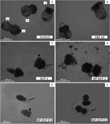
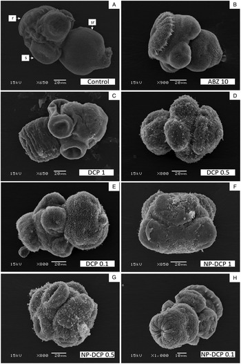
Figure 1 shows the survival of protoscoleces of E. multilocularis after the in vitro exposure to 10 µg mL−1 of ABZ and to different concentrations of DCP and NP-DCP (1, 0.5 and 0.1 µg mL−1). Control protoscoleces remained viable throughout the experimental period and no changes in structure (Fig. 2a) and ultrastructure (Fig. 3a) were observed. Silica nanoparticles (data not shown) and ABZ did not show significant differences compared to DMSO-medium group (P = 0.4698 and P = 0.3505, respectively).
Survival of E. multilocularis protoscoleces after in vitro exposure to different concentrations of DCP and NP-DCP. Each point represents the mean percentage ± s.d. of vital protoscoleces from three different experiments.

Light microscopy of E. multilocularis protoscoleces incubated in vitro with ABZ, DCP, and NP-DCP. (a) Control evaginated protoscoleces, 10 days p.i. (s, sucker; sr, soma region; r, rostellar region; cc, calcareous corpuscles). (b) Protoscoleces treated with 10 µg mL−1 of ABZ during 10 days. (c) Protoscoleces incubated for 2 days with 1 µg mL−1 of DCP. Observe the formation of blebs in the tegument (arrow) and the contraction of the soma region (head arrow). (d) Dead protoscoleces, 2 days p.i. with 1 µg mL−1 of NP-DCP. Observe the loss of the characteristic morphology of the larvae. (e) Protoscoleces with the presence of blebs in the tegument (arrow) and contraction of the soma region (head arrow, 0.5 µg mL−1 of NP-DCP, 2 days p.i.). (f) Protoscoleces incubated with NP-DCP (4 days, 0.1 µg mL−1). Note the presence of blebs (arrow).

Scanning electron microscopy of E. multilocularis protoscoleces incubated in vitro with ABZ, DCP, and NP-DCP. (a) Evaginated control protoscolex (6 days p.i.). Observe the intact microtriches (r, rostello region with hooks; s, sucker; sr, soma region). (b) Protoscolex incubated with 10 µg mL−1 of ABZ for 6 days, with contraction of the soma and alteration of tegument in that region. (c) Protoscolex treated with 1 µg mL−1 of DCP for 2 days, with an intact scolex region. The soma is contracted and the tegument altered. (d) Protoscolex treated 6 days with 0.5 µg mL−1 of DCP. Observe the loss of microtriches and hooks, the contraction of the soma, and the presence of blebs in the tegument. (e) Protoscolex with contraction of soma region and tegumental alterations (6 days p.i., 0.1 µg mL−1 of DCP). The scolex region remains unchanged. (f) Protoscolex showing a total loss of morphology and blebs in the tegument (1 µg mL−1 of NP-DCP, 2 days p.i.). (g) Protoscolex incubated with 0.5 µg mL−1 of NP-DCP (2 days p.i.). Observe the total loss of microtriches, the presence of blebs in the tegument, and the contracted soma region. (h) Protoscolex with an intact scolex region, but with alterations on the tegument of the soma region (0.1 µg mL−1 of NP-DCP, 2 days p.i.).

DCP and NP-DCP produced time and dose-dependent protoscolicidal effect (P < 0.0001). Treatment of protoscoleces with both drugs caused a reduction of larval viability. However, NP-DCP had a stronger protoscolicidal effect compared to DCP.
The greatest protoscolicidal effect was observed with the concentration of 1 µg mL−1 of NP-DCP, causing a rapid decrease in viability and reaching 0% after 6 days. NP-DCP at a concentration of 0.5 µg mL−1 also generated a rapid decrease in the viability of protoscoleces, reaching 0% after 10 days. The concentration of 0.1 µg mL−1 of NP-DCP reduced the viability to 19.67 ± 13.3% at the end of the assay (Fig. 1).
The treatment of protoscoleces with 1 µg mL−1 of DCP caused a marked decrease in their survival after 4 days. Then, the viability decreased slowly. The protoscolicidal effect observed with the concentration of 0.5 µg mL−1 of DCP was similar to that caused by 0.1 µg mL−1 of NP-DCP (P = 0.1377). The decrease in viability was slow and gradual. In addition, 0.1 µg mL−1 of DCP did not show differences compared to DMSO-medium group (P = 0.6316).
At 2 days p.i., the protoscoleces treated with 1 µg mL−1 of NP-DCP showed total loss of their morphology (Figs 2d and 3f) and the concentration of 0.5 µg mL−1 caused a total loss of microtriches and hooks, with the presence of blebs in the tegument (Figs 2e and 3g). Protoscoleces treated with 0.1 µg mL−1 of NP-DCP at 2 days p.i. showed alteration of the tegument of the soma region (Fig. 3h) and the appearance of blebs in the tegument was observed after 4 days (Fig. 2f).
At day 2 p.i. the protoscoleces exposed to 1 µg mL−1 of DCP showed blebs in the tegument, in addition to the loss of rostellar hooks (not shown) and contraction of soma (Fig. 2c). However, after 6 days p.i., loss of microtriches and alteration of the tegument with the presence of small blebs were observed (Fig. 3c). Protoscoleces incubated with 0.5 µg mL−1 of DCP for 2 days did not present ultrastructural alterations. After 4 days, the appearance of blebs in the tegument was detected (data not shown). Protoscoleces incubated for 6 days showed loss of microtriches and blebs in the tegument (Fig. 3d). Finally, DCP at 0.1 µg mL−1 produced soma contraction and alteration of the tegument of the soma region after 6 days p.i. (Fig. 3e). These alterations were similar to the changes caused by 10 µg mL−1 of ABZ (Figs 2b and 3b).
Evaluation of in vitro efficacy of DCP and NP-DCP against metacestode vesicles of E. multilocularis
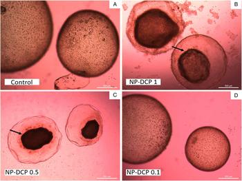
Figure 4 shows the survival of metacestode vesicles after in vitro incubation for 3 and 10 days with different concentrations of DCP and NP-DCP. The viability of control vesicles was 100% throughout the entire assay (Fig. 5a), as well as metacestodes incubated with 10 µg mL−1 of ABZ and 0.1 µg mL−1 of DCP. Treatments with 1 and 0.5 µg mL−1 of DCP and with 0.1 µg mL−1 of NP-DCP produced a decrease in the viability of metacestodes. However, no significant differences were found with respect to the control (P = 0.24, P = 0.22, P = 0.68, respectively; Fig. 5d). NP-DCP at the concentrations of 1 and 0.5 µg mL−1 were the only treatments that had a significant effect on the viability of vesicles (P < 0.0001) causing the collapse of the germinal layer of metacestode vesicles after 1 and 2 days of incubation, respectively (Fig. 5b–c).
Survival of E. multilocularis vesicles after 3 and 10 days of in vitro exposure to different concentrations of DCP and NP-DCP. The criteria for metacestode vesicles viability was the collapse of the germinal layer. Each bar represents the mean percentage ± s.d. of vital vesicles.

In vitro treatment of metacestode vesicles of E. multilocularis with different concentrations of NP-DCP. (a) Control metacestodes after 10 days of incubation with an intact germinal layer. (b) Metacestode vesicles incubated with 1 µg mL−1 of NP-DCP for 1 day. Note the rapid collapse of the germinal layer (arrow). (c) Vesicles with germinal layer detached from the laminar layer (arrow) after 2 days of incubation with 0.5 µg mL−1 of NP-DCP. (d) Metacestodes incubated for 10 days with 0.1 µg mL−1 of NP-DCP.

Therapeutic efficacy study of DCP and NP-DCP against the murine model of AE
All mice belonging to the therapeutic efficacy study developed cysts in the peritoneal cavity. There were no significant differences (P > 0.05) between the weight of the cysts of both control groups. However, oral administration of 25 mg kg−1 of ABZ and 4 mg kg−1 of NP-DCP caused a significant decrease (P < 0.05) in the mean weight of the cysts compared to control groups. The treatment with 4 mg kg−1 of DCP did not cause a decrease in the weight of the developed cysts (Table 1).
Therapeutic and chemoprophylactic efficacy studies

Mean weights (g ± s.d.) of cysts obtained (10 weeks post-infection) from E. multilocularis infected mice. Treatments were performed for 30 days every 24 h.
In the therapeutic study, statistically significant differences were found between:
a The water control group and the following treatment: ABZ group (P < 0.01) and NP-DCP group (P < 0.05).
b The NPs control group and the following treatment: ABZ group and NP-DCP group (P < 0.01).
In the chemoprophylactic study, statistically significant differences were found with:
c The water control group (P < 0.05).
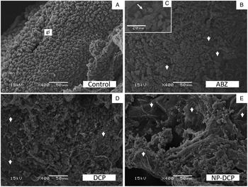
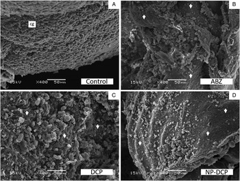
The ultrastructural study by SEM of the germinal layer of metacestodes recovered from control and treated groups is shown in Fig. 6. The germinal layer of cysts obtained from control mice showed the characteristic multicellular structure (Fig. 6a). In contrast, metacestodes developed in mice treated with 25 mg kg−1 of ABZ and with 4 mg kg−1 of DCP showed areas without cells and the presence of altered cells in germinal layer (Fig. 6b–d). However, in the germinal layer of cysts recovered from NP-DCP-treated mice greater extensions of areas without cells as well as altered cells were evidenced (Fig. 6e).
Scanning electron microscopy of E. multilocularis cysts recovered from infected mice treated with 25 mg kg−1 of ABZ, 4 mg kg−1 of DCP or 4 mg kg−1 of NP-DCP, belonging to the therapeutic efficacy study. (a) Control cyst with an intact germinal layer (gl). (b) Cyst recovered from mice treated with ABZ. Observe the altered germinal layer, with loss (arrowhead) and alteration of cells. (c) High magnification image showing altered cells (arrow). (d) Cyst obtained from treatment with DCP. Areas with loss of cells (arrowhead) are observed as well as altered cells. (e) Metacestode from NP-DCP treated group, with extensive areas of the germinal layer without cells (arrowhead). Altered cells and cellular debris can be observed.

Figure 7A shows the viability of protoscoleces obtained from the different experimental groups of the therapeutic efficacy study. The protoscoleces isolated from the control groups had a viability greater than 80% and no changes in ultrastructure were observed (Fig. 7Ba). The treatment with 25 mg kg−1 of ABZ and with 4 mg kg−1 of NP-DCP showed protoscolicidal effect, reducing significantly the viability of protoscoleces (P < 0.05) to 23.92 ± 29.0% and 28.67 ± 25.2%, respectively. The administration of 4 mg kg−1 of DCP reduced the survival of protoscoleces to 36.90 ± 30.8%, but it was not significant with respect to control groups.
(A) Viability and (B) scanning electron microscopy of E. multilocularis protoscoleces obtained from cysts recovered from mice belonging to control and treated groups of the therapeutic efficacy study. (A) Statistically significant differences were found with the water control group: *P < 0.05. (B) a – Evaginated control protoscolex. b – Protoscolex with a total loss of morphology, obtained from mice treated with 25 mg kg−1 of ABZ. c – Protoscolex recovered from mice treated with 4 mg kg−1 of DCP. d – Protoscolex completely altered, obtained from mice treated with 4 mg kg−1 of NP-DCP.

The analysis of the images of protoscoleces by SEM was consistent with the viability data obtained (Fig. 7B). Most of the protoscoleces obtained from mice of the ABZ and NP-DCP groups showed a total loss of morphology (Fig. 7Bb and Bd), while protoscoleces obtained from DCP group showed only some alterations, such as loss of hooks (Fig. 7Bc).
Chemoprophylactic efficacy study of DCP and NP-DCP against the murine model of AE
Cysts development in the peritoneal cavity was observed in all mice belonging to chemoprophylactic efficacy study. No significant differences were found (P > 0.05) between the mean weight of the cysts of the control water and control NPs groups. The treatments with DCP and NP-DCP did not significantly reduce the weight of the cysts, while, administration of ABZ produced a significant decrease compared to the weight of cysts from the water control group (P < 0.05; Table 1).
Metacestodes recovered from the different treatments lost their characteristic multicellular structure. The treatment with ABZ caused a decrease in the number of cells (Fig. 8b) and the presence of altered cells. Although the metacestodes recovered from the mice treated with DCP caused a reduction in cell number of the germinal layer (Fig. 8c), the extension of the damage was lower than with NP-DCP treatment, where only a few cells were observed (Fig. 8d).
Scanning electron microscopy of E. multilocularis cysts recovered from infected mice treated with 25 mg kg−1 of ABZ, 4 mg kg−1 of DCP or 4 mg kg−1 of NP-DCP, belonging to the chemoprophylactic efficacy study. (a) Metacestode recovered from the mice of the control group showing the characteristic ultrastructure of the germinal layer (gl). (b) Cysts from ABZ treated mice. Observe the large areas of the germinal layer without cells (arrowhead). (c) Metacestode of mice belonging to the DCP group. Observe the areas without cells in the germinal layer (arrowhead). (d) Metacestode obtained from mice of NP-DCP group. Note the extensive areas of the germinal layer with loss of cells (arrowhead).

The viability of protoscoleces of the control groups was greater than 90% (Fig. 9A). The treatment of the mice with 4 mg kg−1 of DCP did not reduce the viability of the protoscoleces with respect to the control groups. Protoscoleces recovered from the mice treated with 25 mg kg−1 of ABZ (36.42 ± 30.0%) and with 4 mg kg−1 of NP-DCP (57.28 ± 22.9%) showed a significant decrease of the viability compared to the control groups (P < 0.01).
(A) Viability and (B) scanning electron microscopy of E. multilocularis protoscoleces isolated from cysts obtained of treated mice during the chemoprophylactic efficacy study. (A) Statistically significant differences were found with the water control group: *P < 0.001 and §P < 0.01. (B) a – Invaginated and evaginated control protoscoleces. b – Detail of the scolex of an evaginated control protoscolex. Observe the microtriches (arrow). c – Protoscolex obtained from the treatment with 25 mg kg−1 of ABZ. Observe the rostellar disorganization (arrowhead) and the contraction of the soma (arrow). d – Invaginated protoscolex recovered from treatment with 4 mg kg−1 of DCP. e – Protoscolex isolated from cysts of the NP-DCP group. Note the loss of hooks (arrowhead) and microtriches and the presence of vesicles in the tegument (arrow). f – Evaginated protoscolex obtained from mice treated with 4 mg kg−1 of NP-DCP. Observe the lack of microtriches on the scolex region (arrow).

The ultrastructural analysis of protoscoleces coincided with the percentages of viability obtained from the different experimental groups. The protoscoleces of the control groups were observed unchanged (Fig. 9Ba and Bb), as well as those obtained from the DCP group (Fig. 9Bd). In contrast, protoscoleces isolated from the cystic masses belonging to the mice of the groups ABZ and NP-DCP presented alterations. The protoscoleces of the ABZ group showed rostellar disorganization and contraction of the soma (Fig. 9Bc). The protoscoleces of the NP-DCP group evidenced loss of hooks and microtriches and the presence of vesicles in the tegument (Fig. 9Be and Bf).
Discussion
A perfect agent for the chemotherapeutic treatment of echinococcosis should be selectively toxic for the cestode and present a favourable solubility and absorption to reach a therapeutic concentration in the target site. The lack of undesirable clinical effects and a parasitocidal activity are also desirable (Siles-Lucas et al., Reference Siles-Lucas, Casulli, Cirilli and Carmena2018).
Chemotherapeutic treatment of AE with BMZ has several disadvantages, such as variations in treatment success due to the low and erratic bioavailability of the antiparasitic agent after oral administration, the adverse reactions observed in patients after prolonged treatments and a parasitostatic rather than a parasitocidal effect (Daniel-Mwambete et al., Reference Daniel-Mwambete, Torrado, Cuesta-Bandera, Ponce-Gordo and Torrado2004; Hemphill et al., Reference Hemphill, Stadelmann, Rufener, Spiliotis, Boubaker, Müller, Müller, Gorgas and Gottstein2014; Grüner et al., Reference Grüner, Kern, Mayer, Gräter, Hillenbrand, Barth, Muche, Henne-Bruns, Kratzer and Kern2017; Kern et al., Reference Kern, Menezes da Silva, Akhan, Müllhaupt, Vizcaychipi, Budke, Vuitton, Thompson, Deplazes and Lymbery2017).
In this context, the drug repurposing approach is particularly important for neglected tropical diseases. The time, effort and resources saved in the reuse of already approved drugs with known physicochemical, pharmacokinetic and toxicological properties make this approach an intelligent and ethical option (Panic et al., Reference Panic, Duthaler, Speich and Keiser2014). Consequently, research about novel compounds for the treatment of echinococcosis should focus on drugs or classes of drugs that are already marketed and/or in clinical development (Küster et al., Reference Küster, Stadelmann, Rufener, Risch, Müller and Hemphill2015).
Considerable efforts have been made to improve the chemotherapeutic treatment of AE. Hundreds of anti-infective agents, anti-cancer drugs and antiparasitic compounds were in vitro tested against the larval stage of E. multilocularis (Siles-Lucas et al., Reference Siles-Lucas, Casulli, Cirilli and Carmena2018; Lundström-Stadelmann et al., Reference Lundström-Stadelmann, Rufener, Ritler, Zurbriggen and Hemphill2019). Most of these repurposing initiatives had poor in vivo results. Nitazoxanide (Stettler et al., Reference Stettler, Rossignol, Fink, Walker, Gottstein, Merli, Theurillat, Thormann, Dricot, Segers and Hemphill2004) and mefloquine (Küster et al., Reference Küster, Stadelmann, Rufener, Risch, Müller and Hemphill2015; Rufener et al., Reference Rufener, Ritler, Zielinski, Dick, da Silva, da Silva Araujo, Joekel, Czock, Goepfert, Moraes and de Souza2018) showed efficacy against the murine model of AE. On the other hand, amphotericin B alone or combined with nitazoxanide were the only drugs tested in human patients as an alternative to the treatment with ABZ (Reuter et al., Reference Reuter, Buck, Grebe, Nüssle-Kügele, Kern and Manfras2003; Tappe et al., Reference Tappe, Müller, Frosch and Stich2009).
DCP has demonstrated anthelmintic in vitro and in vivo efficacy, mainly against intestinal parasites. The only study against a systemic parasitic infection was carried out in vitro against the larval stage of E. multilocularis (Sakamoto, Reference Sakamoto1973). However, the low solubility of DCP in water limits its activity against systemic parasitic diseases. Drug repurposing complemented by nanotechnological strategies could be a useful tool to improve the bioavailability and efficacy of repurposed drugs (El-Moslemany et al., Reference El-Moslemany, Eissa, Ramadan, El-Khordagui and El-Azzouni2016). In this context, we evaluated the in vitro and in vivo efficacy of DCP and NP-DCP against the larval stage of E. multilocularis.
The in vitro activity of DCP against E. multilocularis protoscoleces was previously demonstrated by Sakamoto (Reference Sakamoto1973). Although this author used concentrations of DCP 10–100 times higher than those evaluated in this work, the viability results were similar. During our in vitro studies against protoscoleces, both DCP formulations showed time and dose-dependent effect. However, NP-DCP had a stronger effect compared to DCP. The NP-DCP at a concentration of 1 µg mL−1 reduced the viability of protoscoleces to 0% after 4 days. In contrast, 10% of the protoscoleces incubated with the same concentration of DCP were alive at the end of the assay. These results are consistent with the observations reported by Vico et al. (Reference Vico, Arce, Fangio, Gende, Bertran, Mártire and Churio2016). The authors demonstrated an increase in the antimicrobial activity of silica nanoparticles functionalized with gallic acid against Paenibacillus larvae in comparison to free gallic acid.
The results of the viability test coincide with the structural and ultrastructural alterations observed by light microscopy and SEM. Moreover, the ultrastructural alterations agreed to those reported for E. multilocularis protoscoleces incubated with ABZ, thymol, and the combination of both drugs (Albani and Elissondo, Reference Albani and Elissondo2014).
Only the NP-DCP caused a significant decrease of the viability of the E. multilocularis metacestodes during the in vitro assay. Moreover, the activity of the NP-DCP against protoscoleces and cysts was higher than the effect induced by ABZ. This could be explained because the anthelmintic activity of BMZ compounds not only depend on its binding to parasite ß tubulin but also on their ability to reach high and sustained concentrations within the parasite cells, in sufficient time, to cause the therapeutic effect (Thompson and Geary, Reference Thompson, Geary, Harr and Muller1995).
The activity of DCP and NP-DCP was also evaluated in an in vivo murine model of AE. The therapeutic and the chemoprophylactic efficacy of both formulations of DCP were evaluated on mice infected with E. multilocularis. The LD50 value for DCP was 1670 mg kg−1 orally (Yamarik, Reference Yamarik2004). This dose is more than 400 times higher than the dose used in this work. Moreover, Gucklhorn (Reference Gucklhorn1969) demonstrated that 400 mg kg−1 day−1 of DCP for 90 days did not cause a toxic effect in rats.
During the efficacy studies on the murine model of AE, and in accordance to the Guide of Care and Use of Laboratory Animals (National Research Council US, 2011), the appearance and behaviour of mice were normal throughout the experimental period.
In the therapeutic efficacy study, ABZ and the NP-DCP caused a significant decrease in the weight of the metacestodes and in the viability of protoscoleces recovered from the treated mice. All treatments induced ultrastructural alterations on the germinal layer of the murine cysts. However, the extension of damage was greater after NP-DCP-treatment. The treatment with ABZ and the NP-DCP also caused marked ultrastructural changes on protoscoleces recovered from murine cysts. In contrast, DCP only induced slight changes like the loss of hooks.
In the chemoprophylactic efficacy study, both DCP formulations did not cause a significant decrease in the weight of the metacestodes recovered from the treated mice. In contrast, a significant reduction in cyst weight was observed after the administration of ABZ. However, the ultrastructural alterations observed in the germinal layer of the cysts recovered from the DCP and NP-DCP groups were consistent with the changes caused by ABZ. Moreover, the NP-DCP enhanced the efficacy of the drug alone. The damage extension in the germinal layer was greater after the treatment with the nanoparticles and a significant decrease in the viability of the protoscoleces recovered from cysts was observed.
Interestingly, the ultrastructural changes induced by both DCP formulations on cysts and protoscoleces during the in vivo studies were similar to those described for other drugs (Naguleswaran et al., Reference Naguleswaran, Spicher, Vonlaufen, Ortega-Mora, Torgerson, Gottstein and Hemphill2006; Spicher et al., Reference Spicher, Roethlisberger, Lany, Stadelmann, Keiser, Ortega-Mora, Gottstein and Hemphill2008; Albani et al., Reference Albani, Pensel, Elissondo, Gambino and Elissondo2015).
Silica is abundantly distributed in nature, it has good compatibility and is accepted as generally recognized as safe (GRAS) by the FDA. Silica nanoparticles have been widely used in cosmetics and as food additives approved by the FDA (Halas, Reference Halas2008). In addition, silica nanoparticles have potential as drug delivery vehicles for medical and veterinary treatments and as pesticides in the field of agriculture (Barik et al., Reference Barik, Kamaraju and Gowswami2012). This is due to their large and stable surface area and stability in vivo. Moreover, physical and chemical properties of the surface of a nanoparticle determine its capability to adsorb biomolecules and to disrupt cell membranes. The chemical composition of the surface will eventually control the pharmacological effect of a nanomaterial. Therefore, an increase in the hydrophobicity of the surface of silica nanoparticles after being modified with organic groups stimulates the adhesion of the nanoparticles to microorganisms (Arce et al., Reference Arce, Bertolotti, Oliveira, Airoldi, Arques, Santos-Juanes, Gonzalez, Cobos, Allegretti and Mártire2012).
The use of immobilized drugs on silica nanoparticles allows a greater absorption of poorly-water soluble drugs. This could generate an increase in the circulation times. Thus, the improvement in the in vivo efficacy of the NP-DCP could be explained by an increase in the systemic availability of DCP.
In conclusion, our results demonstrated that the NP-DCP had a stronger activity than the drug alone and their therapeutic efficacy was similar to ABZ suspension against the larval stage of E. multilocularis. The manufacture of NP-DCP is scalable, cost-effective and controllable. Therefore, DCP could be a potential repurposed anti-echinococcal drug and this nanotechnology strategy could greatly enhance its efficacy.
Acknowledgments
The authors thank Dr Alejandra Goya and Dr Sonia Ortega (SENASA, Argentina) for their cooperation.
Financial support
This study was financially supported by the PICT 15 No. 0717 (ANPCyT, Argentina) and EXA 769/16 and EXA 871/18 (Universidad Nacional de Mar del Plata, Argentina).
Conflict of interest
None.
Ethical standards
Female CF-1 mice (body weight 25 g ± 5) were used. The animals were housed in a temperature-controlled (22 ± 1 °C), light-cycle (12 h light/dark cycle) room. Food and water were given ad libitum. Animal procedures and management protocols were approved by the Institutional Animal Care and Use Committee (RD 148/15) of the Faculty of Exact and Natural Sciences, National University of Mar del Plata, Mar del Plata, Argentina and carried out in accordance with the revised form of The Guide for the Care and Use of Laboratory Animals (National Research Council US, 2011). Unnecessary animal suffering was avoided throughout the study.