Introduction
The Halo vest is an essential piece of orthotic equipment used to stabilise the cervical spine. We report a notable complication of immobilising the spine in this manner.
Case report
A 16-year-old man presented to the ENT clinic with sudden, profound, left-sided hearing loss. The patient was known to suffer from merosin-deficient congenital muscular dystrophy, resulting in progressive muscle weakness. He had recently undergone corrective spinal surgery for a fixed neck extension deformity. Repositioning of the cervical spine was undertaken via a posterior midline approach, and involved release of extensively fibrosed erector spinae muscles through blunt dissection. A Halo vest was subsequently fitted, with traction, to bring the neck forward into a more normal position.
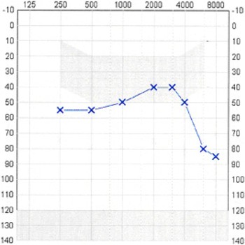
Otological examination was entirely normal. Combined Weber and Rinne tuning fork tests were indicative of left-sided sensorineural hearing loss. This was subsequently confirmed by pure tone audiometry and acoustic reflex testing (Figure 1); bone conduction was difficult to assess due to the presence of the Halo vest. Arrangements were made for a left-sided hearing aid to be fitted.
Pure tone audiogram performed two weeks post-operatively demonstrating profound, left-sided hearing loss.

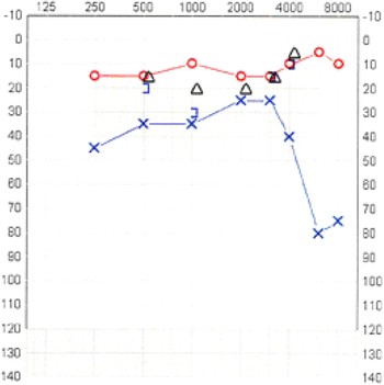
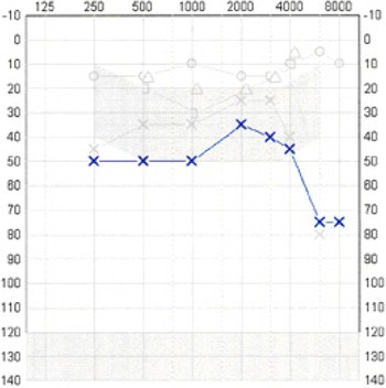
The patient was reviewed in the ENT clinic three weeks later. Pure tone audiometry demonstrated a slight improvement in hearing thresholds (Figure 2). On review a further three weeks later, it was evident that the patient's hearing loss had continued to resolve (Figure 3).
Pure tone audiogram performed five weeks post-operatively demonstrating improvement of left-sided hearing thresholds.

Pure tone audiogram performed eight weeks post-operatively demonstrating further improvement in left-sided hearing thresholds.

The patient was scheduled for a follow-up appointment in three months. However, three weeks after discharge from the ENT clinic, and 11 weeks after the original operation, the patient's Halo vest was disturbed when he hit his head while boarding a bus. The patient noticed a concomitant reduction in his hearing, which was confirmed by repeat pure tone audiometry (Figure 4).
Pure tone audiogram performed eleven weeks post-operatively demonstrating deterioration of left-sided hearing following head trauma.

Discussion
The congenital muscular dystrophies are a heterogeneous group of autosomal recessive myopathies. In 1994, Tomé et al. reported a cohort of congenital muscular dystrophy patients with deficiency in the a2 chain of laminin (or merosin).Reference Tomé, Evangelista, Leclerc, Sunada, Manole, Estournet, Barois, Campbell and Fardeau1 The merosin isoform is specifically expressed in the basal lamina of both striated muscle and peripheral nerves.Reference Leivo and Engvall2 Merosin-deficient congenital muscular dystrophy is one of the most frequent neuromuscular diseases in children of European populations, and accounts for approximately 40 per cent of all classical congenital muscular dystrophies. Features include neonatal hypotonia, joint contracture, debilitation, elevated serum creatine kinase, and alterations in both somatosensory and visual evoked potentials.Reference Philpot, Sewry, Pennock and Dubowitz3 Although most patients have normal intelligence, a minority of children have been reported to show moderate mental retardation and epilepsy.Reference Vainzof, Marie, Reed, Schwartzman, Pavanello and Passos-Bueno4
Sudden sensorineural hearing loss (SNHL) is defined as hearing loss in three contiguous frequencies, of greater than 30 dB, developing over less than three days.Reference Wilson, Byl and Laird5 The incidence of sudden SNHL is approximately 10 per 100 000 of the adult population per year. Clinical and radiological investigations have shown the majority of cases to be idiopathic.Reference Mattox and Lyles6 No specific cause is identified in 85 to 90 per cent of cases,Reference Mattox and Lyles6 although this does not detract from the devastating impact the disorder has upon the patient. Circulatory disturbances, particularly vertebrobasilar occlusive disorders, are an important cause of sudden deafness. However, such cases are typically excluded from sudden SNHL studies, which focus upon idiopathic presentations. For this reason, there is little information in the literature concerning the incidence of sudden SNHL of vascular or ischaemic origin. The diagnosis of vertebrobasilar occlusive disorder is generally made straightforward by the association of other neurological deficits that occur concomitantly at presentation. Conversely, case reports have described initial cochleovestibular symptoms, with other signs suggestive of vertebrobasilar occlusive disorder developing several days later.Reference Lee, Whitman, Lim, Lee and Park7–Reference Toyoda, Hirano, Kumai, Fujii, Kiritoshi and Ibayashi9
The pathophysiology of vertebrobasilar occlusive disorder is focused predominantly on arterial dissection and thrombosis, with embolism being considered a rare occurrence in this territory. The clinical progression of the disease can be explained by several factors.
Firstly, it is logical that anatomical structures supplied by an end artery will be more susceptible to ischaemia. The internal auditory artery, a branch of the anterior inferior cerebellar artery (which in turn is derived from the basilar artery) represents the sole blood supply to the inner ear.Reference Levine and Häussler10 This is in contrast to the multiple source that supply blood to the auditory pathways.
Secondly, symptoms may manifest through hypoperfusion alone. The cochlea has high energy requirements and is consequently more vulnerable to ischaemia than the vestibular organ. Furthermore, the cochlear artery, a branch of the common cochlear artery (which in turn is derived from the internal auditory artery), has been shown to be more prone to occlusion compared to the vestibulocochlear artery. Reference Kim, Lopez, DiPatre, Liu, Ishiyama and Baloh11 Such findings may explain the preservation of vestibular function in the presence of auditory compromise.
Thirdly, platelet aggregation and clot formation can lead to stenosis within the vessel lumen. Stenotic regions have the potential to progressively extend and occlude collateral branches of the afflicted artery. This would involve the anterior internal carotid artery in the case of vertebrobasilar occlusive disorders.
It is well documented that manipulation of the cervical spine can lead to vertebral artery injury. The manoeuvres of chiropractors,Reference Kunkle, Muller and Odom12–Reference Smith and Estridge23 osteopathic physiciansReference Smith and Estridge23 and physiotherapistsReference Parkin, Wallis and Wilson24 have all been reported to cause neurological complications. Crushing, stretching or shearing of the artery by hyperextension or extreme rotation predisposes to the formation of intramural haematoma, with narrowing due to rupture of the vasa vasorum. The result is an intraluminal thrombus that has the potential to extend by direct propagation, embolisation or both.Reference Bladin and Merory16, Reference Parkin, Wallis and Wilson24, Reference Schellhas, Latchaw, Wendling and Gold25 Collateral branches of the stenosed artery may therefore become occluded. Pseudoaneurysms may also form at the site of injury.Reference Davidson, Weiford and Dixon17
Our patient initially presented with severe musculoskeletal extension deformity of the cervical spine. We propose that restoration of the patient's cervical spine to a position deemed more aesthetically normal was not necessarily the best position in terms of vertebrobasilar integrity, as the vertebral circulation had adapted during this young man's lifetime to his abnormal cervical posture. The orthopaedic procedure used to correct the orientation of the cervical spine in this case is unusual; procedures to correct spinal orientation are more commonly performed on patients with ankylosing spondylitis, and typically involve an anterior cervical release.
The proposed vascular cause for our patient's sudden SNHL may not have been solely a result of his initial surgery. The Halo device is the most rigid external prosthesis for immobilisation of the cervical spine, but biometric studies have confirmed that some motion and force transmission to the cervical region does occur. One study demonstrated up to 51° of motion of a patient's cervical spine despite Halo traction,Reference Lind, Sihlbom and Nordwell26 which is equivalent to 70 per cent of the normal range of movement.Reference White and Panjabi27 The greatest degree of motion was observed between C1 and the occiput.Reference Lind, Sihlbom and Nordwell26 It is at this location that the vertebral artery is particularly vulnerable to shearing forces as it pierces the atlanto-occipital membrane, and it may be susceptible to crushing injury from the occipital portion of the skull as it exits the transverse foramen to pass behind the lateral aspect of the atlas. Stretching of the artery between the foramina of the transverse process of the atlas and axis has also been described.Reference Kanshepolsky, Danielson and Flynn14, Reference Schellhas, Latchaw, Wendling and Gold25 Such movement of the cervical vertebra due to Halo vest mobility may explain why our patient's hearing subsequently deteriorated.
Motion of the cervical spine in Halo-immobilised patients has also been observed at the C2–C3 level. Furthermore, when flexion was observed at one segment of the spine, extension was observed at another.Reference Lind, Sihlbom and Nordwell26 Several case reports have documented vertebral artery injury at this level following cervical spine manipulation, the hypothesis being a shearing or impingement of the stretched artery by the anterior articular facet of the third cervical vertebra.Reference Bladin and Merory16, Reference Zimmerman, Kumar, Gadoth and Hodges19, Reference Schellhas, Latchaw, Wendling and Gold25 However, such instances were associated with significant rotational manipulation.
Conclusion
We describe the unique case of a patient who sustained a sudden hearing loss as a result of correction of a cervical spine extension deformity and Halo vest fixation. This case highlights an important complication that is significant to both orthopaedic and ENT surgeons.