Introduction
Systemic lupus erythematosus (SLE) is a multisystem autoimmune disease of unknown aetiology. The course of the disease is chronic, and the manifestations can occur simultaneously or serially. Common presentations involve the cutaneous, renal, neurological and musculoskeletal systems. The presence of serum antinuclear autoantibodies, reduced levels of complement components, and other haematological and immunological alterations are confirmatory. Sensorineural hearing loss (SNHL) is rarely noted in patients with SLE. The mechanism of inner-ear damage in SLE still remains largely unknown.
We present a case of SLE with bilateral SNHL, demonstrate radiological findings and review the literature.
Case report
A 33-year-old woman with SLE presented with acute, right-sided hearing loss, tinnitus and vertigo. Her SLE had first appeared at the age of 24 years. Its course had been marked by: positive antinuclear autoantibodies testing (>1:320); positive speckled anti-double-stranded deoxyribonucleic acid (DNA) antibody testing; symmetrical polyarthritis; pleuritis; Raynaud's phenomenon; and a deep venous thrombosis (DVT) of her right arm. At the time of her right-sided hearing loss, the patient was being treated with warfarin (for the DVT of her right arm) and hydroxychloroquine 200 mg orally twice daily.
The patient's laboratory results revealed an elevated anti-double-stranded DNA antibody level (114 IU/ml); this had been negative one year earlier. She also tested positive for anti-ribonucleoprotein and anti-Ro antibodies. She had normal urinalysis, normal full blood count, erythrocyte sedimentation rate of 22 mm/hour, C-reactive protein of 10 mg/l, and normal complement 3 and 4 levels. Her international normalised ratio was subtherapeutic at 1. Testing for lupus anticoagulant and anticardiolipin antibody was negative on three separate occasions (each at least 12 weeks apart) at the time of her presentation and throughout the clinical course. There was no evidence of active disease in any other organs, and the patient's total systemic lupus erythematosus disease activity index (SLEDAI) score was 12, compared with four one year earlier.
According to the original treating physician, the differential diagnosis of the SNHL aetiology included viral-induced, thrombotic and inflammatory causes secondary to SLE. The patient was treated initially with oral acyclovir, aspirin 81 mg orally daily and low molecular weight heparin. She had received methylprednisolone 500 mg intravenously for three days, followed by Calcort (equivalent of prednisone 5 mg orally daily) for two months, prior to visiting our centre. There had been little effect on her hearing. Her hydroxychloroquine was stopped three months later due to rare reports of hearing loss associated with it. Her right-sided SNHL persisted.
Five months later, the patient complained of bilateral hearing loss, profound on the right and severe on the left. She also complained of dizziness and dysequilibrium. Laboratory results revealed: positive antinuclear autoantibody test (to >1:320); speckled pattern, positive anti-double-stranded DNA antibody test (to 142 IU/ml); positive ribonucleoprotein antibody test; normal complement levels; and negative tests for anticardiolipin antibody and lupus anticoagulant. The patient's total (SLEDAI) score remained 12. Otoscopy revealed normal tympanic membranes. The head and neck examination identified no contributory pathology. Her neuro-otological examination identified a positive head impulse sign bilaterally, as well as an inability to perform Romberg and tandem Romberg testing. The patient's audiogram at this stage revealed a profound neurosensory hearing loss on the right and a moderate to severe neurosensory hearing loss on the left, with a four frequency pure tone average (500, 1000, 2000 and 3000 Hz) of 62.5 dB, a speech reception threshold of 50 dB and a word discrimination score of 16 per cent.
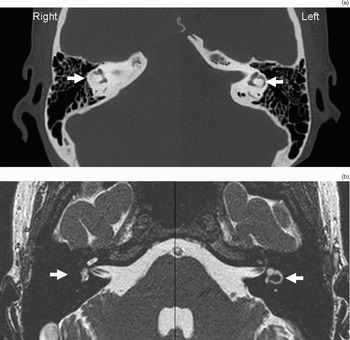
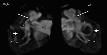
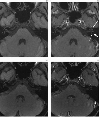
A computed tomography scan of the temporal bones demonstrated partial obliteration of the membranous labyrinth on the right (Figure 1). Magnetic resonance imaging (MRI) of the internal auditory canals with fluid-sensitive sequences revealed constriction of the semicircular canals on the right, with poor visualisation of some of the membranous labyrinth components (Figures 1 and 2). High resolution pre- and post-gadolinium images demonstrated enhancement within the left cochlea and semicircular canals (Figure 3a and b). No definite enhancement was noted on the right.
(a) High resolution, axial computed tomographic scan of the temporal bones, showing poor visualisation of the right lateral semicircular canal, compared with normal appearance on the left. The posterior semicircular canal (not shown) also showed constriction. (b) Axial high resolution fluid-sensitive MRI image at the level of the internal auditory canals (IAC) showing lack of fluid signal intensity within the right lateral semicircular canal and normal appearance of the left lateral semicircular canal. Arrow = lateral semicircular canal.

Three-dimensional, maximum intensity projection obtained by reformatting axial fast imaging employing steady state acquisition magnetic resonance images. The fluid-filled structures within the temporal bones are highlighted and appear as if viewed from above and slightly from behind. The internal auditory canals (IAC) are filled with cerebrospinal fluid. Long white arrow = basal turn of the cochlea (slightly constricted on right); open black arrow = vestibule; short white arrow = lateral semicircular canal (severely stenotic on right); dashed arrow = posterior semicircular canal (showing lack of fluid in inferior limb of right posterior semicircular canal). The superior semicircular canal is not well seen in this projection.

Three-millimetre, axial, T1, fat-saturated MRI images taken (a) pre- and (b) post-gadolinium, showing abnormal enhancement within the cochlea (long arrow), lateral semicircular canal (short arrow) and posterior semicircular canal (dashed arrow) on the left side. No abnormal enhancement in the right membranous labyrinth is seen. Five months later, (c) pre- and (d) post-gadolinium images show improvement. There is subtle, ongoing enhancement in the left cochlea, but the remainder of the membranous labyrinth shows no enhancement.

The patient was commenced on prednisone 60 mg orally daily and azathioprine 200 mg orally daily. Subsequent neuro-otological assessment revealed no further spontaneous nystagmus. Head impulse testing remained positive bilaterally. At three months post-presentation, the patient's follow-up audiogram revealed persistent profound neurosensory hearing loss on the right but a significant improvement in pure tone thresholds on the left. On the left, the four frequency pure tone average had improved to 16.25 dB, the speech reception threshold to 10 dB and word recognition scores to 80 per cent. However, a sharp, sloping, high frequency neurosensory loss persisted above 2000 Hz.
Repeat MRI of the inner ear structures revealed stable constriction of the semicircular canals on the right (not shown). The previously noted enhancement of the left membranous labyrinth had improved (Figure 3c and d).
Discussion
Symptomatic sensorineural hearing loss (SNHL) is uncommon in systemic lupus erythematosus (SLE) patients. A Medline search revealed 17 relevant papers regarding the prevalence of SNHL in SLE.Reference Sheehy1–Reference Roverano, Cassano, Paira, Chiavarini, Graf and Rico17 There are few published studies on SNHL in SLE, and the estimated prevalence varies widely, between 8 and 57.5 per cent, for both symptomatic and asymptomatic SNHL.Reference Bowman, Linthicum, Nelson, Mikami and Quismorio4–Reference Sperling, Tehrani, Liebling and Ginzler7, Reference Kastanioudakis, Ziavra, Voulgari, Exarchakos, Skevas and Drosos12, Reference Roverano, Cassano, Paira, Chiavarini, Graf and Rico17 Recently, a prospective study of 31 patients with asymptomatic SNHL revealed that 21 of 30 (70 per cent) had hearing impairment.Reference Roverano, Cassano, Paira, Chiavarini, Graf and Rico17 The frequency of SNHL in screened SLE patients has been disputed. No hearing deficits were found in one study of 20 SLE patients.Reference Narula, Powell and Davis5
In cases of SLE and SNHL, no correlation has been found between clinical and serological disease activity. A study which audiometrically tested 30 hospitalised patients with exacerbated SLE (defined as active disease in three or more organ systems) found only two patients with asymptomatic SNHL.Reference Bowman, Linthicum, Nelson, Mikami and Quismorio4 A prospective study investigating SLE patients' audiovestibular function found that 23 of 40 patients (57.5 per cent) had asymptomatic hearing impairment.Reference Andonopoulos, Naxakis, Goumas and Lygatsikas6
Antiphospholipid antibodies have been found in patients with SNHL and SLE.Reference Naarendorp and Spiera9, Reference Paira18–Reference Green and Miller20 One study did not observe a statistically significant association between patients with antiphospholipid antibodies and SNHL, when compared with patients with SNHL and no antiphospholipid antibodies.Reference Roverano, Cassano, Paira, Chiavarini, Graf and Rico17 Our patient had bilateral SNHL, negative anticardiolipin antibodies and negative lupus anticoagulant on several occasions, essentially ruling this out as a cause.
Autoimmune SNHL appears to be one of the few causes of reversible SNHL, implicating SLE as a cause, and hence should be promptly identified.Reference Loveman, Comarmond, Cepero and Baldwin16 Treatment is often difficult; there have been so few cases that no one therapeutic approach has been proposed, except that steroids seem to have been included for all treated patients. In fact, in cases of autoimmune SNHL, high dose prednisone is a standard regimen, and its use is part of the criteria proposed for clinical diagnosis of the condition.Reference Loveman, Comarmond, Cepero and Baldwin16
Cyclophosphamide was initially proposed as an immunosuppressive agent which should be used alongside steroids in autoimmunne SNHL.Reference Caldarelli, Rejowski and Corey3, Reference McCabe21–Reference Hughes, Kinney, Barna and Calabrese23 In one study of autoimmune SNHL, 66 patients were treated with cyclophosphamide and prednisolone, resulting in significant improvement in hearing.Reference McCabe22 Autoimmune SNHL patients in whom prednisone was unsuccessful had cyclophosphamide subsequently added to their regimen.Reference Cotter, Singleton and Corman24
Methotrexate has also been proposed as an agent which could be useful in autoimmune SNHL.Reference Sismanis, Wise and Johnson25–Reference Kilpatrick, Sismanis, Spencer and Wise27 The results of a recent prospective, double-blinded, multi-institutional, placebo-controlled study have suggested that the use of methotrexate in autoimmune inner-ear disease after 52 weeks is not as helpful as prior studies had indicated.Reference Harris, Weisman, Derebery, Espeland, Gantz and Gulya28 Sixty-seven patients with an initial response to high dose prednisone were randomised to receive either oral methotrexate (34 patients) in doses of up to 20 mg weekly or a placebo (33 patients). The authors concluded that patients receiving methotrexate did not have a significant difference in maintenance of hearing improvement, compared with those receiving placebo. Another retrospective chart review agreed with these findings.Reference Broughton, Meyerhoff and Cohen29
Results for azathioprine have been favourable: 10 out of 12 autoimmune SNHL patients showed significant improvement with doses of 1 mg/kg daily, when used in combination with prednisone.Reference Saracaydin, Katircioglu, Katircioglu and Karatay30 Another study revealed that six out of 10 patients' hearing loss responded to azathioprine at doses of 25 mg orally thrice daily.Reference Pyykko, Ishizaki and Peltomaa31 However, another study found azathioprine only minimally helpful.Reference Broughton, Meyerhoff and Cohen29
• Sensorineural hearing loss (SNHL) is rarely noted in patients with systemic lupus erythematosis (SLE)
• The mechanism of inner-ear damage in SLE still remains largely unknown
• This case report shows that treatment with azathioprine and imaging follow up can be extremely rewarding in patients with SLE complicated by SNHL
• Magnetic resonance imaging can assist the follow up of patients with autoimmune SNHL
Anticoagulation should be contemplated in those patients with serological evidence of antiphospholipid antibody or lupus anticoagulant.Reference Naarendorp and Spiera9, Reference Hisashi, Komune, Taira, Uemura, Sadoshima and Tsuda32 However, in one case study of patients with anticardiolipin antibodies, high dose prednisone was successful in treating symptoms initially and upon recurrence.Reference Hisashi, Komune, Taira, Uemura, Sadoshima and Tsuda32 Anticoagulation was not contemplated in the current case.
Conclusion
In summary, symptomatic SNHL is rare in patients with SLE. Asymptomatic SNHL appears to be much more common, however. We have shown that treatment with azathioprine, followed up by imaging studies, can be extremely rewarding in patients with SLE and SNHL. This is the first published case to date to show that MRI can assist the follow up of patients with autoimmune SNHL. A multicentre database, with routine audiometric testing, could be a useful tool in the identification of future cases and possible causes. At the present time, in any patient presenting with SLE and aural symptoms, especially in the absence of other organ system involvement, the use of immunological, audiometric and radiological investigations appears to be essential.