Introduction
Strongyloides venezuelensis, a counterpart of the human pathogen Strongyloides stercoralis, naturally infects rodents and is frequently used as an experimental intestinal parasite model (Paula et al., Reference Paula, Sitta, Malta, Gottardi, Corral, Gryschek and Chieffi2013; Rodrigues et al., Reference Rodrigues, Cardoso, Gonçalves, Silva, Massa, Alves, Ueta, Silva and Costa-Cruz2013; Yasuda et al., Reference Yasuda, Matsumoto and Nakanishi2014). Recent research utilizing this model has confirmed the validity of S. venezuelensis larval antigen for the detection of antibody and immune complexes in serum samples (Gonçalves et al., Reference Gonçalves, Silva, Ueta and Costa-Cruz2012c) and bronchoalveolar lavage fluid samples (Gonçalves et al., Reference Gonçalves, Ribeiro, Silva, Ueta and Costa-Cruz2012a). The immune complex is a combination of parasite antigen and rat antibody and can be detected using a capture enzyme-linked immunosorbent assay (ELISA) with anti-parasite antigen and conjugated anti-IgG (Gonçalves et al., Reference Gonçalves, Silva, Ueta and Costa-Cruz2012c).
In the life cycle of S. venezuelensis, filariform larvae reach the lungs and migrate to the small intestine, where they develop into parthenogenetic females (Grove, Reference Grove1996). Humans infected with S. stercoralis typically eliminate larvae in faeces, whereas rodents infected with S. venezuelensis typically eliminate eggs in faeces. Eggs shed in faeces of experimentally infected rodents can be recovered easily and thus serve as a readily available antigen for use in immunodiagnostic tests. Investigating the potential use of parthenogenetic female and egg extracts in immunodiagnosis is practical because these life stages occur in the intestinal mucosa and are thus in contact with host systems that could potentially develop an immune response to them (Gonçalves et al., Reference Gonçalves, Rocha, Gonzaga, Gonçalves-Pires, Ueta and Costa-Cruz2012b).
The aim of the present study was to detect specific IgGs and immune complexes of S. venezuelensis parthenogenetic females and eggs in serum samples for the diagnosis of experimental infection in immunosuppressed rats.
Materials and methods
Parasitological procedures
Male Rattus norvegicus (Wistar) weighing 100–120 g were maintained in the Centro de Experimentação e Utilização de Animais, Universidade Federal de Uberlândia, provisioned with laboratory chow and water ad libitum.
Strongyloides venezuelensis third-stage infective larvae (L3) were obtained from charcoal cultures of infected rat faeces. The cultures were stored at 28°C for 48 h, and the infective larvae were collected and concentrated using the Rugai method (Rugai et al., Reference Rugai, Mattos and Brisola1954). For infection, each rat was inoculated subcutaneously with 1500 S. venezuelensis L3 larvae.
To recover S. venezuelensis parthenogenetic females, rats were anaesthetized (ketamine:xylazine 60:7 mg/kg) and sacrificed at 8 days post-infection (dpi). The small intestines were then removed from the animals, placed in Petri dishes containing saline solution, longitudinally sectioned and incubated at 37°C for 2 h. Parthenogenetic females were then counted following the methods of Sato & Toma (Reference Sato and Toma1990).
To obtain eggs, experimentally infected rats were placed individually on clean, moist, absorbent paper and allowed to defecate over an 18-h period at 5, 8, 13 and 21 dpi. The faeces were collected, diluted in saturated saline and homogenized. The supernatant was placed in a McMaster chamber, and the number of eggs/g of faeces was estimated using the Cornell–McMaster method (Gordon & Whitlock, Reference Gordon and Whitlock1939).
Experimental infections
Rats were divided into four experimental groups: immunocompetent uninfected (n= 6, day 0); immunocompetent infected (n= 24); immunosuppressed uninfected (n= 6, day 0); and immunosuppressed infected (n= 24). Six animals per group were sacrificed at each time point (5, 8, 13 and 21 dpi). Prior to infection, the immunosuppressed rats received 5 μg/ml dexamethasone disodium phosphate in water for 5 days, as described previously by Romand et al. (Reference Romand, Thulliez and Dubey1998). Animals from the infected groups were then inoculated subcutaneously with 1500 L3 S. venezuelensis larvae. At each time point (0, 5, 8, 13 and 21 dpi.), the rats were anaesthetized, and blood samples were collected by cardiac puncture. The blood was centrifuged, and serum aliquots were stored at − 20°C.
Eggs/g of faeces were counted in triplicate on 5, 8, 13 and 21 dpi to verify infection; mean values were used for the analysis.
Immunological protocols
Alkaline extracts were prepared as described previously (Machado et al., Reference Machado, Ueta, Gonçalves-Pires, Oliveira, Faccioli and Costa-Cruz2003). Briefly, 1 ml of 0.15 m NaOH was added to either 500 S. venezuelensis parthenogenetic females or 300,000 eggs, which were gently agitated for 6 h at 4°C. Subsequently, 0.3 m HCl was added until a pH of 7.0 was reached, and the preparations were then centrifuged at 12,400 × g for 30 min at 4°C. The supernatants (alkaline extracts) were analysed for protein content according to Lowry et al. (Reference Lowry, Rosebrough, Farr and Randall1951) and were subdivided into aliquots and stored at − 20°C until use. In a previous study, female extraction analysis using sodium dodecyl sulphate-polyacrylamide gel electrophoresis (SDS-PAGE) showed three major bands, with sizes of 50, 45 and 28 kDa, and one polypeptide group ( < 15 kDa) (Gonçalves et al. Reference Gonçalves, Rocha, Gonzaga, Gonçalves-Pires, Ueta and Costa-Cruz2012b). Egg extracts resulted in somewhat dispersed, faint bands with apparent molecular weights ranging from 27–28 to 32 kDa.
For the production of immune serum, two rabbits were immunized with either S. venezuelensis parthenogenetic females or S. venezuelensis eggs. Briefly, animals were inoculated with 500 μg/ml of both alkaline extract types (parthenogenic females and eggs) emulsified in Freund's complete adjuvant for the first immunization. Inoculations were conducted at intervals of 2 weeks after the first immunization, using Freund's incomplete adjuvant. When ELISA indicated that the antibody count had reached 15,000, 20 ml of blood was collected, and an IgG fraction was prepared using a Protein A Sepharose CL-4B column. After testing IgG activity against the specific antigen, a portion of the IgG fraction was stored at − 20°C for later use for antibody capture by ELISA. The rest of the IgG fraction was conjugated to horseradish peroxidase (Sigma, St. Louis, Missouri, USA) according to Wilson & Nakane (Reference Wilson, Nakane, Knapp, Holuban and Wick1978).
ELISA was performed according to Gonçalves et al. (Reference Gonçalves, Silva, Ueta and Costa-Cruz2012c). Preliminary experiments were performed to determine the optimal conditions for ELISA via the serial dilution of reagents (antigen, control sera and conjugate). Briefly, polystyrene micro-titre plates were coated with 10 μg/ml of S. venezuelensis parthenogenetic female or egg alkaline extracts in 0.06 m carbonate–bicarbonate buffer (pH 9.6) and incubated overnight at 4°C. Then, the plates were washed three times with PBS-T (phosphate-buffered saline plus 0.05% Tween 20), incubated with serum samples (1:10) for 45 min at 37°C and then incubated with the secondary antibody (peroxidase-labelled goat anti-rat IgG (Sigma)) at the ideal dilution of 1:8000 for 45 min at 37°C. The reaction was revealed by adding the enzyme substrate (0.03% H2O2 and o-phenylenediamine (OPD) in 0.1 m citrate–phosphate buffer pH 5.0) and incubating the plate for 15 min at room temperature. Then the reaction was stopped by adding 25 μl/well 2 n H2SO4. The optical density (OD) was measured at 492 nm in a plate reader (Titertek Multiskan, Flow Laboratories, McLean, Virginia, USA). The cut-off value is the criterion that defines the detection limit, and it was calculated as the mean absorbance of three negative sera plus three standard deviations. OD values above the cut-off were considered positive.
Immune complex detection was performed according to Gonçalves et al. (Reference Gonçalves, Silva, Ueta and Costa-Cruz2012c). Preliminary experiments were carried out to determine the optimal conditions for ELISA via serial dilution of reagents (antibody, control sera and conjugate). Briefly, polystyrene micro-titre plates were coated with 50 μl/well of IgG obtained from rabbits immunized with parthenogenetic female or egg alkaline extracts (40 μg/ml) in 0.06 m carbonate–bicarbonate buffer (pH 9.6) and incubated for 18 h at 4°C. After three washes with PBS-T, 50 μl of serum sample (1:10) was added to each well, and the plates were incubated for 45 min at 37°C. Positive (OD values above the cut-off) and negative (OD values below the cut-off) serum samples were used as controls. After incubation for 45 min at 37°C and three additional washes, peroxidase-labelled goat anti-rat IgG (Sigma) was added (50 μl/well) at the ideal dilution of 1:8000, and the plates were incubated for 45 min at 37°C. The reaction was revealed as described above, and the results were determined using the same cut-off value.
Data analysis
Each experiment was performed in duplicate. The results are expressed as the means + SEM. Data were analysed using ANOVA, followed by a Bonferroni test. The level of statistical significance was set at P < 0.05.
Results
For both immunocompetent and immunosuppressed rats, egg shedding in faeces began at 5 dpi, peaked at 8 dpi, and began to decrease at 13 dpi. At 21 dpi, the infection was eliminated in the immunocompetent group, but it persisted in the immunosuppressed group.
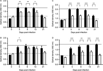
When parthenogenetic female or egg extracts were used as antigens, specific IgGs were not detected in either immunocompetent or immunosuppressed rats. When anti-parthenogenetic-females or anti-S. venezuelensis-eggs were used as antigens, immune complexes were detected for the duration of the infection in immunosuppressed animals, whereas in immunocompetent animals, immune complexes were only detected between 5 and 13 dpi. There were no significant differences between the immunocompetent and immunosuppressed groups throughout the infection when anti-parthenogenetic-female or anti-S. venezuelensis-eggs were used.
Parthenogenetic female extract yielded significant differences between antibody and immune complex detection in immunocompetent rats from 5 to 13 dpi (fig. 1A). In immunosuppressed rats, this difference only occurred at 5 dpi (fig. 1B). Strongyloides venezuelensis egg extract yielded significant differences in both antibody and immune complex detection between immunocompetent and immunosuppressed rats for the duration of the infection (fig. 1C and D).
Detection of antibody (■) and immune complex (□) kinetics, using antigens from parthenogenetic females or anti-parthenogenetic-female antigens, in (A) immunocompetent and (B) immunosuppressed rats; and also, using egg or anti-egg antigens, in (C) immunocompetent and (D) immunosuppressed rats; IgG cut-off values (solid line), immunocomplex cut-off values (dotted line). *, P < 0.05; **, P < 0.01; and ***, P < 0.001.

Discussion
Currently, the diagnosis of strongyloidiasis by immunological testing is typically performed by using total extracts of infective larvae of several species of Strongyloides as the antigen. Recent research has shown that S. venezuelensis filariform larvae total saline extracts can be used to detect IgG antibodies against S. stercoralis (Gonzaga et al., Reference Gonzaga, Ribeiro, Cunha-Júnior, Ueta and Costa-Cruz2011), which demonstrates that different types of antigens can also be used in the diagnosis of infection. Because eggs are eliminated in faeces in experimental rodent models, the use of antigenic egg extract would be of great relevance for diagnosing infection in rodents. Furthermore, antigenic extract from parthenogenetic females could be used for the diagnosis of human strongyloidiasis because this life stage occurs in the intestinal mucosa (Gonçalves et al., Reference Gonçalves, Rocha, Gonzaga, Gonçalves-Pires, Ueta and Costa-Cruz2012b). An interesting result that requires further investigation is the lack of antibody detection when parthenogenetic female or egg extracts were used in both experimental groups in the present study; this is important considering the potential of parthenogenetic females and eggs to elicit an immune response in host tissues.
The results of this study show that parthenogenetic female and egg extracts may not be useful for antibody detection in serum samples because the detection rates were below the cut-off limits for both groups throughout the infection period. In contrast, the immune complex detection in serum samples was encouraging in both groups. The data show that immunocompetent rats are able to accelerate the elimination of immune complexes; in immunosuppressed rats, however, the infection persisted until 21 dpi. These results are in accordance with the fact that the detection of eggs in faeces ceased at 21 dpi in immunocompetent rats but persisted beyond 21 dpi in the immunosuppressed rats. Immune complex detection is an innovative approach for the diagnosis of infection, and this is the first attempt at using this method for strongyloidiasis diagnosis in an experimental animal model.
Immune complex detection was more efficient than specific antibody detection for the duration of the infection for both immune serum types and experimental groups. This finding demonstrates that antigen and antibody complexes may serve as markers for disease activity and that antibody detection may be indicative of a resolved disease with residual antibody presence.
In conclusion, ELISA with alternative antigens may be considered a successful strategy for the identification of immune complexes in serum samples and for the diagnosis of active strongyloidiasis, particularly under conditions of immunosuppression.
Financial support
This study was supported by Conselho Nacional de Desenvolvimento Científico e Tecnológico (CNPq -302426/2012-4), Coordenação de Aperfeiçoamento de Pessoal de Nível Superior (CAPES) and by Fundação de Amparo à Pesquisa do Estado de Minas Gerais (FAPEMIG-CBB-PPM-00 396-13), Brazil.
Conflict of interest
None.
Ethical standards
The authors assert that all procedures contributing to this study comply with the ethical standards of laws of Brazil on the care and use of laboratory animals, in accordance with the ethical principles upon approval of the Comitê de Ética na Utilização de Animais of Universidade Federal de Uberlândia (protocol 096/10).