Introduction
Eurytrema coelomaticum (Trematoda, Dicrocoeliidae) is a digenean flatworm found in ruminants that is the causative agent of eurytrematosis, a disease of veterinary health concern. In Brazil, morphological and molecular data indicate that E. coelomaticum is the only species parasitizing livestock (Travassos, Reference Travassos1944; Travassos et al., Reference Travassos, Teixeira and Kohn1969; Figueira et al., Reference Figueira, Oliveira, Taroda, Alfieri & and Headley2014). The ultrastructural organization of E. coelomaticum cercariae, mother and daughter sporocysts (Franco-Acuña et al., Reference Franco-Acuña, Pinheiro, Oliveira-Menezes, Brandolini, DaMatta and de Souza2011; Pinheiro et al., Reference Pinheiro, Franco-Acuña, Oliveira-Menezes, Brandolini, de Souza and DaMatta2011, Reference Pinheiro, Franco-Acuña, Oliveira-Menezes, Brandolini, DaMatta and de Souza2012), and eggs (Pinheiro et al., Reference Pinheiro, Fanco-Acuña, Oliveira-Menezes, Brandolini, Adnet, Lopes Torres, Miranda, de Souza and DaMatta2015) has been investigated using scanning and transmission electron microscopies. Other works have already used advanced microscopy experiments for investigating nematodes (Adnet et al., Reference Adnet, Gonçalves, de Souza and Attias2013; Lopes-Torres et al., Reference Lopes-Torres, Maldonado, Anjos, de Souza and Miranda2015), cestodes (Almeida et al., Reference Almeida, Oliveira, Neves, Siqueira, Rodrigues-Silva, Daipert-Garcia and Machado-Silva2015) and some trematode species (Machado-Silva et al., Reference Machado-Silva, Pelajo-Machado, Lenzi and Gomes1998; Souza et al., Reference Souza, Garcia, Manso, Neves, Maldonado and Machado-Silva2011, Reference Souza, Garcia, Gomes, Machado-Silva and Maldonado2017; Gonçalves et al., Reference Gonçalves, Oliveira-Menezes, Maldonado, Carvalho and de Souza2016; Marques et al., Reference Marques, Rocha, Manso and D’Ávila2017). Despite the excellent resolution to characterize the surface structures in detail, conventional scanning electron microscopies (SEMs) limit the observation to the external surfaces, missing information about the internal ones. Light microscopy (LM; brightfield and fluorescence) provides information about internal structures, being complemented by SEM and the cryofracture technique (Lopes-Torres et al., Reference Lopes-Torres, Girard-Dias, Mello, Simoes, Pinto, Maldonado, de Souza and Miranda2019).
Currently, most studies on adults of E. coelomaticum are based on conventional LM (Travassos, Reference Travassos1944; Travassos et al., Reference Travassos, Teixeira and Kohn1969). Over recent years, new insights into the morphology and anatomy of parasitic helminths have been generated using advanced imaging technologies, like the fluorescence and confocal microscopies (CMs) associated with electron microscopy experiments (Faulwetter et al., Reference Faulwetter, Vasileiadou, Kouratoras, Thanos and Arvanitidis2013). Fluorescence analysis of large parasites, such as helminths, was neglected until recent advances in fluorescence stereomicroscopy using structured illumination and light sheet fluorescence microscopy, thus opening up new possibilities for the study, with the entire body of helminths in freshly fixed and hydrated samples (Lopes-Torres et al., Reference Lopes-Torres, Girard-Dias, Mello, Simoes, Pinto, Maldonado, de Souza and Miranda2019). These findings show the importance of the association of modern multidimensional techniques for the understanding of helminth morphofunctionality, highlighting the potential of fluorescence microscopy as the basis for other experiments in the parasitology area.
In the present study, different experiments were performed using a combined approach of brightfield LM, fluorescence microscopy, CM and SEM to explore whole and cryofractured samples. This investigation focused on the attachment structures (oral and ventral suckers), reproductive organs (male and female), tegument ultrastructure and excretory apparatus.
Material and methods
Parasite sources
The adult worms were collected from the pancreas of naturally infected cattle that were slaughtered in an industrial slaughterhouse (Matadouro Municipal de Barra Mansa), Barra Mansa, RJ, Brazil, 22°32′39″S, 44°10′17″W) according to the sanitary surveillance rules (Pinheiro et al., Reference Pinheiro, Fanco-Acuña, Oliveira-Menezes, Brandolini, Adnet, Lopes Torres, Miranda, de Souza and DaMatta2015). The parasites were fixed by immersion in AFA solution(the mixture by glacial acetic acid 2%, formaldehyde 3% and 70º ethanol 95%) at room temperature. The parasites were confirmed as E. coelomaticum based on LM according to the size of the suckers and of the body (Travassos, Reference Travassos1944).
Morphological examination
Twenty-five fixed worms were stained and prepared for LM, stereomicroscopy (brightfield and fluorescence) and CM imaging, as previously described (Neves et al., Reference Neves, Pereira, de Oliveira, Gomes and Machado-Silva1998; de Souza, Reference de Souza2011). Briefly, the worms were stained with hydrochloric carmine, dehydrated in a graded ethanol series, cleared with methyl salicylate and mounted on microscope slides with Canada balsam. LM was carried out using a light microscope (Eclipse 80i, Japan) coupled to a digital camera (Nikon DS-Ri1, Japan) at the Rio de Janeiro State University (UERJ), Brazil. The experiments using stereomicroscopy were performed at the Urogenital Research Unit, Department of Anatomy, at the Institute of Biology Roberto Alcantara Gomes, UERJ (Discovery V8, Zeiss, Germany) and in the National Center for Structural Biology and Bioimaging (CENABIO) at the Rio de Janeiro Federal University (UFRJ; Axio Zoom.V16, Zeiss, Germany). The whole-mounted voucher specimen was deposited in the Helminthological Collection of the Institute Oswaldo Cruz, Rio de Janeiro, Brazil, under the number 38879.
CM
Whole mounts were imaged using a Nikon C2 confocal microscope (Nikon Instruments, Inc., New York, USA), operating at 556 nm excitation, 573 nm emission and pinhole at 1.4 (de Souza, Reference de Souza2011), in the Confocal/Fluorescence Microscopy Laboratory at the Institute of Biology Roberto Alcantara Gomes, UERJ.
SEM
Ten specimens were processed for SEM based on a previous protocol (Lopes-Torres et al., Reference Lopes-Torres, de Souza and Miranda2013) and some specimens were frozen and fractured following a simple methodology previously described (Adnet et al., Reference Adnet, Gonçalves, de Souza and Attias2013). Briefly, samples were dehydrated in a graded ethanol series (70–100% Degree Gay Lussac) for 40 minutes at each step, critical-point dried in CO2, mounted on metallic stubs and coated with gold (20–25 nm deposited). After assembly, the samples were examined using the scanning electron microscopes Jeol/EO1.0 (Jeol, Japan) at the Institute of Chemistry, UERJ, and Zeiss EVO-MA10 (Zeiss, Germany) at the CENABIO-UFRJ.
Results
Description
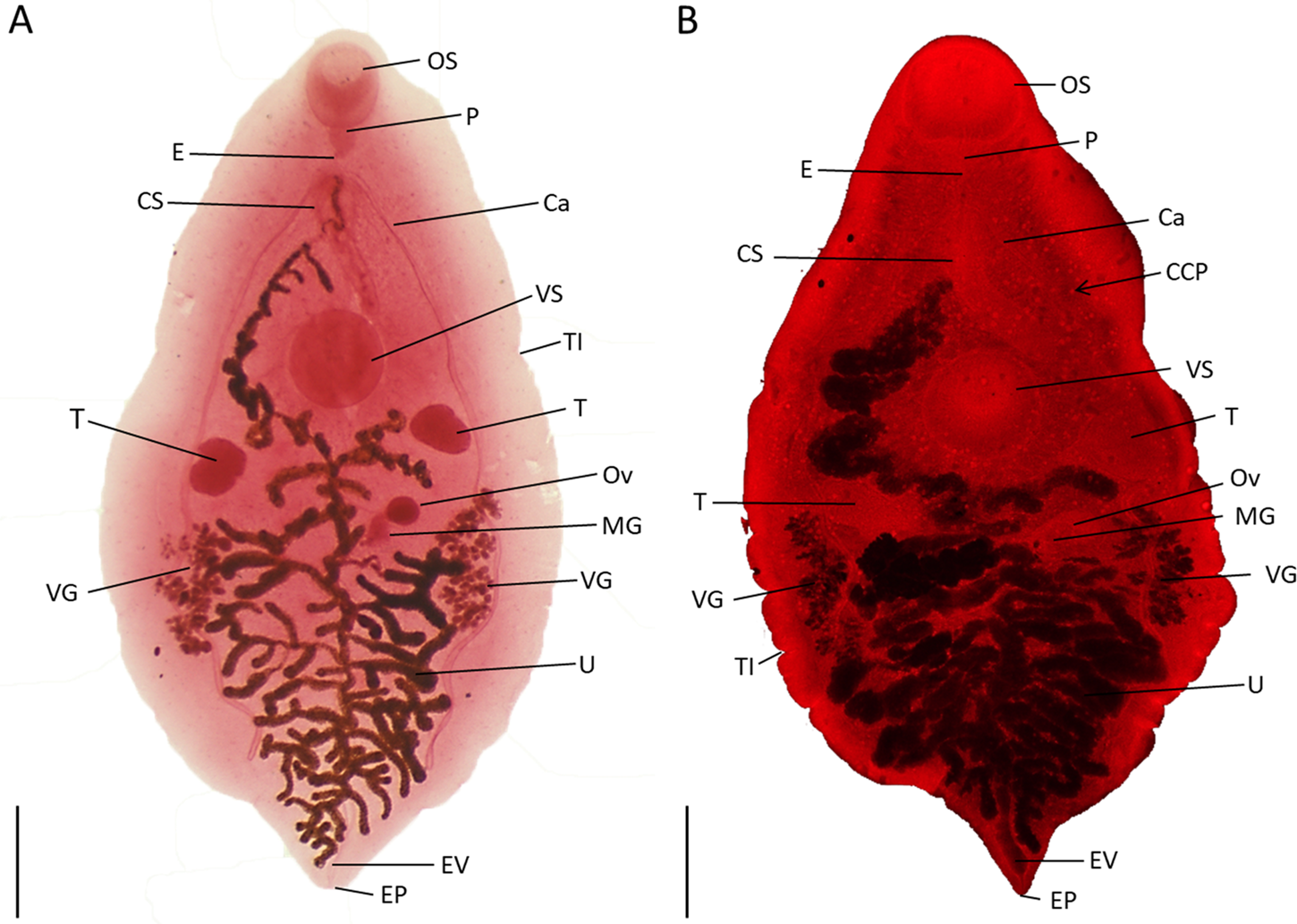
Based on 25 whole-mounted specimens collected from cattle in Brazil and stained with hydrochloric carmine, the morphological and morphometric data of E. coelomaticum were analysed (fig. 1a, b). Specimens had a total body length of 7.07 ± 1.24 (4.43–8.95) mm and width of 3.66 ± 0.69 (2.20–4.91) mm. LM showed the attachment structures (fig. 1a, b), the subterminal oral sucker (0.45–0.96 mm) and ventral sucker (0.46–0.88 mm) placed in the forebody. The pharynx was short (0.17–0.29 mm), and the oesophagus followed by two caeca branches. The male reproductive system showed two globular posterolateral testes and a well-developed elongated cirrus-sac, 1.23 ± 0.18 (0.95–1.66 mm), extending from the intestinal bifurcation to the ventral sucker region (fig. 1a, b). The female reproductive system showed ovaries with a diameter of 0.29 ± 0.08 (0.16–0.48) mm, The Mehlis’ gland complex and the vitelline reservoir were positioned in the mid hindbody, posterior to the testes; the vitelline follicles were arranged in two lateral fields restricted to the mid hindbody; uterus with many brown eggs filling the hindbody, extending to the anterior region and overlapping the ventral sucker. The posterior region presented the excretory pore and eggs overlapping the excretory vesicle (fig. 1a, b). The fluorescence stereomicroscopy highlighted the cubic crystal protein inclusions (fig. 1b).
Overview of Eurytrema coelomaticum adult worm collected from cattle in Rio de Janeiro State (Brazil). Whole-mounted specimen in ventral view showing internal and external structures. (A) Brightfield stereomicroscope; (B) fluorescence stereo zoom microscope. Scale bars: 500 µm. Abbreviations: Ca, caeca; CCP, cubic crystal protein inclusions; CS, cirrus-sac; E, oesophagus; EP, excretory pore; EV, excretory vesicle; MG, Mehlis’ gland complex; OS, oral sucker; Ov, ovary; P, pharynx; T, testes; TI, tegument invagination; U, uterus; VS, ventral sucker; VG, vitelline gland.

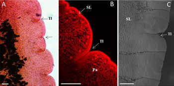
The tegument revealed a typical pattern of indentation of various sizes at irregular intervals (figs 1a, b and 2a). CM showed that the thickened-syncytial layer gives rise to the tegument indentation, invading the parenchyma area (fig. 2b). SEM revealed a spineless surface on the syncytial tegumental layer with a honeycomb pattern (fig. 2c).
External view of Eurytrema coelomaticum adult. (A) Light microscopy of the lateral surface showing (tegument indentation) irregularly distributed tegumental invagination; (B) confocal microscopy showing a thicker syncytial layer, which forms tegument indentation of various sizes, invading the parenchyma area; (C) scanning electron microscopy revealed spineless syncytial layer with a honeycomb pattern and tegument indentation. Scale bars: (A) 100 µm; (B, C) 50 µm. Abbreviations: Pa, pacrenchyma; SL, syncytial layer; TI, tegument identation.

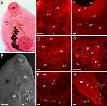
The anterior region of the body is marked by the main fixation structures, the oral and ventral suckers (fig. 3a, b). Observed using SEM, the forebody of the adult worms presented a spineless tegument. The median genital pore was located between the suckers, while typical cubic crystal protein inclusions were originated and projected from the external syncytial layer (fig. 3b). The muscular organization was an important structure for the efficacy of the parasitic strategy, in which the suckers played a critical function in their attachment inside the hosts. Confocal examination revealed that the oral and ventral suckers presented a dense and complex arrangement of the muscle fibre system (fig. 3c–f). The oral and ventral sucker muscles were formed by strong radially arranged fibres overlapping circular, diagonal, transverse and longitudinal fibres (fig. 3c–f). Besides, this tool allowed the visualization of inner parenchymal cells and large cell nuclei around the oral sucker (fig. 3e) and cubic crystal proteins on the outer muscular fibres of the ventral sucker (fig. 3f). The pharynx is an ellipsoid thick-walled organ (figs 3c, g, h) formed, to a large extent, by radial fibres being circumvented on its edges by external and internal longitudinal muscle fibres and presenting parenchymal cells with visible nuclei associated to the pharynx muscular structure (fig. 3g, h). The oesophagus was placed immediately posterior to the pharynx, being thick-walled with longitudinal fibres, with glandular cells around this area (figs 3c, g, h).
Eurytrema coelomaticum forebody. (A) Brightfield showing attachment structures (oral and ventral suckers), digestive (pharynx, short oesophagus and intestinal bifurcation anterior to ventral sucker) and reproductive organs (cirrus and uterus filled with many brown eggs) in the forebody. (B) SEM images showing suckers and genital pore situated between the suckers and cubic crystal protein inclusions situated close to the ventral sucker (inset). (C–H) Confocal images showing typical arrangement of the complex system of the sucker and pharynx muscles. Circular, radial, diagonal transverse and longitudinal fibres within and around the oral sucker (C, D, E), ventral sucker (F) and pharynx (G, H). Parenchymal cells (arrow) and cell nuclei were detected inside the oral sucker (E). Scale bars: (A) 500 µm; (B) 200 µm (inset 20 µm); (D, E, H) 20 µm; (C) 100 µm; (F, G) 50 µm. Abbreviations: Ca, caeca; CCP, cubic crystal proteins inclusions; CF, circular fibres; CS, cirrus-sac; E, oesophagus; GP, gonopore; DF, diagonal fibres; LF, longitudinal fibres; CN, cell nucleus; OS, oral sucker; P, pharynx; Pa, parenchyma; RF, radial fibres; PC, parenchymal cells; TF, transverse fibres; U, uterus; VS, ventral sucker.

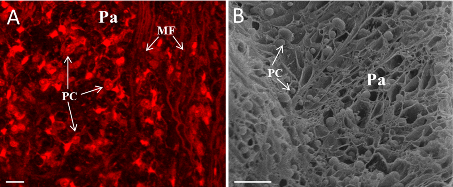
The internal view at the midbody level showed parenchymal cells with visible nuclei and muscle fibres with lower density than in the suckers, which were arranged in dense parenchyma that filled the parasite body (fig. 4a). Freeze-fracture and SEM assays have detailed the parenchymal cell distribution in the meshwork-like parenchyma (fig. 4b).
Internal view of the midbody of Eurytrema coelomaticum adult. (A) Confocal microscopy image showing parenchymal cells and muscle fibres dispersed into the lacunar parenchyma; (B) cryofracture scanning electron microscopy images showing rounded parenchymal cells dispersed into the meshwork-like parenchyma. Scale bars: 20 µm. Abbreviations: MF, muscle fibres; Pa, parenchyma; PC, parenchymal cells.
The whole-mounted specimens examined by LM showed the presence of a cirrus-sac in the forebody (fig. 5a) and the confocal experiments revealed its well-developed wall with a muscular opening to a genital pore (gonopore). The cirrus-sac encompasses the central S-shaped ejaculatory duct enveloped by a well-developed, thick-walled prostatic gland, connected to the seminal vesicle. There were numerous secretory cells external to the ejaculatory duct (fig. 5b). The lumen of the seminal vesicle was filled with spermatozoa (fig. 5c) and, as expected for sexually mature worms, glandular follicles and spermatozoa were visualized inside the testes (fig. 5d). The SEM cryofracture image showed a round-shaped testis with a smooth surface (fig. 5e), while ultrastructure has revealed that male worms present well-developed, extroverted cirrus with a smooth surface (fig. 5f). The scheme of the cirrus-sac was represented as follows: the anterior end represented the protractile cirrus, which emerged out of the gonopore; the genital atrium was small, receiving the female uterus from the left side, laterally, to the cirrus-sac; the ejaculatory duct widened posteriorly to form seminal vesicles filled with spermatozoa; and prostatic glands were densely distributed in the anterior region to the seminal vesicle, surrounding the ejaculatory duct (fig. 5g).
Genitalia of Eurytrema coelomaticum. Ventral view. (A) Whole-mounted specimen observed with brightfield microscopy showing cirrus-sac and uterus; two elliptical nearly symmetrical testes posterior to ventral sucker. (B–D) All stained specimens were also analysed by confocal microscopy. (B) Imaging of cirrus-sac reproductive structures showing gonopore, thick-walled ejaculatory duct and tangled seminal vesicle. The white square details the same region, where secretory cells are also present. (C) Detail of internal seminal vesicle with abundant spermatozoa content. (D) Inside the testes, glandular follicles and spermatozoa were visualized. (E, F) Scanning electron microscopy. (E) Cryofracture image of a single round testis. (F) Image showing everted, conical, smooth cirrus close to the oral sucker. (G) Scheme showing cirrus, uterus with eggs opening at the level of the gonopore, cirrus-sac with ejaculatory duct, prostatic gland and enlarged prolongation of the ejaculatory duct forming seminal vesicle containing abundant spermatozoa. Scale bars: (A) 500 µm; (B) 100 µm (inset 50 µm); (C, D) 20 µm; (E–G) 200 µm. Abbreviations: C, cirrus; CS, cirrus-sac; ED, ejaculatory duct; GF, glandular follicles; GP, gonopore; OS, oral sucker; SC, secretory cells; SV, seminal vesicle; Sp, spermatozoa; T, testes; U, uterus; VS, ventral sucker.

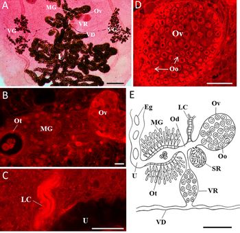
An overview of the whole-mounted worms showed the uterus filled with brown eggs extending from the posterior half of the body to the mid-way between the genital pore and ventral sucker. Ovary was single, small, round and post-testicular; Mehlis’ gland complex and vitelline glands were distributed in two lateral fields (fig. 6a). CM allowed the observation of the Laurer's canal, obscured by eggs, while LM enabled the identification of the intercecal uterus, vitelline reservoir, Mehlis’ gland complex and vitelline duct (fig. 6a). Confocal examination resulted in more detailed images of the reproductive tracts such as the ovary, Mehlis’ gland and cells inside the ootype (fig. 6b). As aforementioned, the Laurer's canal was darkened by eggs. Nevertheless, the CM showed its dense musculature organization (fig. 6c). Inside the ovary, numerous round oocytes were observed (fig. 6d). The scheme of the female reproductive organ is shown in fig. 6e. Oviduct emanating from a single ovary with oocytes, expanding in the proximal portion to form a seminal receptacle. A narrow tube (Laurer's canal) joins the tubular oviduct, together with the seminal receptacle and the vitelline reservoir, before oviduct joins the characteristic thick wall ootype, an expansion of the oviduct surrounded by numerous Mehlis’ gland. This organ contributes to the eggshell formation. The developing eggs leave the ootype to the uterus, through which the eggs are transported to the exterior.
Female genitalia of Eurytrema coelomaticum. Ventral view. (A) Whole-mounted specimen observed with brightfield microscopy showing a small single ovary, Mehlis’ gland complex, vitelline reservoir, vitelline duct, vitelline glands in two lateral fields and thousands of brownish eggs within the uterus. (B–D) All stained specimens were also analysed by confocal microscopy. (B) Site of egg formation showing ovary and ootype surrounded by Mehlis’ gland cells. (C) Thick-walled Laurer's canal and partial view of uterus. (D) Note oocytes within the ovary. (E) Scheme illustrating female genitals. Single ovary with oocytes. Laurer's canal joins the tubular oviduct together with the seminal receptacle and the vitelline reservoir. The oviduct expands in the proximal portion to form the ootype surrounded by Mehlis’ gland cells. The uterus, containing eggs formed, originates at the level of the distal ootype. The vitelline reservoir extends by connecting to the vitelline glands through the vitelline ducts. Scale bars: (A) 500 µm; (B) 20 µm; (C, E) 100 µm; (D) 50 µm. Abbreviations: Eg, eggs; LC, Laurer's canal; MG, Mehlis’ gland; Oo, oocyte; Ot, ootype; Od, oviduct; Ov, ovary; SR, seminal receptacle; U, uterus; VG, vitelline glands; VR, vitelline reservoir; VD, vitelline duct.

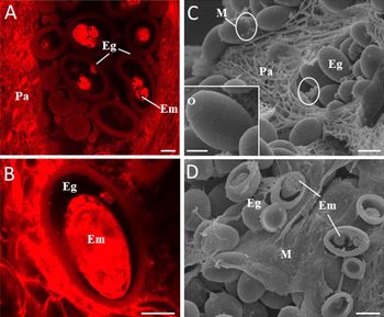
Intrauterine eggs were examined both by the CM and the freeze-fracturing technique (fig. 7). Eggs with thick shells were found in the loops of the uterus (fig. 7a). Eggs containing well-developed embryos were also present (fig. 7b) and images of the fractured eggs showed the eggs inside the uterine lumen in diverse stages of embryonic development (fig. 7c, d). Oval shape, presence of opercula and smooth surface (fig. 7d). In addition, mucus was found in this region, providing protection to the reproductive structures (fig. 7d).
Intrauterine eggs of Eurytrema coelomaticum. (A, B) Confocal microscopy of whole-mounted preparations. (A) Eggs with thick eggshell in the uterus. (B) A single egg with well-developed embryo. (C, D) SEM cryofracture images. (C) Eggshell-enclosing developing embryos in the uterine loops. Inset: note operculum on the anterior end and the apparently smooth tegument. (D) Several thick-shelled eggs in different stages of embryonic development and mucus. Scale bars: (A, B, D) 10 µm; (C) 100 µm (inset 10 µm). Abbreviations: Eg, eggs; Em, embryo; M, mucus; O, operculum; Pa, parenchyma.

An excretory pore and a slice excretory vesicle were visualized at the posterior end of the body (fig. 8a–c). LM revealed an excretory terminal pore (fig. 8a). In addition, CM (fig. 8b) and SEM (fig. 8c) revealed the excretory pore opening, indicating the point of contact with the external environment.
Posterior region of Eurytrema coelomaticum adult. (A, B) Brightfield and confocal microscopies showing the uterus overlying the excretory vesicle and the excretory pore opening. (C) Scanning electron microscopy showing a smooth slightly depressed tegument around the excretory pore. Scale bars: (A) 500 µm; (B) 100 µm; (C) 20 µm. Abbreviations: EP, excretory pore; EV, excretory vesicle; U, uterus.

Discussion
In recent years, there has been significant progress in microscopic technologies, leading to new insights into the morphology and anatomy of parasitic worms (Faulwetter et al., Reference Faulwetter, Vasileiadou, Kouratoras, Thanos and Arvanitidis2013; Lopes Torres et al., Reference Pinheiro, Fanco-Acuña, Oliveira-Menezes, Brandolini, Adnet, Lopes Torres, Miranda, de Souza and DaMatta2015; Gonçalves et al., Reference Gonçalves, Oliveira-Menezes, Maldonado, Carvalho and de Souza2016; Conn et al., Reference Conn, Świderski and Miquel2018). Therefore, the current study aimed to provide new data about the external and internal organization of E. coelomaticum adults, based on a combination of advanced morphological experiments.
Cubic crystal protein inclusions are structures described in E. coelomaticum that probably represent a conservative character, since they were found in E. coelomaticum adults from different countries and were observed in detail using electron microscopy in specific regions of the body (Sakamoto & Oikawa, Reference Sakamoto and Oikawa2007). As expected, SEM images showed cubic crystal inclusions on the tegument surface. In addition, based on our fluorescence stereomicroscopy experiments, a wide distribution of these crystals was detected on the trematode body. There is evidence to support that these inclusions appear in the subtegumental region and are transported to the tegument along cytoplasmic bridges (Sakamoto & Oikawa, Reference Sakamoto and Oikawa2007). Our CM examination revealed that the crystal protein inclusions were also present at the level of the ventral sucker, representing the first report describing such structures by CM. Therefore, this investigation has reinforced the importance of CM to study the inner structures of parasitic flukes (Machado-Silva et al., Reference Machado-Silva, Pelajo-Machado, Lenzi and Gomes1998; Šebelová et al., Reference Šebelová, Stewart, Mousley, Fried, Marks and Halton2004; Neves et al., Reference Neves, de Lamare, Machado-Silva, Carvalho, Branquinho, Lenzi, Hulstijn and Gomes2005, Souza et al., Reference Souza, Garcia, Gomes, Machado-Silva and Maldonado2017). It is likely that cubic crystal protein inclusions present a metabolic function because they were found in adult worms from different countries, showing that they are not associated with a specific condition, such as host diet, fixation or another artefact.
The gonopore is medially situated in the forebody between the suckers; the characteristics of the elongated cirrus-sac and testes herein described are consistent with previous descriptions (Travassos et al., Reference Travassos, Teixeira and Kohn1969; Pinto & de Melo, Reference Pinto and de Melo2016). The confocal results revealed a well-developed cirrus-sac composed of a seminal vesicle, a well-developed prostatic gland and secretory cells surrounding the S-shaped ejaculatory duct, which ended in the cirrus exposed through the genital pore, as found in Fasciola hepatica and Trichobilharzia regenti (Terasaki et al., Reference Terasaki, Itagaki, Shibahara, Noda and Moriyama-Gonda2001; Skírnisson et al., Reference Skírnisson, Kolářová, Horák, Ferté and Jouet2012). In addition, SEM showed that both cirrus and testes have smooth margins, similar to the other Dicrocoeliidae species (Casanova & Ribas, Reference Casanova and Ribas2004; Hildebrand et al., Reference Hildebrand, Okulewicz and Popiolek2007).
Although earlier LM-based studies (Travassos, Reference Travassos1944; Travassos et al., Reference Travassos, Teixeira and Kohn1969) have shown the lateral margin of E. coelomaticum adult, no information is currently available regarding its characterization. Confocal and SEM examination enabled us, for the first time, to show that it is composed of invaginations of various sizes at irregular intervals, as reported in digenean flatworm parasites of Brazilian catfishes (Cohen et al., Reference Cohen, Kohn and Baptista-Farias2001).
Given that digenean trematodes utilize suckers for dislodging, attachment and feeding within their host tissues, the muscular organization of these suckers has attracted much interest, especially due to the increase of knowledge about the organization of the fibres that might be new targets for drugs (Kumar et al., Reference Kumar, McGeown, Reynoso-Ducoing, Ambrosio and Fairweather2003; Krupenko, Reference Krupenko2019). Consistent with previous investigations also using CM, a typical arrangement formed by diagonal, circular, radial, transverse and longitudinal (=meridional) muscle fibres was observed (Mair et al., Reference Mair, Maule, Shaw and Halton1998; Kumar et al., Reference Kumar, McGeown, Reynoso-Ducoing, Ambrosio and Fairweather2003, Šebelová et al., Reference Šebelová, Stewart, Mousley, Fried, Marks and Halton2004; Krupenko, Reference Krupenko2014; Borges et al., Reference Borges, Costa, Mantovani, Barros, Santos, Mafra and Santos2017; Krupenko, Reference Krupenko2019). These types of muscle fibres may vary in organization and amount across different helminth groups, suggesting an evolutionary character associated with the behaviour of the parasite and the site they occupy within the host (Mair et al., Reference Mair, Maule, Day and Halton2000; Krupenko & Dobrovolskij, Reference Krupenko and Dobrovolskij2015; Krupenko, Reference Krupenko2019).
Previous works showed in detail the female reproductive system, and our results using LM have confirmed the morphological and morphometric patters presented in these classical descriptions (Travassos, Reference Travassos1944; Travassos et al., Reference Travassos, Teixeira and Kohn1969). Nevertheless, confocal images showed a single ovary filled with numerous oocytes, a conserved character among digenean trematodes (Neves et al., Reference Neves, de Lamare, Machado-Silva, Carvalho, Branquinho, Lenzi, Hulstijn and Gomes2005; Souza et al., Reference Souza, Garcia, Manso, Neves, Maldonado and Machado-Silva2011; Hanna, Reference Hanna2015; Marques et al., Reference Marques, Rocha, Manso and D’Ávila2017), highlighting that reproductive efficiency is an important adaptation of the organisms to parasitic life. Laurer's canal is a structure present in digenean species. However, its detection is very difficult using only brightfield microscopy, mainly because of its overlapping position with the uterus. In the present study, the confocal series images have overcome this limitation, enabling us to detail the Laurer's canal, showing that it is a thick-walled tube (Bullard et al., Reference Bullard, Williams and Bunkley-Williams2012; Skírnisson et al., Reference Skírnisson, Kolářová, Horák, Ferté and Jouet2012; Womble et al., Reference Womble, Cox-Gardiner, Cribb and Bullard2015; Roberts et al., Reference Roberts, Orelis-Ribeiro, Dang, Halanych and Bullard2016).
Limited data are currently available describing the egg-forming complex in E. coelomaticum (Pinheiro et al., Reference Pinheiro, Fanco-Acuña, Oliveira-Menezes, Brandolini, Adnet, Lopes Torres, Miranda, de Souza and DaMatta2015). The present study has revealed that the ootype is surrounded by the Mehlis’ gland, as previously shown for F. hepatica using transmission electron microscopy (Colhoun et al., Reference Colhoun, Fairweather and Brennan1998) and for Echinostoma paraensei using CM (Souza et al., Reference Souza, Garcia, Manso, Neves, Maldonado and Machado-Silva2011). Among digeneans, it is well-established that egg formation takes place in a thick-walled chamber (ootype), where the eggshell is formed from material derived from the vitelline cells combined with secretions of the Mehlis’ gland (Cogswell et al., Reference Cogswell, Kommer and Williams2012).
Knowledge about the uterine development of E. coelomaticum eggs is scarce (Pinheiro et al., Reference Pinheiro, Fanco-Acuña, Oliveira-Menezes, Brandolini, Adnet, Lopes Torres, Miranda, de Souza and DaMatta2015). As observed in the current confocal study, the uterus contains numerous thick-walled spherical-to-ellipsoid eggs, free within the uterine loops, as previously described (Pinheiro et al., Reference Pinheiro, Fanco-Acuña, Oliveira-Menezes, Brandolini, Adnet, Lopes Torres, Miranda, de Souza and DaMatta2015). Over the last decade, the cryofracture technique has been proved a simple method used to prepare biological samples by exposing internal structures for visualization using SEM (Taylor, Reference Taylor2008). Indeed, previous studies have provided valuable new information about parasitic flukes (Adnet et al., Reference Adnet, Gonçalves, de Souza and Attias2013). As mentioned above, in order to better characterize the uterine eggs observed by CM, a detailed examination using the cryofracture technique was performed. The main advantage was the visualization of embryos inside the maternal uterine tissues, which may greatly facilitate the analysis of the embryogenesis. This investigation has shown uterine eggs surrounded by mucus with embryo in different developmental stages (Conn et al., Reference Conn, Świderski and Miquel2018) and eggs with a well-defined operculum (Pinheiro et al., Reference Pinheiro, Fanco-Acuña, Oliveira-Menezes, Brandolini, Adnet, Lopes Torres, Miranda, de Souza and DaMatta2015).
In recent years, increasing drug resistance worldwide has been observed in gastrointestinal helminths that parasitize livestock (Radio et al., Reference Radio, Fontenla and Solana2018). Hence, strategies are necessary to develop and test new and effective anthelmintic drugs (Srivastava & Misra-Bhattacharya, Reference Srivastava and Misra-Bhattacharya2015). The detection of intrauterine eggs found in adult parasites, as performed in the present study, might also contribute to a better analysis of the action of relevant compounds on digenean flukes of human and veterinary importance, as determined by histology and light and electron microscopies (Hanna, Reference Hanna2015; Abdelaal et al., Reference Abdelaal, Brennan, Abdel-Azi and Fairweather2016; Hanna et al., Reference Hanna, Moffetta, Forstera, Trudgettb, Brennanb and Fairweatherb2016; Pereira et al., Reference Pereira, Oliveira, Coaglio, Santos, Cezar, Mendes, Oliveira, Conzensa and Lima2016). Although a previous study has investigated the action of praziquantel and triclabendazole against Eurytrema pancreaticum adults (Jiraungkoorskul et al., Reference Jiraungkoorskul, Sahaphong, Tansatit, Kangwanrangsan and Pipatshukiat2005), the treatment for E. coelomaticum remains unknown (Ilha et al., Reference Ilha, Loretti and Reis2005).
Brightfield microscopy has shown the body terminating into a terminal excretory pore at the posterior end of the body, although the anterior region of the excretory vesicle was hidden by the uterine eggs (Bray & Justine, Reference Bray and Justine2013). However, detailed information on its morphology is limited. In our confocal results we have noted that the opening of the excretory pore presents a well-defined border and the SEM images enabled a detailed analysis of this structure, revealing a smooth tegument and depressions in the wrinkled body wall, as seen in other digenean flukes (Machado-Silva et al., Reference Machado-Silva, Lanfredi and Gomes1997; Maldonado et al., Reference Maldonado, Vieira and Lanfredi2003).
Considering the results mentioned above, this work has conclusively demonstrated that the combination of advanced morphological experiments might yield complementary data that are not provided by a single isolated technique. Thus, our understanding was enhanced about the structural organization of E. coelomaticum adult stages. Finally, this work contributes new morphological criteria to evaluate the effects of antiparasitic drugs on flukes of medical and veterinary importance.
Acknowledgements
The authors would like to thank Dr Jairo Pinheiro da Silva, Department of Physiological Sciences, Institute of Biology and Department of Animal Parasitology, Veterinary Institute, Federal Rural University of Rio de Janeiro, Seropédica, RJ, for providing the Eurytrema samples. Our thanks to the Department of Anatomy, Confocal Microscopy/Fluorescence Laboratory and Institute of Chemistry, Rio de Janeiro University State, and also to the National Center for Structural Biology and Bioimaging, Rio de Janeiro Federal University, for providing technical assistance.
Financial support
This work was supported by the Conselho Nacional de Desenvolvimento Científico e Tecnológico (CNPq) and Fundação Carlos Chagas Filho de Amparo à Pesquisa do Estado do Rio de Janeiro (FAPERJ). KGL received scholarship from Coordenação de Aperfeiçoamento de Pessoal de Nível Superior (CAPES); (JCNE E-26/202.660/2018).
Conflicts of interest
None.