Introduction
In the last decade, we have seen tremendous progress in three-dimensional (3D) cell culture technologies, and the development of culture systems exhibiting more complex cellular interfaces than a conventional flat petri dish. Reference Bhatia and Ingber1,Reference Liu, Gill and Huang2 These culture systems have been used for landmark developments in mini-organ models, such as organoids with realistic microanatomy, Reference Sato, Vries, Snippert, Van de Wetering, Barker, Stange, Van Es, Abo, Kujala, Peters and Clevers3 and organ-on-a-chip systems in simulating tissue/organ-level physiology. Reference Huh, Leslie, Matthews, Fraser, Jurek, Hamilton, Thorneloe, McAlexander and Ingber4 Combined with advances in stem cell technologies, these culture systems are envisaged to fulfill roles in bridging gaps between preclinical and clinical models in the drug development pipeline, and ultimately, reducing costly failures in clinical trials. Reference Pampaloni, Reynaud and Stelzer5,Reference Esch, Bahinski and Huh6
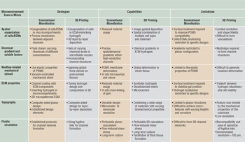
While it is important to harness in vitro biological models to address specific questions, simple two-dimensional cultures cannot capture many of the key microenvironmental factors ( Table I ) known to influence cell fates. Culturing cells in a complex configuration, however, is labor intensive and costly. Recent advances in bioprinting and biofabrication technologies offer promising new strategies to create tissue scaffolds and tissue models.
Reproducing microenvironmental cues using established lithography-based microfluidics and three-dimensional (3D) bioprinting. Comparison of engineering strategies, capabilities, and limitations of both techniques.

Note: ECM, extracellular matrix; PDMS, poly(dimethylsiloxane); 2D, two-dimensional.
Bioprinting can provide the potential capability to repeatedly build small-scale tissue systems, minimizing human intervention and improving standardization and accuracy. Reference Murphy and Atala7,Reference Mandrycky, Wang, Kim and Kim, D.H8 Second, due to its ability to automate and program the deposition of cells and materials in 3D, Reference Ozbolat and Hospodiuk9,Reference Gudapati, Dey and Ozbolat10 bioprinting also provides new possibilities to construct a cell niche with prescribed complexity and physiological resemblance. These two general attributes may address some of the critical steps toward the adaption of complex culture systems. These include, for example, the necessity for standardization, validation, and reproducibility, and meeting investigators’ desires to create complex co-culture systems with more than four cell types, in a predefined spatial configuration (please see Reference Reference Liu, Gill and Huang2 for survey results). The latter attribute may be regarded as the key advantage not easily facilitated by microfabrication and lithography-based approaches.
In this article, direct comparisons between 3D bioprinted and microfabricated organ-on-a-chip models are presented. Further, we propose how a fit-for-purpose bioprinting process can be designed to construct cellular microenvironments for in vitro tissue and organ models.
Comparison between bioprinted and microfabricated models
A number of organ- and disease-on-a-chip models have been developed as a result of advanced microfluidic technologies. Reference Bhatia and Ingber1,Reference Huh, Kim, Fraser, Shea, Khan, Bahinski, Hamilton and Ingber11 Mini-organ models have been established for various organs, including the lung, Reference Huh, Leslie, Matthews, Fraser, Jurek, Hamilton, Thorneloe, McAlexander and Ingber4 heart, Reference Agarwal, Goss, Cho, McCain and Parker12 kidney, Reference Jang, Mehr, Hamilton, McPartlin, Chung, Suh and Ingber13 and liver. Reference Toh, Lim, Tai, Xiao, van Noort and Yu14 Simultaneously, disease models such as local cancer invasion and cancer transendothelial migration through the vessel barrier have also been demonstrated. Reference Jeon, Bersini, Gilardi, Dubini, Charest, Moretti and Kamm15 Among these, there are simple systems comprising a simple cell type within a microfluidic channel, Reference Tsai, Kita, Leach, Rounsevell, Huang, Moake, Ware, Fletcher and Lam16 to complex designs containing multicellular components coupled with 3D gels and circulation. Reference Maschmeyer, Lorenz, Schimek, Hasenberg, Ramme, Hübner, Lindner, Drewell, Bauer, Thomas and Sambo17 Huh et al. Reference Huh, Hamilton and Ingber18 and Sackmann et al. Reference Sackmann, Fulton and Beebe19 have summarized the progress to date.
In comparison to the microfabricated platforms, the capability of 3D bioprinting has yet to be extensively exploited, though selected functional tissue and organ models on a chip were recently demonstrated. Reference Mandrycky, Wang, Kim and Kim, D.H8 With the main purpose of recreating aspects of microenvironmental cues, it is important to identify how a bioprinting approach resembles or differs from a microfabrication approach. Table I lists the main operating mechanisms associated with the respective technique in achieving the desired microenvironmental cues. We further present a side-by-side comparison between similar organ-on-a-chip and screening models generated by the two techniques in Table II . Reference Huh, Leslie, Matthews, Fraser, Jurek, Hamilton, Thorneloe, McAlexander and Ingber4,Reference Agarwal, Goss, Cho, McCain and Parker12,Reference Toh, Lim, Tai, Xiao, van Noort and Yu14,Reference Wufuer, Lee, Hur, Jeon, Kim, Choi and Lee20–Reference Lind, Busbee, Valentine, Pasqualini, Yuan, Yadid, Park, Kotikian, Nesmith, Campbell, Vlassak, Lewis and Parker31 We highlight the key results shown in the two tables here.
Side-by-side comparison of microengineered tissue/organ models via established microfabrication versus three-dimensional (3D) bioprinting techniques.

Note: hMSC, human mesenchymal stem cell; hiPS-CM, human-induced pluripotent stem cell-derived cardiomyocytes; PU, polyurethane; ECM, extracellular matrix; HiPSC, human-induced pluripotent stem cell; μBBB, microfluidic blood–brain barrier; DBB, droplet-based bioprinting; EBB, extrusion-based bioprinting; DLP, digital light processing; LIFT, laser-induced forward transfer; GelMA, gelatin methacryloyl; GMHA, glycidyl methacrylate-hyaluronic acid; RGD, arginine-glycine-aspartic acid; PDMS, poly(dimethylsiloxane).
Although both microfluidics and bioprinting allow spatial organization of cells and extracellular matrix gels, the mechanisms facilitating these differ greatly. In microfluidic chips, materials are organized by flow paths in the channels, and therefore, compartmentalization and special flow geometries need to be designed to allow localized cell material seeding. Reference Zheng, Fu, Cheng, Wang, Zhao and Gu32,Reference Shin, Han, Jeon, Yamamoto, Zervantonakis, Sudo, Kamm and Chung33 On the other hand, 3D printing can provide a more direct deposition approach through the control of nozzle positions. Reference Truby and Lewis34 However, in order to create vessel and tubule-like features, both techniques require flow-directed cell-seeding within a channel geometry. Reference Miller, Stevens, Yang, Baker, Nguyen, Cohen, Toro, Chen, Galie, Yu and Chaturvedi35
Nonetheless, the convenience of bioprinting to integrate multiple cells, hydrogels, and even sensors has enabled the creation of liver, Reference Matsusaki, Sakaue, Kadowaki and Akashi23,Reference Ma, Qu, Zhu, Li, Yuan, Zhang, Liu, Wang, Lai, Zanella and Feng24 heart, Reference Lind, Busbee, Valentine, Pasqualini, Yuan, Yadid, Park, Kotikian, Nesmith, Campbell, Vlassak, Lewis and Parker31 and skin Reference Lee, Singh, Trasatti, Bjornsson, Xu, Tran, Yoo, Dai and Karande21,Reference Koch, Deiwick, Schlie, Michael, Gruene, Coger, Zychlinski, Schambach, Reimers, Vogt and Chichkov22 models with improved biological functions and read-out abilities than conventional culture systems. For example, Lind et al., Reference Lind, Busbee, Valentine, Pasqualini, Yuan, Yadid, Park, Kotikian, Nesmith, Campbell, Vlassak, Lewis and Parker31 in a recent heart model, reported that soft strain gauge sensors could be directly printed by a multimaterial printer and embedded in a printed cardiac tissue, enabling long-term and facile digital readouts of contractile stresses in the engineered tissue. As a comparison, such one-step integration of on-chip sensors in tissue constructs is difficult to achieve by a microfabrication-based approach. Agarwal et al. Reference Agarwal, Goss, Cho, McCain and Parker12 developed a heart-on-a-chip system and showed that multistep assembly procedures were required to form the muscular thin film (thin elastic films on which muscle cells can grow) within the chip. Although such a system only has a single-layered configuration, it has benefits of being an autoclavable and reusable device.
As shown in Table II, current bioprinting approaches offer lower planar resolution than microfluidics. For example, the creation of channel features is important for many purposes, including improving long-term cell viability by providing nutrient/waste exchange, imposing flow-induced shear, and incorporating dynamic concentration gradients. Lithography-based templating offers established protocols that facilitate the creation of these microchannel features. Reference Sackmann, Fulton and Beebe19 In contrast, achieving channels in bioprinting involves the use of fugitive inks (inks that liquefy when exposed to temperature changes) and intricate ink removal procedures, also accompanied by poorer feature resolution of ∼100 µm at the present. Reference Lee, Lanzi, Ngo, Yoo, Vincent and Dai29
For screening applications, one important practical consideration is the amount of cells and extracellular matrix materials required for forming a tissue model, and also, the quantity of compounds needed for a drug test. So far, processes inherent in low-resolution cell printing lead to larger tissue sizes compared to the microfabricated chip systems; thus, larger quantities of cells and testing compounds, which can often be expensive or sparse, are required. On the other hand, if materials restrictions are not imposed, bioprinting provides the unique capability to create thick tissues by incorporating 3D interconnected channels or vessels for nutrient/waste exchange. Reference Kolesky, Homan, Skylar-Scott and Lewis30,Reference Kang, Lee, Ko, Kengla, Yoo and Atala36
As shown in Table II, a recent 3D vasculature printed from fugitive inks by Kolesky et al. Reference Kolesky, Homan, Skylar-Scott and Lewis30 was able to support a large bioprinted multicellular tissue (>1 cm in thickness) under long-term perfusion (>6 weeks). In comparison, in established microfluidic chips, although the channel resolution and the resultant vasculature are finer, the cell layout is usually much thinner in the planar fashion (<1 cm in thickness). Reference Wang, Phan, Sobrino, George, Hughes and Lee28 Therefore, bioprinted thick tissue has advantages in capturing a higher degree of tissue heterogeneity and complexity Reference Kolesky, Truby, Gladman, Busbee, Homan and Lewis37 and possibly providing closer physiological relevance in simulating realistic drug transport from circulation to the targeted tissues.
Finally, non-flow related mechanical stimuli can be seen as one of the most demanding microenvironmental factors to be integrated within a microphysiology device. In soft-lithography-based microfluidics, the majority of the published work has harnessed the deformability of poly(dimethylsiloxane) (PDMS) to form pillars, valves, and stretchable membranes. Reference Agarwal, Goss, Cho, McCain and Parker12 Huh et al. Reference Huh, Leslie, Matthews, Fraser, Jurek, Hamilton, Thorneloe, McAlexander and Ingber4 provide a well-known example in the membrane-based double-layer lung-on-a-chip system. To mimic the mechanical deformation of the lung alveoli during the breathing motion, a vacuum pump was connected to two empty side channels to generate a cyclic stretching motion on the PDMS membrane sandwiched between two layers of microchannels. Such a deformation mechanism imposes stringent demands on the robustness of the materials and device packaging, not yet achievable in a bioprinted construct at the microarchitectural level. Hölzl et al. Reference Hölzl, Lin, Tytgat, Van Vlierberghe, Gu and Ovsianikov38 and Arslan-Yildiz et al. Reference Arslan-Yildiz, El Assal, Chen, Guven, Inci and Demirci39 show in their studies how mechanical forces were exerted on the bulk of bioprinted tissues utilizing a separate mechanical rig, which, however, is only viable for larger tissue constructs at a reduced test throughput. Nonetheless, continued improvements in print technology and the potential combination of microfabrication and bioprinting approaches can help address some of these technical challenges.
Material integration for printing microenvironmental cues
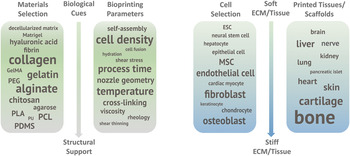
The integration among printed cells, extracellular matrices, and a miniature bioreactor can potentially lead to more precise and flexible recreation of the microenvironmental cues in vitro. To facilitate such a “bioprinted organ-on-a-chip” device, materials with diverse properties, from natural to synthetic in origin, need to be combined together to perform different roles in the device. Figure 1 summarizes different materials that have been used in bioprinting (not specific to organ-on-a-chip). The proposed materials functionality can be largely defined as being biologically focused or structurally focused. For example, biologically active hydrogels can provide a matrix for cell encapsulation, cell binding, and a reservoir for growth factor release. The mechanical weakness of the biologically active hydrogels can be potentially overcome by combining with other robust hydrogels, or by designing mechanical supports from thermoplastic or thermoset structures. Reference Hinton, Jallerat, Palchesko, Park, Grodzicki, Shue, Ramadan, Hudson and Feinberg40 Examples include the creation of core–shell hydrogel structures between alginate and cell-embedded collagen, Reference Ahn, Lee and Kim41 and the incorporation of nano- and microfibers Reference Li, Li, Wang, Ma, Meng, Pritchard, Gill, Liu and Huang42 within hydrogels. The design of multiwell compartments using PDMS can also be seen as a strategy to provide structural support to the printed bioinks. Reference Kolesky, Truby, Gladman, Busbee, Homan and Lewis37
Word cloud diagrams illustrating the statistics of literature reports on materials selection, bioprinting parameters, and the bioprinted cells and tissues. The relative size of each word/phrase is an indication of the relative abundance of the reported subject from Scopus and Web of Science (February 2017), in comparison to the other subjects in the same category. The stiffness ranking of the cells as well as tissue and organs are based on their reported in vivo native extracellular matrix (ECM)/tissue stiffness. Note: GelMA, gelatin methacryloyl; PLA, polylactic acid; PCL, poly(caprolactone); PEGDA, polyethylene glycol diacrylate; PU, polyurethane; PDMS, poly(dimethylsiloxane); ESC, embryonic stem cell; MSC, mesenchymal stem cell; PEG, poly(ethylene glycol).

The diverse materials properties inevitably require different processing techniques for these materials. This means that print techniques based on direct material dispensing mechanism may find broader applications with their cross-technique compatibility. Based on this concept, a number of commercial bioprinters have already established multinozzle deposition platforms. Ozbolat et al. Reference Ozbolat and Hospodiuk9 and Gudapati et al. Reference Gudapati, Dey and Ozbolat10 provide overviews of the different direct deposition printing mechanisms and their material suitability. Regardless of the technique, specific attention must be paid to the mismatched processing parameters between different materials. For example, thermoplastics are normally printed under elevated temperatures in a molten form. Reference Truby and Lewis34 However, this processing condition may affect the hydrated biological system if the solidified thermoplastic structure cannot be cooled rapidly. Any solvents or excess cross-linking reagents contained in the thermoset material can also disturb biological behaviors. Reference Mandrycky, Wang, Kim and Kim, D.H8 Further, it is well known that process-induced thermal, mechanical, or photo stresses can dramatically decrease cellular viability. Reference Wang, Huang, Grujicic and Chrisey43 The processing time (i.e., the duration for which cells are taken out of the incubator environment) needs to be minimized.
Finally, it is worth noting that many of the bioprinted constructs demonstrated so far consist of simple components aimed at musculoskeletal applications that can create robust, macroscopic tissues for implantation. Reference Kang, Lee, Ko, Kengla, Yoo and Atala36 The bioprinting techniques established from these experiences may need to be adjusted for printing soft, complex tissues for organ-on-a-chip devices. Considerations should be given to the self-assembly capability of different cell types; the matrix synthesis and remodeling ability of cells; and also the phase segregation phenomena of different material mixtures. These factors should be accounted for in designing a chip device to accommodate the tissue dynamics over long-term culture.
Conclusion
Advances in bioprinting are accelerating progress toward organ-on-a-chip devices for modeling tissue behaviors with enhanced physiological relevance. Integrating multiple printing techniques, materials, and cells will provide new opportunities for making complex tissue models supporting long-term cultures; but one should be cautious in designing the fabrication strategy to accommodate the processing tolerances for different materials/cells.
Continued developments in this area could bring higher print resolutions, incorporate dynamic mechanical stimulation that better mimic what cells/tissues experience in vivo, and integrate optical and electronic materials for in situ sensing and activation. Harnessing these functionalities will lead to smart organ-on-a-chip devices for high-content pharmaceutical screening with low-reagent usage. It is important to note that the examples illustrated in this article stem from an engineering technology development prospective. The successful creation and implementation of an in vitro model relies heavily on in-depth understanding of the biological pathways and physiological systems; and this will require significant collaborative efforts between engineers and biomedical researchers to fine-tune and optimize the culture conditions. As an intermediate step, 3D bioprinting can be seen as an invaluable toolkit to facilitate easy customization of the culture conditions, enabling systematic evaluation of the different microenvironmental cues (and their combinations) to realize a particular biological phenotype. Like all in vitro model systems, for bioprinted organ-on-a-chip to have a real impact on drug development, one should design the level of biological model complexity, corresponding to the stage of the preclinical testing required. Ongoing system validation, in vivo–in vitro correlation, and regulatory approvals should be sought.
Acknowledgments
D.Z. gratefully acknowledges support from the China Scholarship Council. Y.L. acknowledges the Schlumberger Foundation and Trinity College Cambridge. Y.Y.S.H. thanks EPSRC, The Royal Society London, and the Isaac Newton Trust for funding support.

Yan Yan Shery Huang has been an assistant professor of bioengineering, in the Department of Engineering at the University of Cambridge, UK, since 2013. She obtained her PhD degree in physics in 2011 from the University of Cambridge. She was a joint Oppenheimer Research Fellow and a Homerton College Junior Researcher. Her research focuses on translational bioengineering research, 3D bioprinting/biomicrofabrication, and developing biomimetic organ-on-chips for high-throughput drug testing. Huang can be reached by email at yysh2cam@gmail.com.

Duo Zhang is a doctoral candidate in the Department of Engineering at the University of Cambridge, UK. He earned his BEng degree in electronics engineering from The University of Hong Kong in 2013, and received a China Scholarship Council scholarship for his graduate study. His research interests focus on 3D biofabrication techniques and development of in vitro mimetic tumor models for characterizing tumor–host interactions. Zhang can be reached by email at dz301@cam.ac.uk.

Ye Liu is a doctoral candidate in the Department of Engineering at the University of Cambridge, UK. She received her BEng degree in biomedical engineering and her MSc degree in biochemistry from Xi’an Jiaotong University, China, and a MEng degree from École Centrale de Lille, France. She is funded by the Schlumberger Foundation and Trinity College Cambridge. Her research focuses on understanding and reproducing the microenvironment of human stem cells. Liu can be reached by email at yl558@cam.ac.uk.