Introduction
Heterocarpy, or fruit heteromorphism on a single plant (McDonough, Reference McDonough1975; Imbert, Reference Imbert2002), is often associated with differential dispersal mechanisms (heterodiaspory) (Lu et al., Reference Lu, Tan, Baskin and Baskin2010; Baskin et al., Reference Baskin, Lu, Baskin and Tan2013) and ecological behaviour (Picó and Koubek, Reference Picó and Koubek2003). This strategy increases the adaptability of the species to highly variable environments (Lloyd, Reference Lloyd1984; Venable et al., Reference Venable, Burquez, Corral, Morales and Espinosa1987), such as the Mediterranean seasonal climate with highly variable temperature and precipitation regimes (Skordilis and Thanos, Reference Skordilis and Thanos1995; Doussi and Thanos, Reference Doussi and Thanos2002; Carta et al., Reference Carta, Bedini, Müller and Probert2013). Fruit heteromorphism is particularly common in Asteraceae, occurring in several tribes and genera (Tanowitz et al., Reference Tanowitz, Salopek and Mahall1987; Imbert, Reference Imbert2002). Moreover, a number of studies describe fruit heteromorphism in association with differences in germination behaviour, e.g. in Bidens, Senecio, Picris and Crepis spp. (McEvoy, Reference McEvoy1984; Venable et al., Reference Venable, Burquez, Corral, Morales and Espinosa1987; Rocha, Reference Rocha1996). Central cypselae have a thinner pericarp and usually exhibit higher dispersal ability, germinating over a wider range of temperatures immediately after favourable conditions occur, whereas peripheral ones are characterized by limited dispersal ability and more restricted germination conditions (Forsyth and Brown, Reference Forsyth and Brown1982; Corkidi et al., Reference Corkidi, Rincon and Vazquez-Yanes1991; Brändel, Reference Brändel2007). As a consequence, the pericarp structure could be, in part, responsible for the different germination responses between central and peripheral cypselae (McEvoy, Reference McEvoy1984; Tanowitz et al., Reference Tanowitz, Salopek and Mahall1987). In Garhadiolus papposus the thick pericarp and phyllary of the peripheral cypselae, as well as the thick pericarp of the intermediate cypselae, were theorized to be the reason why these two cypsela types are more dormant than the central cypselae, by means of imposing a mechanical restraint on the embryo (Sun et al., Reference Sun, Lu, Tan, Baskin and Baskin2009). Similarly, the morphological and anatomical variability of the thickness and structure of the pericarp in dark cypselae of Anthemis chrysantha, belonging to the same tribe as Glebionis coronaria, was hypothesized to effect differences in dormancy and germination behaviour, leading to variations in imbibition time, oxygen exchange and mechanical restriction (Aguado et al., Reference Aguado, Martínez-Sánchez, Reig-Arminana, García-Breijo, Franco and Vicente2011). In Raphanus raphanistrum, a Brassicaceae species growing in disturbed environments and bearing persistent fruits, the fruit wall was shown to restrict water uptake (Cousens et al., Reference Cousens, Young and Tadayyon2010). Seed dormancy, which spreads seed germination over time and enables synchronization of germination and seedling growth with seasons of the year when conditions are favourable (Baskin and Baskin, Reference Baskin and Baskin1998; Schütz et al., Reference Schütz, Milberg and Lamont2002), is another effective adaptive mechanism (Venable et al., Reference Venable, Burquez, Corral, Morales and Espinosa1987; Mandák, Reference Mandák2003), especially in highly disturbed environments (Hawes et al., Reference Hawes, Begg, Squire and Iannetta2005; Toorop et al., Reference Toorop, Cuerva, Begg, Locardi, Squire and Iannetta2012). G. coronaria Cass. ex L. Spach (Asteraceae) is a heterocarpic annual weed with dormant cypselae. Although native to the Mediterranean region (Meusel et al., Reference Meusel, Jäger and Weinert1965; Pignatti, Reference Pignatti1982), nowadays it is naturalized far beyond its ‘native’ range and, due to its abundance and persistence, it is reported as an invasive outside Europe (Wang et al., Reference Wang, Chen and Chen2009; Cook and Talley, Reference Cook and Talley2014). Knowledge of its germination mechanisms is important in limiting the distribution of this species in habitats where its presence could alter the ecosystem equilibrium (Cook and Talley, Reference Cook and Talley2014).
G. coronaria grows in cultivated fields, along roadsides and in disturbed sites on a variety of soil types. Its heteromorphic cypselae differ in dispersal behaviour, pericarp size and level of dormancy (Bastida and Menéndez, Reference Bastida and Menéndez2004; Bastida et al., Reference Bastida, Gonzáles-Andújar, Monteagudo and Menéndez2010). In previous studies, it was hypothesized that G. coronaria cypselae display physical dormancy imposed by the pericarp tissue (Bastida and Menéndez, Reference Bastida and Menéndez2004; Bañón et al., Reference Bañón, Martinez-Sánchez, Vicente, Conesa, Franco and Fernández2009). However, according to the classification system proposed by Baskin and Baskin (Reference Baskin and Baskin2004) physical dormancy is defined as the impermeability of the seed (or fruit) covers to water, and it is frequently associated with the presence of one or multiple layers of, usually, Malpighian palisade cells isolating the embryo (or seed) from water uptake. However, even though fresh matured cypselae of G. coronaria display an evident inhibition of germination, physical dormancy is not known to occur in the Asteraceae family (Baskin et al., Reference Baskin, Baskin and Li2000). Therefore, the aims of this study were to investigate: (1) whether physical dormancy plays a role in the germination of G. coronaria seeds; and (2) whether physiological dormancy plays a role in the germination of G. coronaria seeds. The two heteromorphs were both evaluated separately in this investigation.
Materials and methods
Plant material
Freshly matured cypselae were collected from G. coronaria plants growing in natural populations in an abandoned field at road margins in the province of Catania (37°32′49.65″N, 14°56′45.60″E), 252 m above sea level in June 2013 and used within 1 month. After cleaning, cypselae were separated into two collections stored at different conditions: one seed lot, used only for afterripening experiments, was kept in paper bags in the laboratory at ambient conditions [19–24°C, 25–50% relative humidity (RH)], while the other lot was maintained at 15% RH and a temperature of 15°C and it was used for all the other tests.
Cypsela germination tests
Germination experiments consisted, where not otherwise specified, of three samples of 50 cypselae each sown on two layers of moist filter paper (Whatman No. 1) in 90-mm-diameter Petri dishes. Central and peripheral heteromorphs were always tested separately. To reduce mould proliferation during germination tests, cypselae were washed with 1% commercial chlorine and then rinsed three times in distilled water before sowing. Filter papers were soaked in double-distilled water, and extra water was added when needed during the experiments to maintain the moisture of the paper and cypselae. Experiments were conducted in temperature ( ± 1°C) and light (40 μmol m− 2s− 1) controlled conditions using a 8/16 h daily thermo- and photo-period ( = light hereafter).
For analysing the germination response of fresh matured cypselae, the two heteromorphs were immediately cleaned after collection and imbibed at constant temperatures of 5, 10, 15, 20 or 25°C and alternating temperatures of 15/5 or 20/10°C in light for 100 d. Germination in constant darkness was examined by wrapping Petri dishes in two aluminium-foil layers, kept inside a thick-textured black bag for 100 d.
The effect of afterripening on germination response was tested by storing cypselae at ambient conditions (see above) for 0, 4, 12 or 18 weeks and then imbibing them in light at constant temperature regimes of 5, 10, 15 or 20°C, or alternating temperatures of 15/5 or 20/10°C. Cold stratification treatment was carried out by sowing fresh matured central and peripheral cypselae at 5°C for 0, 4, 8 or 12 weeks in constant darkness. After each cold stratification period, cypselae were incubated in light at the constant temperature regimes of 5, 10, 15, 20 or 25°C or at an alternating temperature regime of 15/5 or 20/10°C. Warm stratification treatment was applied by imbibing fresh matured central and peripheral cypselae at 35/20°C for 0, 3, 5 or 8 weeks in constant darkness with 8/16 h of alternating temperatures. After each warm stratification period, cypselae were incubated in light at the constant temperature of 5, 10, 15 or 20°C or alternating temperatures of 15/5 or 20/10°C.
In all experiments, dishes were examined for germinated seeds daily during the first 2 weeks and with an interval of at least 2 d subsequently. For dark experiments, germination was checked once at the end of the test. Seeds of Asteraceae species are characterized by a thin seed coat and a reduced or absent endosperm within a thick pericarp (Boesewinkel and Boumann, Reference Boesewinkel, Bouman, Kigel and Galili1995; Debeaujon et al., Reference Debeaujon, Lepiniec, Pourcel, Routaboul, Bradford and Nonogaki2007). Thus, protrusion of the radicle >1 mm through the seed coat was the criterion for seed germination. All emerged seedlings were counted and removed from the dish. At the end of the test, seeds from ungerminated cypselae were cut and checked for viability on the basis of embryo appearance, paying special attention to colour and turgor. Those cypselae that looked viable were tested further for viability using the tetrazolium test with a 1.0% (w/v) solution; defective seeds (i.e. empty, damaged and infected) were excluded from all calculations. Seed coat rupture was always observed after the rupture of the fruit coat (FCR), which disclosed the seed inside (see supplementary Fig. S1). This event was also recorded during germination tests and used for subsequent data analysis. To examine the influence of high temperatures on any physiological dormancy, intact cypselae were sown in continuous darkness at constant (35, 40, 45 or 50°C) and 8/16 h alternating (35/20, 40/20, 45/20 or 50/20°C) regimes for 5 d and the FCR and seed death rate were recorded.
Water uptake and pericarp scarification
Water imbibition of G. coronaria cypselae was monitored under six different conditions: (1) intact cypselae, in order to test whether the fruit coats were permeable to water; (2) excised seeds (from dry cypselae) to examine seed coat permeability; (3) only pericarp tissue (from dry cypselae), to analyse the influence of fruit coat during imbibition; and (4–6) different blocking conditions, where either (4) basal, (5) apical or (6) the entire pericarp surface except for the basal region was blocked with Super Glue® (de Souza et al., Reference de Souza, Voltolini, Santos and Silveira Paulilo2012), in order to localize the fruit coat portion that contributes most to water uptake. In all the conditions, the weight increase of four samples of 25 tissues per group [cypselae in (1), (4), (5) and (6); fruit coats in (3); and seeds in (2)] was monitored with a Sartorius A200S four-place electronic precision balance (Sartorius, Göttingen, Germany). Weight measurements were taken of dry seeds and, subsequently, after various imbibition times at 15°C (8/16 h photoperiod) at regular time intervals up to 72 h. Water uptake (%) was calculated as [(final mass – initial mass)/initial mass] × 100.
To further investigate the permeability of pericarp structure, intact cypselae from both heteromorphs were immersed for 1, 2, 4, 8, 16, 24 or 48 h in a 1% methylene blue solution (Sigma–Aldrich, Milan, Italy) at 15°C (8/16 h photoperiod), as previously described by Orozco-Segovia et al. (Reference Orozco-Segovia, Márquez-Guzmán, Sánchez-Coronado, De Buen, Baskin and Baskin2007). Afterwards, at each time interval, central and peripheral cypselae were dissected and the staining pattern of intact seeds (still enveloped by intact testa) was compared with that of longitudinally sectioned seeds under a dissecting microscope (Olympus szx12) equipped with a digital camera (Olympus u-tv0.5xc-3; Olympus, Tokyo, Japan), to monitor any internal staining of seeds.
To determine whether the pericarp inhibits seed germination mechanically, for both heteromorphs three samples of 50 seeds were excised from the pericarp and imbibed in light at constant temperatures (5, 10, 15, 20, 25 or 30°C). A control experiment was carried out by sowing intact cypselae under the same conditions. Due to the higher speed of germination in this test, dishes were examined twice a day until radicle protrusion. At the end of the test period, the number of days to reach 50% of the total germination (t 50) was calculated. Furthermore, to analyse the role of pericarp integrity, the germination response was tested after different degrees of pericarp scarification. Four samples of 25 cypselae of both heteromorphs were scarified at the apical end (where cotyledons are located), at the basal end (where the radicle is located) or by removing a lateral half of the pericarp tissue. Scarified cypselae were subsequently imbibed and incubated in light at 10°C for 3 weeks.
Pericarp characterization
To estimate the relevant contribution of the pericarp tissue and the seed mass to the total cypsela mass, 100 randomly selected dry cypselae of each type were weighed, with a Sartorius A200S four-place electronic precision balance, before and after pericarp removal. To investigate the anatomical structure of the pericarp tissue, 50 randomly selected cypselae from each cypsela type were cut longitudinally and transversely. Afterwards the samples were dehydrated through a graded series of ethanol and then placed on an aluminium stub, sputter-coated with gold plate (100 Å) and observed by a Zeiss EVO10-LS environmental scanning electron microscope (Zeiss, Göttingen, Germany). To study the pericarp tissue composition, longitudinal and transverse sections were made from 50 randomly selected cypselae from each cypsela type. Pericarp sections were stained in a solution of 1% phloroglucinol, and 1N HCl was added until tissue sections acquired a pink/red-purplish colouring. Images were taken with a camera mounted on a Primo Star – Carl Zeiss microscope at 40 × magnification.
Data analysis
We analysed the effects of treatments on final germination, fruit coat rupture (FCR) and seed mortality by fitting factorial generalized linear models (GLMs, logit link function, binomial distribution) to the germination data (or total FCR events or number of dead seeds) with incubation conditions (mean temperature and temperature regimes) and pre-treatments (dry afterripening, cold stratification, warm stratification), where present, as predictors. We fitted full models with all variables pooled together and then we excluded some of them based on the Akaike Information Criterion (AIC). Alternating and constant temperatures were contrasted. Mean temperature was included as a continuous variable. Significant differences in germination of scarified cypselae were tested with the χ2-test. Analysis of variance (ANOVA) was used to analyse the cypsela, pericarp and seed mass. All measurements were obtained from viable seeds and analysed using the R environment for statistical computing (R Development Core Team, 2013).
Results
Effect of temperature and light on the germination of fresh cypselae
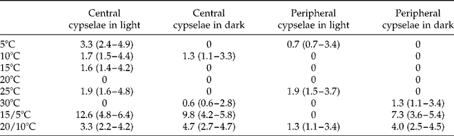
Central and peripheral cypselae did not display a significant difference in the germination response; they occasionally reached 12% and 7% germination respectively, at 15/5°C, but generally germinated to less than 5% (Table 1). The GLM model showed a significant improvement (P< 0.001) of germination at alternating regimes compared with constant temperatures, especially with cooler mean temperatures (15/5°C; P< 0.01). Overall, the presence of light produced a small but significant (P< 0.05) increase in germination of central cypselae. In contrast, peripheral cypselae germinated better in the dark (P< 0.001). In all germination tests conducted, FCR at the basal end of the cypsela was always observed to precede seed coat rupture. Seed viability was always above 90% for fresh matured cypselae in all of the tested conditions.
Mean final germination (%) of fresh matured peripheral and central cypselae incubated in light (8/16 h photo- and thermo-period) and in darkness for 100 d under different temperature regimes. Upper and lower 95% binomial confidence intervals are reported in parentheses

Effect of afterripening and stratification treatments on dormancy release
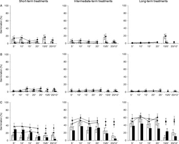
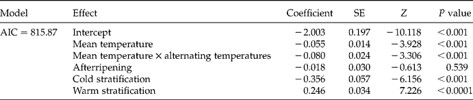
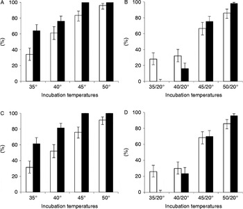
GLM analysis revealed that, both for central and peripheral cypselae, afterripening did not produce any significant improvement of germination, reaching 15.3, 15.6 and 17.3% for central cypselae and 1.2, 2.0 and 1.9% for peripheral cypselae respectively, after 4, 12 and 18 week-long treatments across the temperature treatments (Fig. 1A, Table 2). Cold stratification had a negative coefficient in the GLM model since it significantly inhibited (P< 0.001) germination of cypselae (Fig. 1B, Table 2). Cypsela germination reached 7.1, 3.1 and 3.2% for central cypselae and 2.0, 2.0 and 0.67% for peripheral cypselae after 4, 8 and 12 week-long treatments, respectively. In contrast, warm stratification treatment had a highly significant positive effect (P< 0.0001). Indeed, after warm stratification, cypsela germination reached 14.3, 46.6 and 57.6% respectively, after 3, 5 and 8 week-long treatments for central cypselae, whereas for peripheral morphs 29.0, 37.2 and 44.2% germination was obtained respectively, after the same time intervals (Fig. 1C, Table 2). Warm stratification led to a highly significant (P< 0.0001) promotion of fruit-coat rupture for both heteromorphs, with the FCR percentage significantly increasing (P< 0.0001) with the applied stratification time period. Incubation at higher temperatures for 5 d, regardless of the analysed heteromorph, produced a highly significant increase of FCR events and seed mortality with higher temperatures (P< 0.0001; Fig. 2).
Mean final germination (%) of central and peripheral cypselae (white bars and dark bars respectively) and mean final FCR (fruit coat rupture) (%) of central and peripheral cypselae (circles and triangles respectively). (A) Afterripened cypselae for short-, intermediate- and long-term treatments (4, 12, 18 weeks, respectively) and imbibed in light (8/16 h photoperiod) at constant and alternating temperatures. (B) Cold-stratified cypselae for short-, intermediate- and long-term treatments (4, 8, 12 weeks, respectively) and imbibed in light (8/16 h photoperiod) at constant and alternating temperatures. (C) Warm-stratified cypselae for short-, intermediate- and long-term treatments (3, 5, 8 weeks, respectively) and imbibed in light (8/16 h photoperiod) at constant and alternating temperatures. Error bars indicate upper and lower 95% binomial confidence intervals. Germination control data (no treatment) are provided in Table 1.

Generalized linear model fitted to germination data comparing mean temperature, alternating temperature contrasted with mean temperature, afterripening, cold stratification and warm stratification treatments. AIC, Akaike Information Criterion; SE, standard error; Z, Wald statistic

Mean final fruit-coat rupture (white bars) and seed mortality (dark bars) of central (A, B) and peripheral (C and D) cypselae incubated in continuous darkness, at constant (A, C) and alternating regimes (B, D), for 5 d (no germination was observed). Error bars indicate upper and lower 95% binomial confidence intervals.

Water uptake
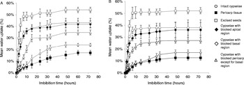
Imbibition of intact cypselae showed a significant (P< 0.001) mass increase after 1 h for both heteromorphs. After 48 h of imbibition the weight increase attained a stable plateau, while after 72 h it reached 43.0 and 38.0% for central and peripheral cypselae respectively (Fig. 3). Excised seeds showed a significantly (P< 0.0001) higher capability to take up water compared with intact cypselae, with increased water content of 54.1 and 52.1% respectively, in central and peripheral seeds (Fig. 3). Pericarp tissues retained a significantly (P< 0.0001) lower amount of water compared with intact cypselae, increasing their weight by 17.2 and 12.7% respectively, for pericarp derived from central and peripheral cypselae (Fig. 3). Blocking the apical region of pericarp surface resulted only in a slightly significant (P= 0.036 for central and P= 0.029 for peripheral cypselae) weight decrease after 72 h (reaching 41.7% for central and 36.0% for peripheral cypselae; Fig. 3). However, cypselae of both heteromorphs that were blocked in the basal region of the pericarp showed a highly significant (P< 0.0001) limited water uptake, achieving only 24.0 and 17.0% respectively, in central and peripheral cypselae (Fig. 3). On the other hand, blocking the entire pericarp surface except for basal region produced an increase in weight of 34.7 and 27.0% for central and peripheral cypselae respectively, which was significantly higher (P< 0.0001) than the cypselae with the blocked basal region, but at the same time significantly lower (P< 0.0001) than the intact cypselae (Fig. 3).
Mean water uptake during 72 h at 15°C (8/16 h photoperiod) after sowing central cypselae/seeds (A) and peripheral cypselae/seeds (B) with different parts of the pericarp blocked. Data are means of four samples of 25 seeds; error bars indicate standard deviation.

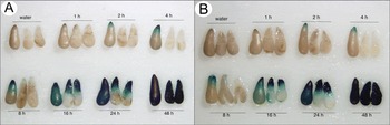
Excised seeds from both cypsela heteromorphs stained with methylene blue showed seed coat coloration after 1 h at the basal tip. This spread progressively towards the apical end (Fig. 4), showing no evident difference in the colouring between the two heteromorphs. Embryo coloration, as shown in longitudinally sectioned seeds, showed a considerable delay compared with seed coat coloration, as demonstrated in intact seeds. Methylene blue penetration into the seed coat was observed at the radicle end of seeds 8 h after onset of imbibition, while in the embryo after 16 h of imbibition, and it was complete after 48 h (Fig. 4).
Intact seeds (enveloped by the seed coat) and two seed halves, after manual seed extraction from central (left figure) and peripheral (right figure) cypselae that were stained in methylene blue solution (1%) for different time intervals, as indicated in the figure. For each sampling time point, one intact seed and two longitudinal seed halves are shown, the latter two demonstrate the stain penetration into the embryo.

Effect of pericarp on germination response
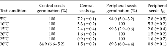
Regardless of the morphotype, excising seeds from fruit tissue resulted in a highly significant (P< 0.0001) improvement of germination percentage (Table 3). In particular at 5, 10, 15, 20 and 25°C cypsela germination always exceeded 90%. At 30°C seeds excised from central and peripheral morphs germinated to 84.9% and 89.3% respectively. Values of germination speed (t 50) indicated a faster response at the temperature range between 20 and 30°C (Table 3).
Mean final germination (%) and days to reach 50% of total germination (t 50) of excised seeds from central and peripheral cypselae incubated in light under different temperature regimes. For germination results upper and lower 95% binomial confidence intervals, and standard error for time to achieve 50% of the final germination level, are reported in parentheses

Scarification of the pericarp at the apical end for both cypsela heteromorphs did not produce any significant increase of germination with respect to intact cypselae. In contrast, after removal of a lateral portion of the pericarp, a highly significant improvement (P <0.0001) in cypsela germination was observed, up to 59% for central and 67% for peripheral cypselae. Scarification at the basal end induced only a moderately significant (P< 0.01) germination increase in both central and peripheral cypselae to 9 and 4% respectively.
Analysis of the masses of seed, pericarp tissue and whole cypsela in both heteromorphs revealed that the pericarp tissue is the main component of the total cypsela mass. Furthermore, ANOVA analyses showed that, even though the cypsela weight of the two heteromorphs was highly significantly different (P <0.001), the weight of seeds excised from central and peripheral morphs (1.10 ± 0.26 mg and 1.90 ± 0.46 mg respectively) was not significantly different. On the other hand, the pericarp mass of the two heteromorphs showed a highly significant (P <0.001) difference, 3.49 ± 0.41 mg and 8.53 ± 0.62 mg for central and peripheral cypselae respectively.
Anatomical characterization of cypsela pericarp
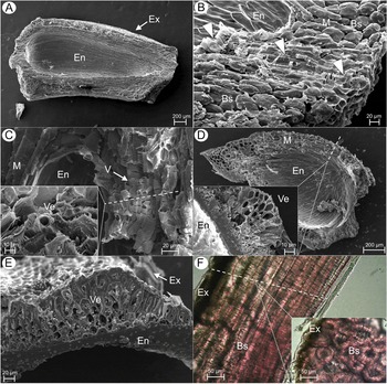
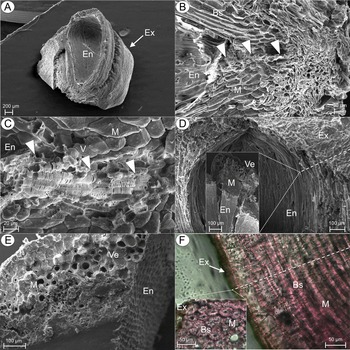
In both morphotypes, the exocarp consisted of flat epidermal cells with an indistinctly reticulate ornamentation, which presents glands irregularly spread in the intercostal gaps (exocarpic region between two longitudinal prominences). The inner endocarp surface of the two heteromorphs generally showed a similar structure (Figs 5A and 6A). In contrast, remarkable differences were observed in the mesocarp tissue thickness between the two morphotypes. The central cypselae mesocarp is formed, in the lateral portion, by 8–10 layers of flattened sclereid cells with a total thickness from 200 μm up to 550 μm (in the winged area), whereas in the basal part the mesocarp is 200–250 μm thick and it is mainly made up of isodiametric cells. Peripheral cypselae presented a broader mesocarp that consisted of 12–18 layers of flattened sclereid cells with a lateral thickness from 300 μm up to 1.8 mm (in the wing area), and 400–500 μm in the basal area, which is formed of isodiametric cells (Figs 5B and 6B). High-magnification images of the pericarp tissue showed the presence of xylem vessels with a lateral pseudoscalariform wall pitting (Carlquist, Reference Carlquist2001) (Figs 5C and 6C), which form a channel-like structure at the basal end, running from the endocarp through the external mesocarp (Figs 5B and 6B). At the apical end, bundles of vessel elements were found but they remained in the internal part of the mesocarp (Figs 5D and 6D). In the median portion of the cypsela pericarp, only tangential vessel elements were observed (Figs 5E and 6E). Transverse sections of the above-mentioned pericarp region confirmed that vessels remain in the mesocarpic tissue without entering into the seed cavity. Further anatomical characterization by light microscopy highlighted the presence of brachysclereid-lignified cells in the mesocarp area, more flattened at the internal portion (Figs 5F and 6F), while transverse sections showed the presence of lumen and pits in these brachysclereid-lignified cells.
Anatomical characterization of central cypselae of G. coronaria observed by scanning electron microscopy (A, B, C, D, E) and by light microscopy (F). (A) Micrograph of a longitudinal section of a central cypsela. (B) Magnification of the basal end of the pericarp. Arrowheads indicate xylem-vessel bundles running through the pericarp tissue into the seed cavity. (C) Detail of a longitudinal section of a vessel bundle; insert: transverse section of vessels. (D) Inside view of apical cypsela pericarp; insert: transverse section of apical portion of pericarp showing only tangential vessels in mesocarp. (E) Transversal section of pericarp median portion showing only tangential vessels in mesocarp. (F) Longitudinal section of pericarp stained with phloroglucinol, showing lignified brachysclereid cells in the mesocarp; insert: detail of transverse section stained with phloroglucinol, showing lignified brachysclereid cells in the mesocarp. Abbreviations: Ex, exocarp; M, mesocarp; Bs, brachysclereid cells; En, endocarp; V, vessels; Ve, vessel element.

Anatomical characterization of peripheral cypselae of G. coronaria observed by scanning electron microscopy (A, B, C, D, E) and by light microscopy (F). (A) Micrograph of a longitudinal section of a peripheral cypsela. (B) Magnification of the basal end of the pericarp. Arrowheads indicate xylem-vessel bundles running through the pericarp tissue into the seed cavity. (C) Detail of a longitudinal section of a vessel bundle (arrowheads). (D) Inside view of apical cypsela pericarp from a longitudinal section; insert: transverse section of apical portion of pericarp showing only tangential vessels in mesocarp. (E) Transversal section of pericarp median portion showing only tangential vessels in mesocarp. (F) Longitudinal section of pericarp stained with phloroglucinol, showing lignified brachysclereid cells in the mesocarp; insert: detail of transverse section stained with phloroglucinol, showing lignified brachysclereid cells in the mesocarp. Abbreviations: Ex, exocarp; M, mesocarp; Bs, brachysclereid cells; En, endocarp; V, vessels; Ve, vessel element.

Discussion
Freshly matured cypselae of G. coronaria with high viability demonstrated very limited germination when sown at a range of temperature conditions. Obviously, some form of dormancy is present. Even though heterocarpy in Asteraceae has often been associated with differences in germination behaviour (McEvoy, Reference McEvoy1984; Brändel, Reference Brändel2007), in this study the germination response of the two morphotypes was not significantly different under the tested conditions. Afterripening and cold stratification treatments did not considerably improve germination, whereas warm-stratified cypselae displayed a marked improvement, with a higher response after longer treatments. A similar trend was observed for fruit-coat rupture frequency, which had an even higher value at supra-optimal stratification temperatures of 35°C or higher, where, conversely, extensive cypsela mortality was observed. Regardless of the cypsela morphotypes, progressive scarification of the pericarp tissue resulted in germination promotion that is likely to be a consequence of the loss of mechanical constraint against radicle protrusion exerted by the pericarp. In this perspective, warm stratification seems to act as a scarification agent, loosening the pericarp constriction and allowing radicle emergence after incubation at cooler regimes.
Anatomical analyses of central and peripheral cypselae revealed the absence in seed covers of a palisade layer(s) of macrosclereid cells, which is a characteristic trait of physical dormancy (sensu Baskin and Baskin, Reference Baskin and Baskin2004), whereas a point of water entry was found, located at the basal end of the cypselae. Both morphotypes exhibited a considerable and rapid mass increase after sowing in water, with the latter entering mainly through the basal tip, as shown in Fig. 3. The staining experiments with methylene blue solution confirmed the direction of water penetration, and showed that the seed coat delays embryo imbibition, which is complete 48 h after sowing seeds in water.
Further examination revealed that the seed mass is similar in the two heteromorphs and the difference in fruit weight is mainly due to the pericarp tissue. In fact, anatomical analyses revealed a similar composition of mesocarp, which is thicker in peripheral cypselae than in the central ones. Moreover both morphotypes share the presence of a channel-like structure composed of a xylem-vessel system, which connects the seed within the cypsela with the external environment. The channel-like structure runs from the basal end, where vessel bundles allow water entrance throughout the pericarp towards the seed part where the radicle is located. In addition, vessel elements located in the apical and lateral portions and brachysclereid cells, spread across the mesocarp, allow a further water supply through lateral parts of the pericarp. This system forms a gateway through the pericarp and allows a similar degree of seed imbibition for both cypselae types. Consequently, a similar germination response is observed under laboratory conditions. Nevertheless, unlimited water availability and full imbibition of cypselae are very unusual in the natural environment of this species (Doussi and Thanos, Reference Doussi and Thanos2002; Aguado et al., Reference Aguado, Martínez-Sánchez, Reig-Arminana, García-Breijo, Franco and Vicente2011; Carta et al., Reference Carta, Bedini, Müller and Probert2013). Under natural soil conditions, with limited and unstable water soil saturation, central morphs bearing a thinner pericarp may germinate earlier, while viable peripheral ungerminated cypselae are more prone to be seasonally incorporated in the soil seed bank (Bastida et al., Reference Bastida, Gonzáles-Andújar, Monteagudo and Menéndez2010). Rapid loss of viability at 35°C or higher under non-limiting water availability suggests that exclusion of water in the summer is essential for survival of cypselae in the soil. Therefore, the hard pericarp that restricts the water passage represents an essential tool in controlling seed germination and survival upon occasional rains, thus providing an ecological advantage.
We conclude that in G. coronaria the pericarp plays a decisive role in controlling seed germination by exerting a mechanical constraint against radicle protrusion. Warm stratification, mimicking the environmental conditions experienced by cypselae at the end of summer, overcomes mechanical (physiological) dormancy by weakening the hard pericarp scaffold, resulting in a higher number of fruit-coat rupture events and, as a consequence, in higher germinability. Warm-stratified cypselae respond to a broad range of germination temperatures, although the optimum seems to be 10°C. Even though physical dormancy was not observed, water uptake through the lateral part, as well as the apical end, of the pericarp was very limited. A point of water entry was found located only at the basal end of the pericarp, resulting in longer times for water to reach the apical end of the seed within the intact cypsela and thus contributing to temporal spread in germination.
Supplementary material
To view supplementary material for this article, please visit http://dx.doi.org/10.1017/S0960258515000148
Acknowledgements
We are grateful to Dr Girolamo Fichera and Professor Antonia Cristaudo (Department of Biological, Geological and Environmental Science, University of Catania) respectively, for scanning electron microscopy analyses and for support in light microscopy. Moreover, we would like to thank Professor Cristina Salmeri (University of Palermo) for her advice on anatomical features.
Financial support
This work was financed with founding provided by Department of Biological, Geological and Environmental Science, University of Catania and supported by the Millennium Seed Bank Project, Kew.
Conflicts of interest
None.