Perioperative implantation of stents is technically feasible, and has been shown to date to have a favourable outcome in a limited number of patients.Reference Rosales, Lock, Perry and Geggel1–Reference Zahn, Dobrolet, Nykanen, Ojito, Hannan and Burke3 In infants with functionally univentricular physiology, stenoses in the pulmonary arterial system may be encountered at several locations, such as the site of ductal insertion, at the pulmonary arterial bifurcation, from external compression of the pulmonary arteries by the neo-aorta after a Damus type of reconstruction of the aortic arch, or at the site of anastomosis of the superior caval vein to pulmonary artery. Such stenoses may compromise the pulmonary circulation in the immediate postoperative period, with increasing cyanosis and superior caval venous congestion, the so-called superior caval vein syndrome, as important clinical consequences.
Concomitant with improved surgical outcomes, there has been remarkable progress in techniques of interventional catheterization, which potentially allow some of these stenoses to be successfully treated. Proximal pulmonary arterial lesions, in particular, have been shown to be highly compliant, and therefore amenable to balloon angioplasty. These lesions also have a higher degree of elastic recoil, which may necessitate implantation of stents as the primary therapeutic measure.Reference Bergersen, Gavreau, Lock and Jenkins4, Reference Bergersen and Lock5. While this approach has been shown to be efficacious, with a low rate of complications, there is a paucity of data on its efficacy in the perioperative and immediate postoperative period, particularly in critically ill infants.Reference Rosales, Lock, Perry and Geggel1–Reference Zahn, Dobrolet, Nykanen, Ojito, Hannan and Burke3 We report on our experience with our implantation of stents in this population of patients.
Patients and methods
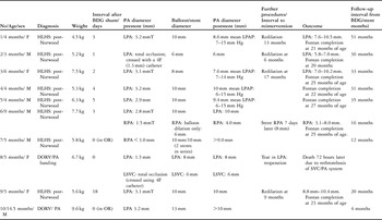
Our comments are based on experience with 10 consecutive patients, 6 male, with functionally univentricular physiology in whom we implanted stents for relief of clinically relevant pulmonary arterial stenoses. The patients were treated at multiple institutions, but always by one of the authors. The majority, 8 in all, had hypoplastic left heart syndrome, and had undergone a standard Norwood type of palliation, with placement of shunts from the right ventricle to the pulmonary arteries in the neonatal period. The remaining patients had other variants of functionally univentricular physiology (see Table 1). They ranged in age from 3 months to 14.5 months at the time of creation of a shunt from the superior caval vein to pulmonary arteries, and weighted from 4.5 to 9.6 kilograms. In six of them, preoperative cardiac catheterization had identified significant stenoses in one or other pulmonary artery, and in both pulmonary arteries in two of them. In 4 infants, the stenoses had been dilated percutaneously using standard Tyshak II balloons (NuMed, Canada), the ratios of diameters of balloon to stenosis varying between 2.4 and 3.0. In the two remaining patients, we decided to implant stents in the operating room, at the time of surgery. In the 4 undergoing balloon dilation, the previously stenotic areas were augmented with a patch at the time of constructions of the shunt from the superior caval vein to the pulmonary arteries.

Abbreviations used in Table: PA = pulmonary artery; HLHS = hypoplastic left heart syndrome; SCV = superior caval vein; LSCV = left superior caval vein; DORV/PA = double outlet right ventricle with pulmonary atresia; LPA = left pulmonary artery; LPAP = mean left pulmonary pressure; RPA = right pulmonary artery. OR = operating room. The symbol T refers to vessels that were dilated during preoperative cardiac catheterization, and where patch angioplasty was performed at the time of the superior caval vein to pulmonary artery shunt.
The remaining 8 patients all presented in the immediate postoperative period with persistent or increasing hypoxaemia, with saturations of arterial oxygen persistently less than 70%, and clinical evidence of the superior caval vein syndrome, mean pressures in the superior caval vein ranging from 18 to 25 millimetres of mercury. In this clinical setting, cardiac catheterization was undertaken at between 1 and 18 days, with a median of 3 days, after the surgical procedure. All patients were mechanically ventilated, and receiving appropriate inotropic and vasodilator therapy.
Technique of catheterization and implantation of stents
Via a percutaneous puncture of the internal jugular vein, a 4 French endhole catheter was advanced to the junction of the superior caval vein with the pulmonary artery. Angiography at this site was performed to demonstrate the surgical anastomosis and the distal pulmonary arterial system. Newly created anastomotic sites and surgical suture lines were crossed using an 0.035 inch floppy guidewire (Terumo Corporation Japan), followed by the endhole catheter. In one infant with complete occlusion of the left pulmonary artery, and another with occlusion of the left superior caval vein, this combination was used successfully to cross the thrombosed segments. We measured the diameters of the right and left pulmonary arteries, and of the stenoses, offline from magnified images of the biplane angiograms, using the known diameter of the catheter as a reference. We chose balloons so as to achieve a final diameter of the treated segment which was at least equal to, or slightly greater than, the maximum measured diameter of either pulmonary artery. Where possible, the mean pulmonary arterial pressure was recorded distal to the stenosis, both before and following placement of the stent.
Results
In 2 patients, we placed one or two stents in series across the stenotic segment in the operating room, and dilated them to between 10 and 13 millimetres. In the infant undergoing placement of a stent in the right pulmonary artery, the stent extended to the suture line between the pulmonary artery and the superior caval vein. In both cases, an excellent result could be documented directly by inspection.
In the remaining 8 infants, angiograms taken prior to the procedure demonstrated a minimum diameter of the stenotic segments ranging from 1.3 millimetres, the nominal diameter of the 4 French catheter, in the infant with total occlusion of the left pulmonary artery, to 3.2 millimetres. The stenoses in the left pulmonary artery were located at the original ductal insertion in 2 cases, at the bifurcation of the pulmonary trunk, immediately posterior to the neo-aorta, in 3, or at the anastomosis between the superior caval vein and the pulmonary arteries in the remaining 3. The stenosis of the right pulmonary artery in 1 infant was at the anastomosis.
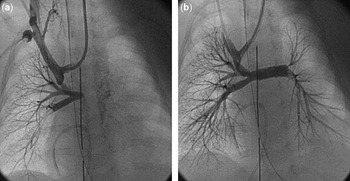
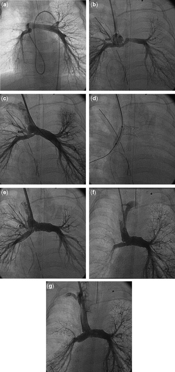
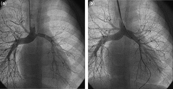
We implanted one or more stents, 8 in all, in the left pulmonary artery in 7 patients (Figs 1–3). In 1 patient with bilateral superior caval veins who had undergone anastomoses of both superior caval veins to the pulmonary arteries, and in whom angiography demonstrated occlusion of the left superior caval vein, in addition to stenosis and thrombosis of the left pulmonary arterial system, we implanted a stent in the left superior caval vein (Fig. 4). In the sixth patient, who had bilateral stenoses, a stent was implanted in the right pulmonary artery at the site of its anastomosis with the superior caval vein (Fig. 2). At the first procedure, a premounted 10 millimetre Palmaz-Genesis stent (Palmaz-Genesis stents on OptaPro balloons, Cordis Europe, Roden, the Netherlands) was implanted in the left pulmonary artery, reaching the suture line, while the right pulmonary artery was only dilated with a 6 millimetre diameter balloon, as it was felt that placing two adjacent stents across the freshly created anastomosis might carry a risk of tearing the anastomosis. The infant failed to show symptomatic improvement after the first procedure, so a second stent was implanted successfully 7 days later in the proximal right pulmonary artery. In this series of patients, 7 of the stents crossed a newly created surgical anastomotic site, including one stent which had been implanted in the operating room under direct vision. We used stents either premounted on 6, 8 or 10millimetre diameter balloons (Palmaz-Genesis stents on OptaPro balloons, Cordis Europe, Roden, the Netherlands), or hand-mounted (Palmaz PG1910P or P188 stents on Cordis Powerflex balloons, Cordis Europe, Roden, the Netherlands). The final diameters achieved subsequent to treatment are shown in the Table 1.
Superior caval venous angiogram (a) in the frontal projection in our second patient demonstrating occlusion of the left pulmonary artery. (b) shows the result of implantation of the stent.

Pulmonary angiogram (a) prior to surgery in our sixth patient, demonstrating a stenosis at the bifurcation. This was dilated, and augmented by a pericardial patch during subsequent surgery. (b) shows the superior caval venous angiogram 3 days post-surgery, demonstrating bilateral pulmonary arterial stenoses. (c) shows the result of implantation of a stent in the left pulmonary artery. A severe stenosis at the origin of the right pulmonary artery is unmasked. (d) shows balloon dilation of the right pulmonary artery with a balloon of 6 millimetres diameter, with (e) showing the acute result following dilation. Repeat catheterization 7 days later (f) demonstrates recurrence of stenosis at the origin of the right pulmonary artery. The final result is shown in (g) after implantation of a stent in the right pulmonary artery.

Left pulmonary arterial stenosis (a) in the fifth patient, with the acute result (b) following implantation of a stent.

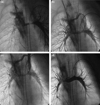
Thrombotic obstruction (a) of the left pulmonary artery in our 8th patient. There was complete occlusion of the left superior caval vein (b) in the same patient, who also has a patent bridging vein. Following balloon dilation of the left superior caval vein (c), there is residual obstruction at the junction of the caval vein with the pulmonary artery. Implantation of a stent in the left superior caval vein at its junction with the left pulmonary artery (d) produced a seemingly good result, but the patient had loss of blood into the pericardium. At reoperation, a tear was identified on the undersurface of the left pulmonary artery at the location of the stent.

Complications
We created a small tear in the left pulmonary artery in our 8th patient (Fig. 4), having placed multiple stents to recanalise an occluded left superior caval vein and a stenotic left pulmonary artery. We returned this infant directly to the operating room, where the stents in the left pulmonary artery were removed, and the vessel repaired. The infant returned to the intensive care unit in a stable condition, but over the next 24 hours redeveloped progressive cyanosis and superior caval vein syndrome, and unfortunately died 72 hours later. At postmortem examination, multiple thrombus was found in the pulmonary arterial system. We subsequently established a family history of a hitherto undiagnosed coagulopathy, which was probably responsible for occlusion of the anastomosis between the left-sided caval vein and the pulmonary arteries in the immediate postoperative period. All other infants, apart from the 6th patient in whom we implanted a second stent as described above, showed rapid clinical improvement, with resolution of the superior caval vein syndrome, and a sustained improvement in systemic saturations of oxygen to levels of between 76 and 85%. All of them survived their surgical or catheterization procedures, and were discharged from hospital in stable clinical condition.
Follow-up
All 9 surviving patients have remained under routine clinical follow-up. The median duration of follow-up after implantation of the stents is 31 months, with a range from 4 to 51 months. During this period, we have redilated the stents in the left pulmonary arteries of 4 patients during routine follow-up catheterizations at intervals ranging from 6 to 17 months after initial implantation. We achieved a limited increase in the diameter of the treated segments (see Table 1). All patients have shown normal neurologic development, comparable to their peers.
The Fontan circulation has successfully been completed in 7 of the survivors by placement of an extracardiac conduit. To date, none of the implanted stents has been surgically removed, albeit that it proved necessary to incise 2 stents crossing the anastomosis of the superior caval vein to the pulmonary arteries so as to create the necessary space to attach a 16 millimetre extracardiac Goretex conduit from the inferior caval vein to the pulmonary artery. This was accomplished easily in both patients.
Discussion
Transcatheter intervention in the perioperative period, particularly in young infants, has been perceived to carry a high risk.Reference Zahn, Dobrolet, Nykanen, Ojito, Hannan and Burke3 As a result, there are few studies which address the efficacy of these procedures in this setting. There is, however, an evolving tendency toward performing hybrid procedures, in which surgical repair and transcatheter dilation, implantation of stents, or placement of occlusion devices may safely be combined whilst the patient is supported by cardiopulmonary bypass. Such an approach is feasible where stenoses have been well defined preoperatively, and are relatively easily accessible to the surgeon. For stenoses in the pulmonary arterial system, which are only recognised clinically in the early postoperative period, balloon dilation alone appears to be less favourable, with reported acute rates of success of approximately 50%, and a procedure-related mortality approaching 20%.Reference Rosales, Lock, Perry and Geggel1 The deaths from balloon dilation have, in the majority, resulted from rupture of vessels.Reference Rosales, Lock, Perry and Geggel1 In contrast, more favourable results have been reported with primary implantation of stents in this clinical setting.Reference Rosales, Lock, Perry and Geggel1–Reference Zahn, Dobrolet, Nykanen, Ojito, Hannan and Burke3 In two recent publications, not dealing exclusively with perioperative pulmonary arterial stenoses, it was shown that proximal pulmonary arterial lesions tend to be more compliant, and therefore amenable to balloon angioplasty.Reference Bergersen, Gavreau, Lock and Jenkins4, Reference Bergersen and Lock5 These lesions also demonstrate more elastic recoil, and hence implantation of stents may be optimal.Reference Bergersen, Gavreau, Lock and Jenkins4, Reference Bergersen and Lock5 Given the improvements in design, and the availability of premounted models with a lower profile, the currently available stents demonstrate more flexibility, less foreshortening, increased ease of implantation, and offer the feasibility for redilation.Reference Bergersen, Gavreau, Lock and Jenkins4, Reference Bergersen and Lock5 All of these characteristics are of practical importance when considering implantation in infants. An added observation from or study is that, when required, the stent may safely be incised surgically, without risk of tearing or perforation of the pulmonary artery, at a subsequent reoperation.
Surprisingly, when stents are implanted, rupture of the vessel has rarely been reported, even though in the majority of patients the ratio of the balloon to the stenosis when stents were implanted was much higher than for balloon angioplasty alone. In some instances, ratios of 5.0 have been reached during implantation, without untoward effects.Reference Rosales, Lock, Perry and Geggel1 The reasons for this observation are unclear. In our own series, we created a tear in the left pulmonary artery in 1 patient remote from any surgical suture site. In this infant, although the stenosis measured only 1.3 millimetres, this was due to thrombosis in the vessel, the distal left pulmonary artery measuring between 7 and 8 millimetres in diameter. We deployed an 8 millimetre stent. As shown in other studies, implantation of stents across a freshly repaired surgical site was also a safe procedure.
One of the potentially worrying aspects of implanting stents at a young age is that none of the patients thus treated will have completed their somatic growth. The final diameters of pulmonary arteries achieved at implantation, and following subsequent redilation, cannot be considered to be appropriate for adult life. Smaller stents also have only a limited capacity to increase further in diameter with serial redilation, and it is unavoidable that a substantial majority of these patients will require surgical removal of the stent in later life, with patch enlargement of the vessel, with or without implantation of a larger stent. Careful clinical follow-up, and serial catheterization, therefore, is mandatory. This does not detract from the potentially life-saving role of implanting stents in the acutely ill infant, without recourse to further surgery.
Our limited experience, therefore, confirms the beneficial clinical effects of implanting stents in the peri- and postoperative treatment of pulmonary arterial stenoses in infants. Successful accomplishment of such procedures in infancy requires careful and close collaboration between cardiologists, surgeons and intensive care staff to ensure that the infant is kept haemodynamically stable during transport to and from the catheterization room, and during the procedure. Skilled cardiac surgical standby, with facilities for immediate reoperation in the event of unforeseen complications, are mandatory.