Introduction
Obstructive lung diseases (OLDs), encompassing asthma, chronic obstructive pulmonary disease (COPD), bronchiectasis and chronic bronchitis, represent a significant global health challenge characterised by airflow limitation and chronic inflammation. These conditions manifest through symptoms such as dyspnea, wheezing and persistent cough, driven by airway narrowing from smooth muscle contraction, mucosal inflammation and structural remodelingReference Kim and Criner1–Reference MacNee3 While sharing symptomatic overlap, OLDs differ markedly in their epidemiology, progression and therapeutic needs. Asthma often emerges in younger populations with reversible airway obstruction, whereas COPD, typically linked to smoking or environmental exposures, progresses irreversibly, predominantly affecting older adults.Reference Sarkar, Bhardwaz, Madabhavi and Modi4, Reference Burkhardt and Pankow5 Globally, COPD affects an estimated 65 million individuals and ranked as the third leading cause of death in 2019, claiming 3.23 million lives.Reference Lakiang, Nair, Ramaswamy and Singhal6, 7 In India, COPD prevalence reaches 19.4 per cent, chronic bronchitis 5 per cent and asthma varies widely from 2 per cent to 23 per cent, reflecting regional environmental and genetic influences.Reference Sharma, Kumar and Chandel8, Reference Kumar, Roy, Subitha and Sahu9
The socioeconomic burden of OLDs is substantial, akin to a 0.11 per cent annual tax on global gross domestic product (GDP), driven by healthcare costs, reduced workforce productivity and diminished quality of life.Reference Chen, Kuhn, Prettner, Yu, Yang and Bärnighausen10 In India, the average annual cost of COPD management is 29,885 ± 11,995.33 rupees (Rs), straining resource-limited settings.7 Inhalational therapies, particularly fixed-dose combinations (FDCs) of corticosteroids and bronchodilators, are cornerstone treatments, enhancing adherence and symptom control by targeting inflammation and bronchoconstriction simultaneously.Reference Cazzola, Hanania, Page and Matera11, Reference Ronchetti, Ayroldi, Ricci, Gentili, Migliorati and Riccardi12 These agents, delivered via metered-dose inhalers (MDIs) or dry powder inhalers (DPIs), offer rapid relief and long-term disease stabilisation, yet their prolonged use raises concerns about local adverse effects, particularly on the larynx and voice.Reference Dos Santos, Echeveste and DCGM13–Reference Menvielle, Luce, Goldberg, Bugel and Smoking16
Laryngeal involvement in OLD stems from both the disease process—respiratory decline and coughing—and medication side effects. Inhaled corticosteroids, while reducing airway inflammation, can deposit in the oropharynx and larynx, causing mucosal erythema, oedema, vocal fold thickening, granulations, leukoplakia and candidiasis.Reference Saha, Saha and Chatterjee17–Reference Abumossalam, Ahmed, Ibrahim, Algreisy and Al-Shenqiti19 Dysphonia, a common complaint, affects 5–58 per cent of patients on inhaled corticosteroids, varying with dosage, durat and delivery method.Reference Saha, Saha and Chatterjee17 Such vocal changes not only impair communication but also signal underlying structural damage, necessitating greater awareness and preventive measures to curb severe laryngeal dysfunction.Reference Abumossalam, Ahmed, Ibrahim, Algreisy and Al-Shenqiti19
Acoustic voice analysis provides an objective tool to quantify these changes. PRAAT®, a freely available software, measures parameters like jitter (pitch stability), shimmer (intensity variation), HNR (voice clarity), mean pitch and MPT (phonation duration), offering precision comparable to advanced systems like Computerised Speech Labs.Reference Saeed, Riad, Osman, Khattab and Mohammed20 Despite the widespread use of inhalational therapies, few studies have longitudinally tracked their laryngeal effects in OLD patients from treatment onset, particularly with combined formulations. This study aims to fill this gap by assessing structural and acoustic laryngeal changes in OLD patients over 12 months, exploring how different medications and delivery systems influence outcomes and elucidating the underlying pathophysiology to guide clinical practice.
Methods
Study design
This prospective observational study, approved by the Institutional Ethics Committee, was conducted collaboratively by the Departments of ENT and Pulmonary Medicine at a tertiary care hospital in South India from January 2022 to January 2024. The design allowed for real-time monitoring of laryngeal and vocal changes following the initiation of inhalational therapy.
Participants and procedures
Eligible participants were adults over 18 years, newly diagnosed with OLD (bronchial asthma, COPD or bronchiectasis) via standard pulmonary function tests and clinical criteria and starting combination inhalational therapy. The formulations included MDI formoterol fumarate and budesonide, MDI beclomethasone and levosalbutamol, MDI fluticasone propionate and formoterol, DPI formoterol fumarate and budesonide and DPI fluticasone propionate and formoterol. Isolating individual agents (e.g., corticosteroids vs. bronchodilators) was impractical due to ethical concerns of withholding standard care, limited device availability and the predominance of FDCs in this setting. A sample size of 112 was determined using a 5.4 per cent prevalence of laryngeal nodules from prior research,Reference Abumossalam, Ahmed, Ibrahim, Algreisy and Al-Shenqiti19 with alpha and beta errors set at 5 per cent and 20 per cent, respectively, and convenient sampling applied due to logistical constraints.
Exclusion criteria were stringent to minimise confounders: patients with baseline laryngeal symptoms, endoscopic abnormalities, acute severe asthma, occupational asthma, inhalational injury, trauma, surgery, gastroesophageal reflux disease (GERD), laryngopharyngeal reflux (LPR), recent intubation, pregnancy, lactation, uncontrolled diabetes or hypertension, immunocompromised states or anticancer therapy were excluded.
Methodology
At baseline, participants underwent a comprehensive evaluation: detailed history (e.g., smoking, alcohol use and symptom onset), clinical examination and direct video laryngoscopy using a 70-degree rigid endoscope to assess laryngeal oedema, hyperaemia, vocal fold mobility, nodules or polyps. Those with structural abnormalities were excluded. Voice analysis was performed using PRAAT® (version 6.2.14), recording mean pitch (Hz), jitter (per cent), shimmer (dB), HNR (dB) and MPT (seconds) in a quiet room with standardised microphone placement. Patients were instructed to sustain the vowel /a/ at a comfortable pitch and loudness for MPT measurement.
Interventions involved standard inhalational therapy prescribed by a pulmonologist, with adherence monitored via self-report and prescription refill checks. Follow-ups at 3, 6 and 12 months included repeat laryngoscopy and acoustic analysis, alongside assessment of throat-related complaints (e.g., dysphonia, harsh cough phonasthenia). Only patients adherent for at least 12 months were included in the final analysis.
Data collection and analysis
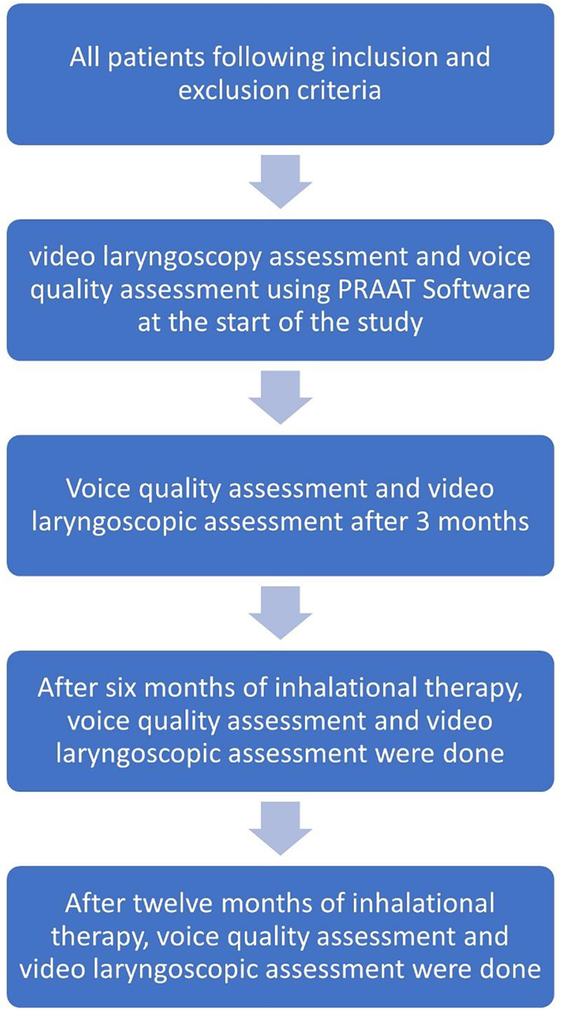
Data were collected via a clinical platform, entered into Microsoft Excel, and analysed using IBM SPSS version 23. Descriptive statistics summarised demographics (e.g., age, sex) and clinical variables (e.g., diagnosis, laryngeal findings). Acoustic parameters were reported as means ± standard deviations, compared using unpaired t-tests (e.g., smokers vs. non-smokers) and repeated-measures analysis of variance (RMANOVA) for longitudinal changes, with p less than 0.05 denoting significance. Confidentiality was ensured, and informed oral and written consent was obtained. The study methodology is illustrated in Figure 1.
Methodology of the study.

Results
Baseline characteristics
Amongst the 112 participants, the mean age was 43.05 plus or minus 12.707 years (range: 19–67), with 54.5 per cent female (n = 61) and 45.5 per cent male (n = 51). Urban residents comprised 54.5 per cent (n = 61), reflecting potential pollution exposure, while 45.5 per cent (n = 51) were rural. Smoking history was reported by 30.4 per cent (n = 34), and 64.3 per cent (n = 72) consumed alcohol. Diagnoses included COPD (45.5 per cent, n = 51), bronchial asthma (34.8 per cent, n = 39) and bronchiectasis (19.6 per cent, n = 22). Treatment distribution was: MDI formoterol fumarate with budesonide (47.3 per cent, n = 53), MDI beclomethasone with levosalbutamol (19.6 per cent, n = 22), MDI fluticasone propionate with formoterol (8 per cent, n = 9), DPI formoterol fumarate with budesonide (11.6 per cent, n = 13) and DPI fluticasone propionate with formoterol (6.2 per cent, n = 7).
Structural changes
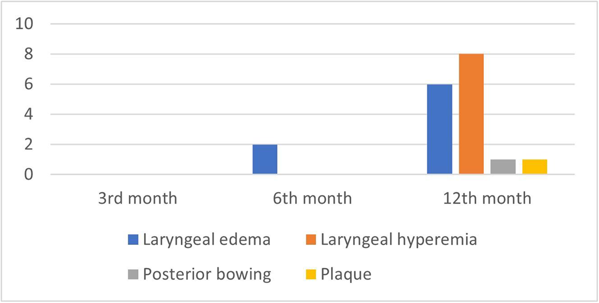
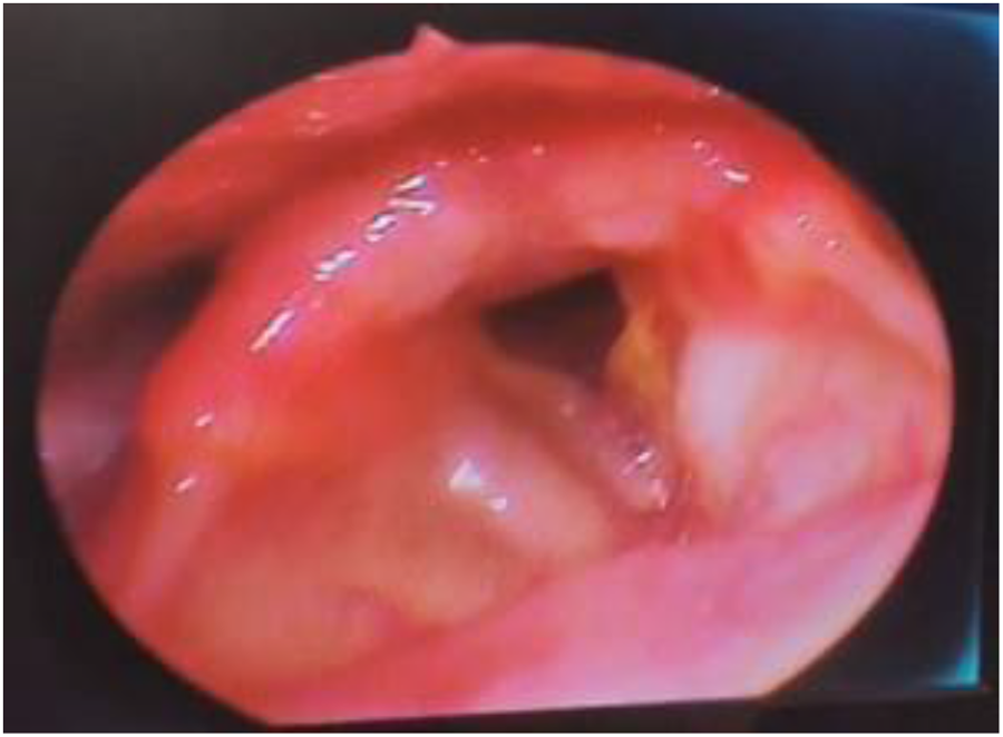
No laryngeal abnormalities were observed at baseline. By 6 months, 1.8 per cent (n = 2) developed laryngeal oedema. At 12 months, oedema increased to 5.3 per cent (n = 6), hyperaemia to 7.1 per cent (n = 8), with one case each of vocal fold plaques and posterior bowing (Figures 2 and 3).
Distribution of patients based on development of structural changes.

Development of laryngeal oedema at 6th and 12th month follow-up.

Voice acoustic analysis
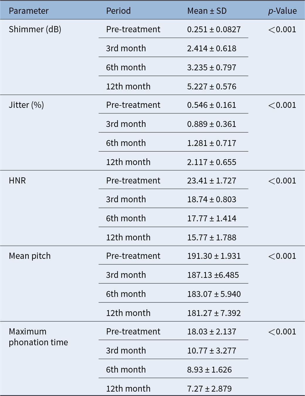
Significant changes were observed over time (p < 0.001). Shimmer increased progressively from 0.251 ± 0.083 dB (baseline) to 2.414 ± 0.618 dB (3 months), 3.235 ± 0.797 dB (6 months) and 5.227 ± 0.576 dB (12 months). Similarly, jitter rose from 0.546 ± 0.161 per cent to 0.889 ± 0.361 per cent (3 months), 1.281 ± 0.717 per cent (6 months) and 2.117 ± 0.655 per cent (12 months). HNR declined from 23.41 ± 1.727 dB (baseline) to 18.74 ± 0.803 dB (3 months), 17.77 ± 1.414 dB (6 months) and 15.77 ± 1.788 dB (12 months). Mean pitch showed a gradual reduction from 191.30 ± 1.931 Hz to 181.27 ± 7.392 Hz over 12 months (p = 0.001). MPT decreased markedly from 18.03 ± 2.137 s at baseline to 7.27 ± 2.879 s at 12 months (Table 1).
Voice parameters during various periods of treatment (n = 112)

Inhaler type comparison
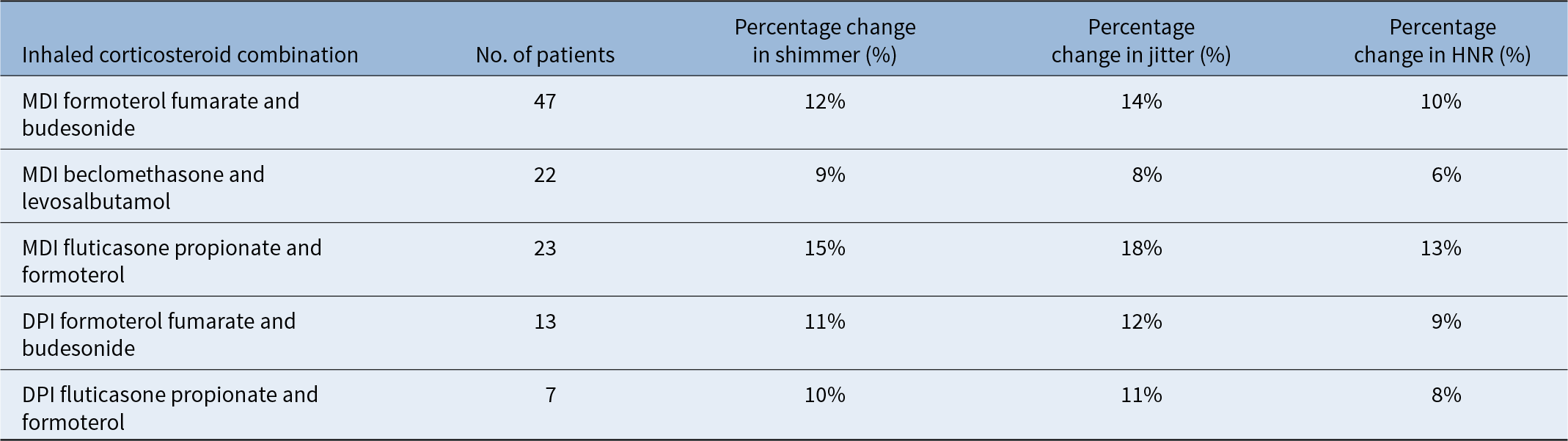
MDIs showed greater acoustic impact than DPIs. MDI fluticasone propionate with formoterol had a 15 per cent shimmer increase and 18 per cent jitter increase, versus 8 per cent HNR reduction with DPI fluticasone propionate with formoterol. MDI beclomethasone with levosalbutamol showed the least change (9 per cent shimmer, 8 per cent jitter) (Table 2).
Percentage changes in the acoustic parameters (shimmer, jitter and HNR) for different inhaled corticosteroid combinations

Smokers vs. non-smokers
Smokers (30.4 per cent) exhibited more severe changes: 50 per cent shimmer increase, 45 per cent jitter increase and 40 per cent HNR reduction, versus 40 per cent, 35 per cent and 30 per cent in non-smokers (69.6 per cent). At 12 months, smokers had higher oedema (3.5 per cent vs. 1.7 per cent), hyperaemia (5.3 per cent vs. 0 per cent) and unique findings (0.8 per cent plaques, 0.8 per cent bowing) (Table 3).
Acoustic and structural changes in smokers vs. non-smokers

Discussion
OLDs impose a dual burden of respiratory compromise and systemic impact, managed effectively with inhalational therapies. However, their long-term effects on laryngeal health and voice quality remain underexplored. This study provides robust evidence of progressive structural and acoustic changes in OLD patients over 12 months, offering insights into medication effects, delivery systems and underlying mechanisms.
The mean age of 43.05 years reflects a relatively young cohort, consistent with Saeed et al.Reference Saeed, Riad, Osman, Khattab and Mohammed20 for asthma but younger than typical COPD populations.Reference Holm, Plaufcan, Ford, Sandhaus, Strand and Strange21, Reference Divo, Marin, Casanova, Cabrera, Pinto-Plata and Marin-Oto22 This may stem from early-onset OLD in India due to smoking (30.4 per cent) or pollution (54.5 per cent urban).Reference Neptune23 The slight female predominance (54.5 per cent) aligns with findings by Han et al.,Reference Han, Postma, Mannino, Giardino, Buist and Curtis24 possibly linked to indoor air pollution or hormonal factors, contrasting male-dominated COPD cohorts.Reference Chapman, Tashkin and Pye25 Studies by Laniado-LaborínReference Laniado-Laborín26 and Song et al.Reference Song, Chen and Liu27 support the association between smoking and COPD development because of its damaging effects on the defense mechanisms and structure of the lung. Alcohol use (64.3 per cent) may exacerbate inflammation, though its role remains debated.Reference Yeboah-Kordieh, Bene-Alhasan, Mensah, Patel and Malik28–Reference Barnes30
Laryngeal changes emerged gradually, with oedema (5.3 per cent) and hyperaemia (7.1 per cent) by 12 months, less frequent than reported in Abumossalam et al.Reference Abumossalam, Ahmed, Ibrahim, Algreisy and Al-Shenqiti19 Acoustic deterioration was marked: shimmer rose to 5.227 dB, jitter to 2.117 per cent, HNR fell to 15.77 dB, pitch to 181.27 Hz and MPT to 7.27 s (all p < 0.001), paralleling findings by BarnesReference Barnes30 and Lavy et al.Reference Lavy, Wood, Rubin and Harries31 These reflect altered vocal fold dynamics and reduced phonatory capacity.
Variations across inhalers suggest differential laryngeal impacts. MDI fluticasone propionate with formoterol showed the greatest vocal deterioration (15 per cent shimmer, 18 per cent jitter, 13 per cent HNR reduction), while MDI beclomethasone with levosalbutamol was least affected (9 per cent shimmer, 8 per cent jitter). DPI formulations (e.g., 8 per cent HNR reduction with fluticasone propionate and formoterol) exhibited milder effects. No single inhaler is universally superior—efficacy in OLD control must be balanced against laryngeal risks. Fluticasone’s potency may amplify local effects, whereas beclomethasone’s milder profile could benefit voice preservation.Reference Saeed, Riad, Osman, Khattab and Mohammed20 Clinicians might prioritise DPIs or lower-impact MDIs for patients prone to dysphonia, though larger studies are needed to confirm clinical significance. Patient-specific factors, such as occupation (e.g., teachers, singers) or symptom severity, could further guide inhaler choice, emphasising personalised therapy.
Corticosteroids likely drive these changes, with fluticasone showing stronger effects than budesonide or beclomethasone due to higher tissue affinity and potency. This aligns with its greater acoustic impact in our study and prior reports of increased dysphonia risk.Reference Saeed, Riad, Osman, Khattab and Mohammed20 Bronchodilators (e.g., formoterol, levosalbutamol) may contribute via mucosal dryness or irritation, though their isolated role is obscured by combination use. The corticosteroid component’s anti-inflammatory benefits must be weighed against local toxicity, suggesting a need for dose optimisation or alternative agents (e.g., biologics for asthma). Future trials could explore whether lower-potency corticosteroids or non-steroidal anti-inflammatories reduce laryngeal effects while maintaining OLD control, addressing a critical gap in current practice.
MDIs consistently outperformed DPIs in adverse vocal effects, with shimmer and jitter increases nearly double in some MDI groups. This aligns with findings by Melani et al.,Reference Melani, Bonavia, Cilenti, Cinti, Lodi and Martucci32 attributing higher oropharyngeal deposition in MDIs to propellant use, versus DPIs’ reliance on inspiratory flow, which directs more drug distally.Reference Chrystyn and Price33
However, DPI efficacy hinges on patient technique and lung capacity—compromised in severe COPD—limiting their universal applicability. MDI-related dysphonia might be mitigated with training or device adjustments, such as optimising inhalation timing or breath-hold duration. Comparative trials assessing patient proficiency and long-term outcomes could clarify whether DPIs offer a safer profile for laryngeal health, particularly in vulnerable subgroups like the elderly or those with advanced disease.
Spacer use, unassessed here, could reduce MDI side effects by enhancing lower airway delivery and minimising laryngeal deposition.Reference Newman and Clarke34 Roland et al.Reference Roland, Bhalla and Earis35 reported a 50 per cent dysphonia reduction with spacers, suggesting a practical intervention for our MDI cohort (e.g., 15 per cent shimmer increase with fluticasone). Spacers alter aerosol particle size and trajectory, reducing oropharyngeal impaction—a key driver of mucosal irritation. Barriers like cost, portability and patient education limit spacer adoption in resource-constrained settings like India, necessitating implementation studies. Clinicians could advocate spacer use as standard practice with MDIs, particularly for high-potency corticosteroids, while research quantifies its impact on laryngeal outcomes in OLD.
Corticosteroids induce mucosal inflammation (oedema, hyperaemia), increasing vocal fold mass and reducing stiffness, elevating jitter and shimmer.Reference Acharya, Anuradha and Balasubramanium36 Prolonged exposure may cause muscle weakness or atrophy via steroid myopathy, impairing tension control and lowering MPT and pitch. Histological studies suggest corticosteroid-induced thinning of vocal fold epithelium, reducing vibratory efficiency. In smokers, oxidative stress and chronic inflammation amplify these effects, evidenced by plaques and bowing—rare but severe findings. Breathiness from reduced subglottic pressure in OLD further degrades HNR,Reference Acharya, Anuradha and Balasubramanium36 reflecting impaired glottal closure. These direct (mucosal) and secondary (muscular) effects highlight a complex interplay, potentially reversible with therapy cessation—a hypothesis for future exploration. Cumulative dose and duration likely exacerbate damage, suggesting a threshold effect that warrants investigation.
These findings underscore the need for routine laryngeal screening in OLD patients on inhalational therapy, using laryngoscopy and acoustic tools like PRAAT®. Preventive measures—spacer use, post-inhalation rinsing or safer formulations (e.g., lower-potency corticosteroids)—could mitigate risks. Smokers require heightened monitoring given their susceptibility, possibly integrating smoking cessation support into OLD management. Further discussion is warranted on long-term outcomes (e.g., beyond 12 months), reversibility of changes and optimal device-medication pairings. Extended studies with stroboscopy, larger cohorts and spacer comparisons are critical to refine management strategies, addressing whether early intervention can halt progression or if alternative therapies (e.g., biologics) offer a safer profile.
Limitations of the study
This study provides valuable insights into the effects of combined inhalational therapies on laryngeal health, although it has several limitations. The relatively small sample size of 112 patients may limit generalisability; and a larger cohort would enhance the statistical power and subgroup analysis. The 12-month follow-up period may not capture long-term effects, highlighting the need for extended studies. While acoustic analysis offers objective data, it may not fully represent voice quality, and factors such as patient anxiety could influence the results. Additionally, smoking and comorbidities may act as confounders. Owing to the unavailability of stroboscopy at our institute, the use of videolaryngoscopy may introduce some subjectivity compared with stroboscopic examination.
• Effective management:
Combined inhalational therapies are effective in managing obstructive lung diseases
• Notable adverse effects on laryngeal health and voice quality observed:
• Increased shimmer and jitter values indicate vocal instability
• Reduced harmonics-to-noise ratios suggest a decline in voice quality
• Decreased maximum phonation times indicate impaired vocal function
• Structural changes in the larynx noted over 12 months:
• Laryngeal oedema increased from 1.2 per cent at 6 months to 5.3 per cent at 12 months
• Observed cases of laryngeal hyperaemia, posterior vocal fold bowing and plaque-like changes highlight potential risks to laryngeal integrity from inhalational therapies
• Early detection:
• Regular voice analysis using free software like PRAAT® can aid in identifying laryngeal pathology
• Combining voice analysis with traditional laryngeal evaluation methods (videolaryngoscopy or stroboscopy) enhances early detection
• Value in resource-limited settings:
• Voice analysis can reveal subclinical changes that precede overt signs of laryngeal pathology
• This approach is particularly valuable in resource-limited settings with limited access to advanced diagnostic tools
• Clinical integration:
• Integrating routine voice analysis into clinical practice can significantly enhance the monitoring and identification of potential laryngeal issues in at-risk patients
By employing these strategies, healthcare providers can better monitor and manage the impacts of inhalational therapies on laryngeal health and voice quality.
Financial support
This research received no specific grant from any funding agency, commercial or not-for-profit sectors.
Competing interests
The author(s) declare none.
Ethical approval
The Institute Ethics Committee of the Mahatma Gandhi Medical College and Research Institute (Registration number, (MGMCRI/Res/01/2021/27/IHEC/77) was obtained before commencement of the study.