INTRODUCTION
The Syllidae (Polychaeta: Syllidae) from Indonesia have been recently studied, revealing the presence of at least 30 different taxa (Aguado et al., Reference Aguado, San Martín and ten Hove2008); the genus Haplosyllis Langerhans 1887, was not considered in such study. Four species of Haplosyllis have been reported from nearby areas: H. uncinigera (Grube, 1878) from the Philippines, H. basticola Sardá, Ávila & Paul, 2002 from Guam, Micronesia, and H. crassicirrata Aguado, San Martín & Nishi, Reference Aguado, San Martín, Nishi, Sardá, San Martín, López, Martin and George2006 and H. anthogorgicola Utinomi, 1956 from Japan. The reports on H. spongicola (Grube, 1855) from Japan and Korea (Imajima, Reference Imajima1966; Lee & Rho, Reference Lee and Rho1994) must be regarded as doubtful unless material from these localities can be examined. The specific biogeographical distribution of the type species, as well as that of most currently known species of the genus, suggest that the Japanese and Korean populations may belong to different, likely new species (Lattig et al., in preparation).
Recent studies have demonstrated that Haplosyllis is more diverse than traditionally thought. The genus is variable in its morphology, which leads us to postulate the existence of a high number of species still not described. Magnino & Gaino (Reference Magnino and Gaino1998) discussed the morphological differences (i.e. cirri length pattern and chaetal shape) of two H. spongicola populations from Tanzania, collected inside the sponges Theonella swinhoei Gray 1868 and Liosina paradoxa Thiele 1899. Like the Japanese and Korean populations mentioned above, these materials need further analyses, as they may also be two different new species. Recently, Martin et al. (Reference Martin, Núñez, Riera and Gil2002) studied the morphology, ecology and biology of two gorgonian associated species: H. chamaeleon (Laubier, 1960) and H. anthogorgicola, and described a third one, H. villogorgicola Martin, Núñez, Riera & Gil, Reference Martin, Núñez, Riera and Gil2002, from the Canary Islands. Since then, several new species have been described (Aguado et al., Reference Aguado, San Martín, Nishi, Sardá, San Martín, López, Martin and George2006; Paola et al., Reference Paola, San Martín and Martín2006; Lattig et al., Reference Lattig, San Martín and Martin2007), and the type species, H. spongicola has been formally re-described (Lattig et al., Reference Lattig, San Martín and Martin2007).
More recently, a taxonomic revision of Haplosyllis defined its key morphological terms, and provided a general introduction to the genus, as well as re-descriptions of all poorly known species (Lattig & Martin, in press). The present paper contributes with more reports on the genus Haplosyllis from Indonesia, including the description of four new species. Comparative tables summarize the key morphological characters for the Indonesian species and all known species from the nearest areas are provided.
MATERIALS AND METHODS
The studied materials were collected during The Snellius II Expedition at 30–500 m deep, and at the Spermonde Archipielago (south-western Sulawesi, Indonesia), by N.J. de Voogd, B.W. Hoeksema and A. Janssen. The specimens were fixed in a 4% commercial formalin–seawater solution and preserved in 70% alcohol. The material used to describe the new species was loaned by the Zoologish Museum, Amsterdam (ZMA) and by the National Museum of Natural History, formerly called Rijksmuseum van Natuurlijke Historie (RMNH), Leiden. The type material of Haplosyllis basticola Sardá, Ávila & Paul, 2002 (MNCN 16.01/8439), H. uncinigera (Grube, 1878) (ZMB 1977), and H. hainanensis Sun, 1996 (syntypes, ZMF) were loaned for taxonomic comparisons from the following museums: Museo Nacional de Ciencias Naturales, Madrid (MNCN); Zoologisches Museum, Humboldt Universität, Berlin (ZMB) and Senckenberg Museum, Frankfurt (ZMF). All new type materials were deposited in the (RMNH).
An Olympus SZ30 stereomicroscope and an Olympus CH30 high power microscope were used for the identifications. Drawings were made to scale, with a camera lucida attached to a Nikon optic microscope equipped with interference contrast optics (Nomarsky).
Scanning electron microscope (SEM) images were taken at the Servicio Interdepartamental de Investigación (SIDI) of the Universidad Autónoma de Madrid (UAM) and at the Servei de Microscopia Electrònica of the Institut de Ciències del Mar of Barcelona (ICM, CSIC). All specimens were prepared using standard SEM procedures.
The definition and explanations of terms are found in Martin et al. (Reference Martin, Britayev, San Martín and Gil2003, see figure 11) and Lattig & Martin (in press). The terms used to describe the chaetae are: length of main fang (LMF); chaetal width (SW); upper side (US) of main fang (MF); and mid-joining point (MJP) between teeth and MF.
For histological observations, the worms were dehydrated through a series of increasing ethanol concentrations up to absolute ethanol (10 minutes each), then through xylol and ethanol absolute mixtures (1:2, 1:1, 2:1 in volume, 2 minutes each), and through pure xylol (three times, 2 minutes each) and embedded in paraffin. The histological sections were made with a Reichert–Jung 2040 ultramicrotome, and stained with haematoxylin/eosin. Microphotographs were taken with a ProgRes C10 plus digital camera (Jenoptics, Jena) attached to a Zeiss Stemi 2000–C compound microscope.
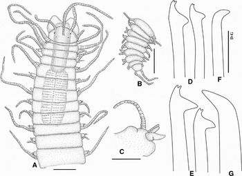
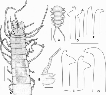
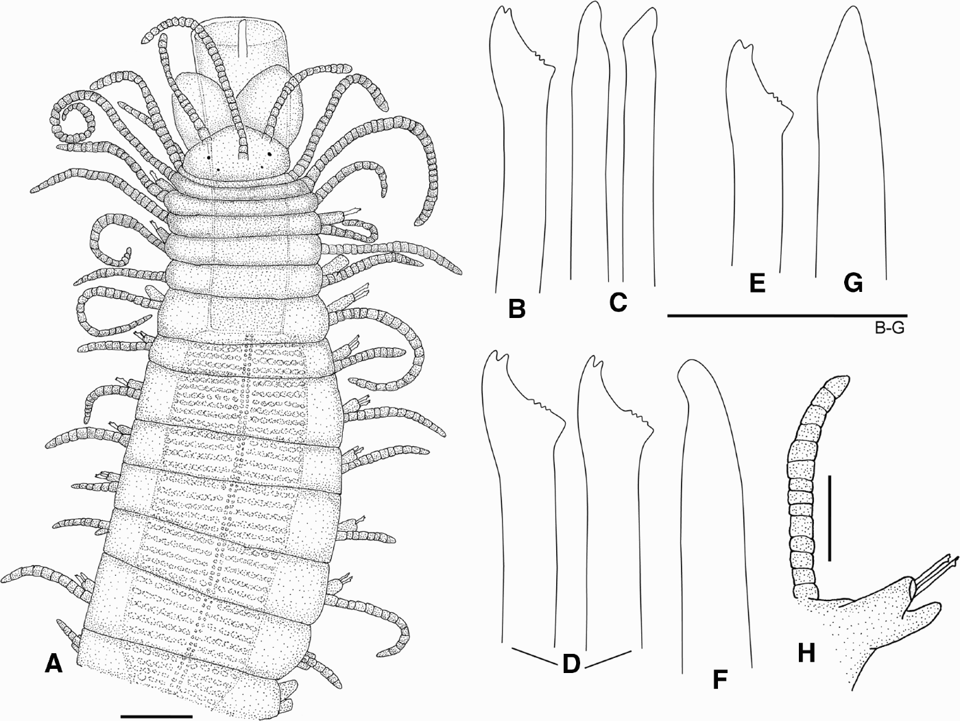
Haplosyllis aciculata sp. nov. (A) Anterior end, dorsal view; (B) posterior end, dorsal view; (C) midbody parapodium, ventral view; (D) chaetae, fourth chaetiger; (E) midbody chaetae; (F) acicula, fourth chaetiger; (G) posterior acicula. Scale bars: A–B = 250 µm; C = 200 µm; D–G = 20 µm.

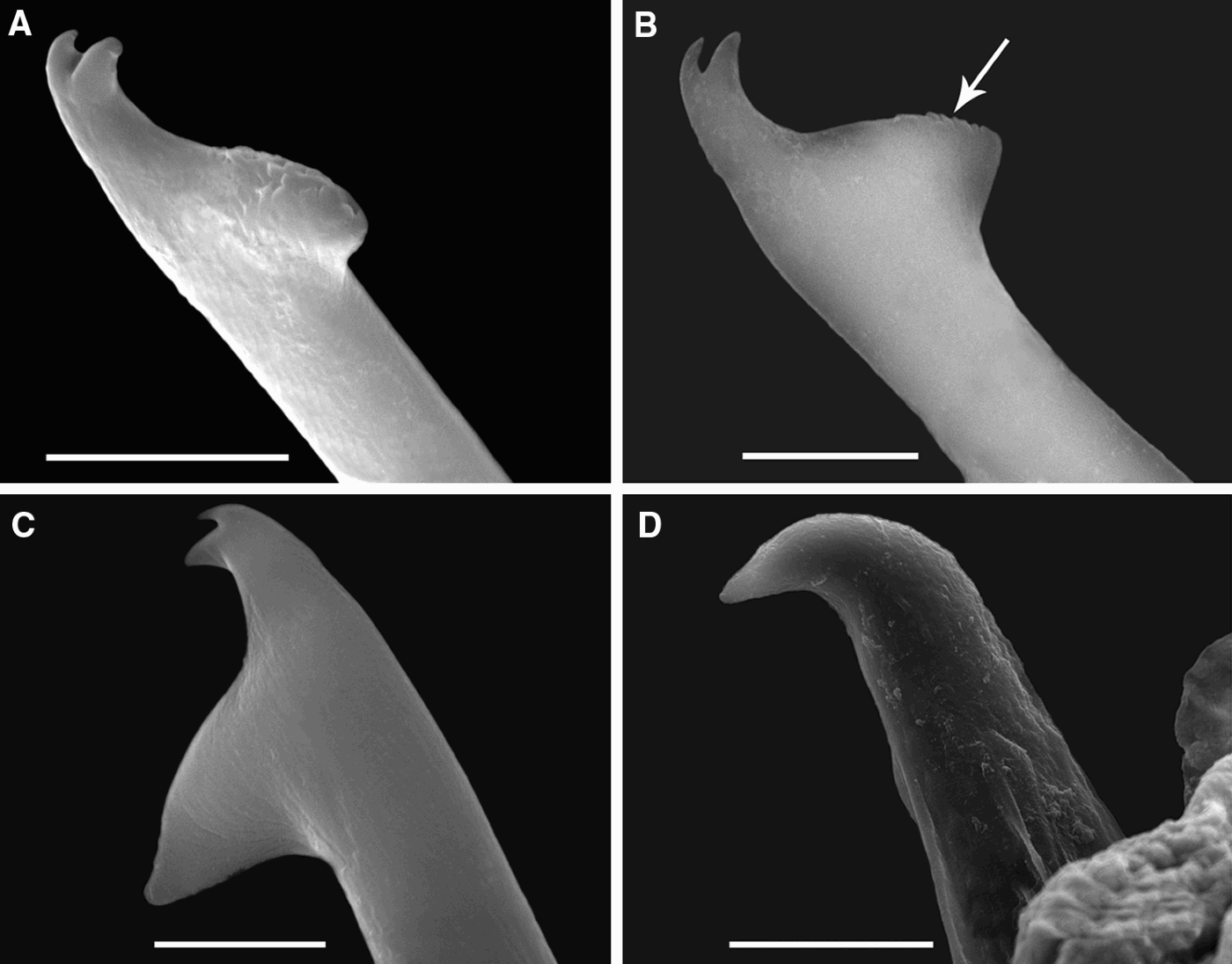
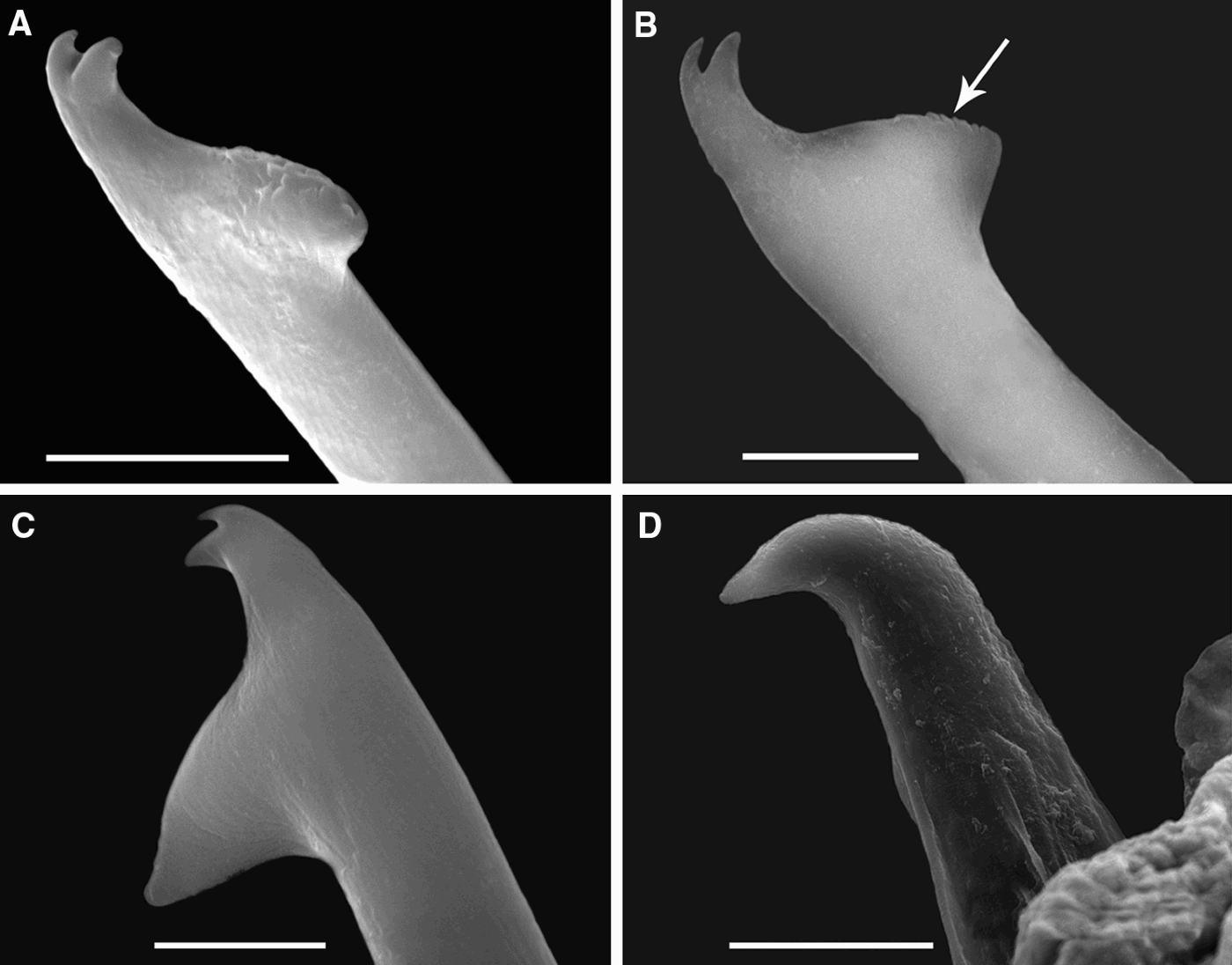
Haplosyllis aciculata sp. nov. (A, B) Anterior chaetae, arrow pointing the short serration of main fang; (C) midbody chaeta with a long main fang; (D) aciculae, posterior parapodium. Scales bars: A–C = 5 µm; D = 20 µm.
TYPE MATERIAL
Holotype (RMNH 21105), collected by Snellius II expedition; Indonesia, north-eastern coast of Sumba, 09°57′S 120°49′E, 45 m, 16 September 1984. 55 paratypes (RMNH 21106), same information as in holotype. 1 paratype (RMNH 21107), collected by Snellius II Expedition Indonesia, north-eastern coast of Sumba, 09°57′S 120°48′E, 50 m, 16 September 1984.
DESCRIPTION
Body robust, gradually tapering from midbody to posterior end, 3–9 mm long for 30–52 segments; about 0.5 mm wide (excluding parapodia). Holotype length 7.6 mm, with 49 segments. Body pale-cream. Prostomium sub-pentagonal (Figure 1A), with two pairs of small eyes in trapezoidal arrangement, some specimens apparently without eyes. Antennae similarly wide from base to tip; median antennae inserted on medium of prostomium, long, with 15–34 articles (less than 15 in small individuals); lateral ones inserted on anterior margin, with 8–20 articles. Palps slightly longer than prostomium, broadly triangular, fused at their bases but clearly divergent all along their length (Figure 1A). Sensory organs of palps and nuchal organs not seen. Pharynx orange, extending through 4–5 segments, usually everted, with a short anterior tooth surrounded by a crown of 10–12 soft papillae. Proventricle long, cylindrical, extending through 5–7 segments (occasionally 4), with 32–38 muscular cell-rows. Peristomium well defined, shorter than subsequent ones. Dorsal tentacular cirri twice as long as ventral ones with 15–32 articles. Dorsal cirri similar to antennae and tentacular cirri, long, whip-shaped. Anterior six pairs of dorsal cirri with a distinct length pattern: first cirri longer than remaining ones (19–53 articles), second short (4–24 articles), third and fourth gradually longer (10–29 and 12–40 articles, respectively), fifth short (3–17 articles), sixth long (19–40). Subsequent dorsal cirri slightly decreasing in size, alternating long (18–37 articles, less than 15 in small individuals) and short (9–22 articles). Posterior dorsal cirri short, with 1–4 articles (Figure 1B). Ventral cirri digitiform; anterior ones broad, triangular, slightly longer than parapodial lobes (Figure 1C); midbody and posterior ventral cirri, shorter. Chaetae all bidentate, anterior ones small; 2–3 per parapodia, all similar in shape and different in size. US of MF with short spines, mostly in anterior parapodia, only visible under SEM (Figure 2A–B); MJP straight, relatively small; LMF progressively longer, on midbody chaetae longer than SW (Figures 1D–E; 2C); apical teeth equal in length, clearly separated, distal tooth slightly shorter than proximal one. Aciculae broad, with long, pointed, curved tip upwards directed or 90° bent (Figures 1F–G; 2D), some protruding out of parapodial lobe, one on each parapodium. Pygidium with two long anal cirri, usually with 8–10 articles (Figure 1B). Stolons not seen.
ECOLOGY
Collected in sandy bottoms among sponges and gorgonians, from 45 to 50 m.
DISTRIBUTION
North-eastern coast of Sumba, Indonesia.
ETYMOLOGY
The name refers to the characteristic wide and long aciculae of the medium and posterior body regions.
REMARKS
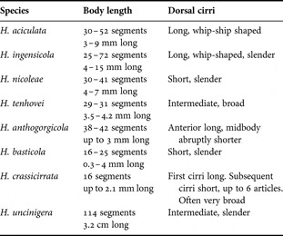
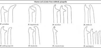
Haplosyllis aciculata sp. nov. is characterized by its robust body, long, whip-shaped cirri, and strong aciculae with curved tip. It can be distinguished from H. spongicola by the shape and size of aciculae and because the chaetae are similar all along the body, while H. spongicola has the anterior chaetae very different from midbody and posterior ones. H. aciculata sp. nov. resembles Haplosyllis ingensicola sp. nov. (described below) in the chaetae and acicular shape (for differences see remarks on H. ingensicola sp. nov. and Tables 1–2) and differs from H. uncinigera in its long pharynx (extending through 14–17 segments), straight aciculae and chaetal shape (see Table 2). Haplosyllis aciculata sp. nov. can be easily distinguished from Haplosyllis basticola and Haplosyllis crassicirrata by their smaller bodies (2–3 mm long) and shorter MJP of chaetae (Table 2).
Comparison of body length and dorsal cirri between the newly described Haplosyllis species from Indonesia and the Indo-pacific Haplosyllis species.

Graphical comparison of chaetae and aciculae between the newly described Haplosyllis species from Indonesia and the Indo-Pacific ones.

Haplosyllis ingensicola sp. nov. (A) Anterior end, dorsal view; (B) anterior parapodium, showing broad ventral cirrus and long dorsal cirrus; (C) posterior end, dorsal view; (D) anterior chaetae; (E) midbody chaetae; (F) anterior aciculae; (G) posterior acicula. Scale bars: A = 350 µm; B = 100 µm; C = 320 µm; D–G = 20 µm.

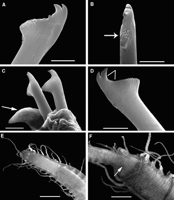
Haplosyllis ingensicola sp. nov. (A, B) Anterior chaetae, arrow pointing spines of main fang; (C) midbody chaetae and one acicula (pointed by the arrow); (D) posterior chaeta, bars showing the mid-joining point (MJP); (E) anterior end, dorsal view; (F) small specimen inside the pharynx (pointed by the arrow) of a big one. (A–D) SEM micrographs. Scale bars: A–B, D = 5 µm; C = 10 µm; E–F = 600 µm.

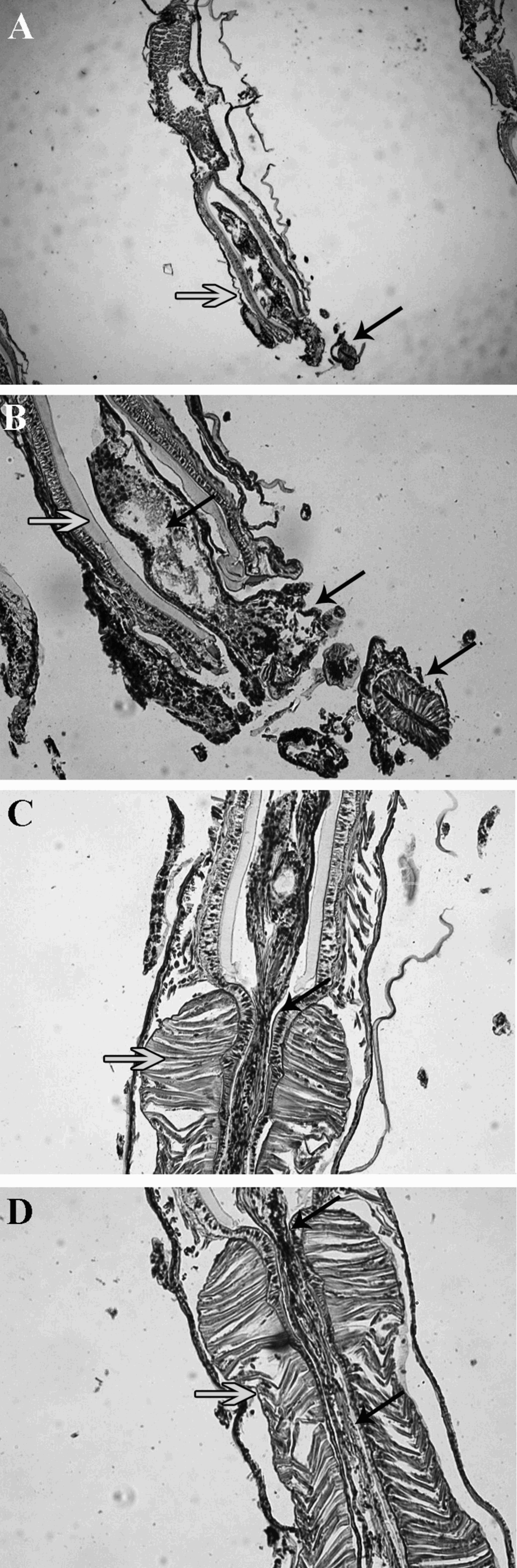
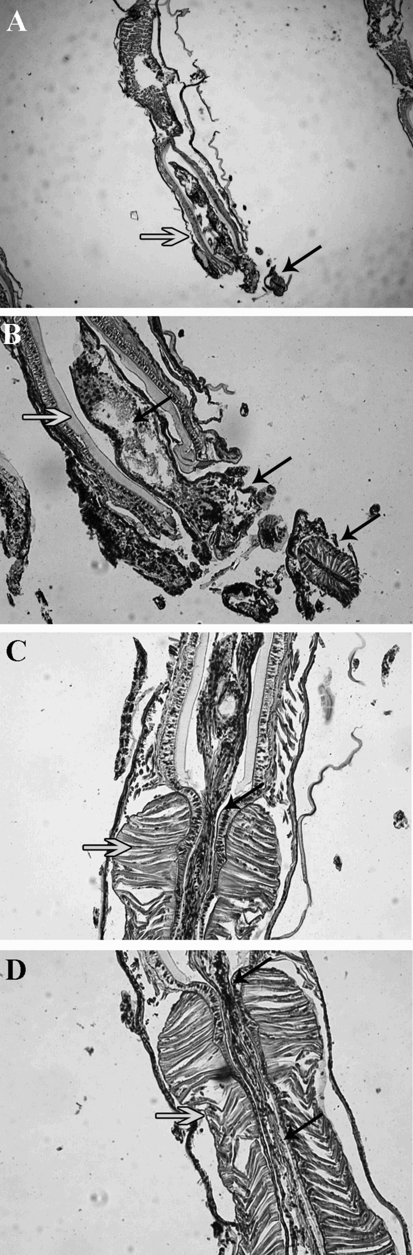
Haplosyllis ingensicola sp. nov. Histological sections showing a small individual inside the digestive tract of a larger one. (A, B) The small individual is inside the pharynx of the large one, but its proventricle (pointed by the arrow) in still outside the pharynx of its predator; (C, D) traces of the small individual inside the proventriale of its predator. Black arrows points the small individual, white arrows points the larger one.
TYPE MATERIAL
Holotype (RMNH 21103) and >500 paratypes (RMNH 21101): Indonesia, north-western Java. Collected inside the sponge Acanthostrongylophora ingens (Thiele, 1899), 9 m, 9 September 2005. 200 paratypes (RMNH 21104): Indonesia, south-western Sulawesi, Spermonde Archipelago, collected inside the host sponge A. ingens, 14 m, 29 April 1998.
DESCRIPTION
Body long, fragile, slender, mid-sized to long specimens, 4–15 mm long, for 45–72 segments (25–35 in small specimens), 0.5 mm wide, excluding parapodia, wider at proventricular segments, abruptly tapering from proventricle to posterior end. Holotype length 1.2 cm long, for 68 segments. Dark brown pigmentation in palps and prostomium as well as in anterior segments (until the beginning of proventricular segments), thereafter gradually disappearing, becoming pale-cream (Figures 3A, 4E). Prostomium sub-pentagonal with two pairs of small eyes in trapezoidal arrangement (Figure 3A). Median antenna inserted on medium of prostomium, 2–3 times longer (17–54 articles) than lateral ones (11–23 articles); lateral antennae inserted on anterior margin. Palps long, broad, fused at their bases but distally divergent (Figure 3A), slightly longer than prostomium. Sensory organs of palps and nuchal organs not seen. Pharynx brown, extending through 6–8 segments (4–5 in small specimens); with a large anterior tooth (Figure 3A) and a crown of about 10 soft papillae; pharyngeal ciliary ring not seen. Proventricle cylindrical, dark-brown, extending through 5–6 segments (3 in small specimens). Peristomium well defined, shorter than subsequent ones. Antennae, tentacular cirri and dorsal cirri long, slender, whip-shaped, gradually tapering from base to tips. Dorsal tentacular cirri twice as long (11–38 articles) as ventral one. Dorsal cirri of six anterior chaetigers with a distinct length pattern: first cirri longer than remaining ones (28–59 articles), second short (4–23 articles), third intermediate (12–28 articles), fourth long (20–54 articles), fifth short (6–25 articles), sixth long (17–40). Subsequent cirri alternating long (18–47 articles) and short (9–20 articles), but very difficult to observe; longest cirri exceeding body width; posterior cirri short (3–8 articles) (Figure 3C). Ventral cirri digitiform; six anterior ones slightly longer than parapodial lobes (Figure 3B), on midbody and posterior cirri shorter and thinner. Chaetae all bidentate, broad; anterior ones smaller; 2–3 on each parapodium (sometimes one on posterior chaetigers), similar in shape but different in size; with 2–3 rows of short spines on US of MF (Figure 4A, B); MJP short and curved (Figures 3E, 4D); LMF similar or longer than SW, progressively longer from anterior to posterior end (Figure 3D, E); apical teeth long, clearly separated; distal tooth smaller than proximal one (Figure 4D). Anterior parapodia with 1–2 aciculae, one enlarged with pointed tip, another with curved tip, almost 90° bent (Figure 3F); midbody parapodia with one broad acicula, with curved tip almost 90° bent (Figures 3G, 4C). Pygidium with two long anal cirri (14–15 articles) (Figure 3C).
REPRODUCTION
Haplosyllis ingensicola. sp. nov. reproduces by means of acephalous stolons having one pair of blackish parapodial ocular spots per segment.
ECOLOGY
More than 500 specimens were found inside the host sponge Acanthostrongylophora ingens (Thiele, 1899) between 9–14 m. Some large specimens were observed with a small con-specific individual inside the pharynx (Figure 4F), which were swallowed by the large worm, as traces of the small specimen were observed inside its proventricle (and even deeper into its gut) (Figure 5 A–D), suggesting a cannibalistic behaviour.
Cannibalism was described as the process of killing and eating an individual of the same species, with the larger (older) individuals apparently being more voracious cannibals than smaller (younger) animals (Polis, Reference Polis1981). When removed from their host sponges or in case of abandoning the host due to low oxygen availability, some Caribbean and Hawaiian Haplosyllis were observed to rapidly remain attached to any available organism through the pharynx, which is automatically everted and retracted (Leslie Harris, personal communication).
Taking into account the observations above and the fact that all specimens of H. ingensicola sp. nov. were sent to us once extracted from their host sponge, we could not confirm that our observations corresponded to cannibalism (i.e. as a common behaviour in non-disturbed specimens) or to an automatic stress response generated by the enormous quantity of specimens and the lack of oxygen or the sample manipulations. Although the latter are probable explanations, only small worms were captured by larger ones, within the hundreds of specimens studied. Therefore, this could correspond to true cannibalistic behaviour, but further studies should be addressed, either in situ or in experimental conditions, to assess the real meaning of this peculiar observation.
DISTRIBUTION
Indonesia, north-western Java (Thousand Islands Archipelago) and south-western Sulawesi Island, (Spermonde Archipelago).
ETYMOLOGY
The species name refers to the specific name of the host sponge, Acanthostrongylophora ingens.
REMARKS
Haplosyllis ingensicola sp. nov. is characterized by its long and fragile body, whip-shaped cirri, dark-brown pigmentation of the anterior end, and its broad, strong aciculae. Haplosyllis ingensicola sp. nov. differs from H. spongicola in body shape and in having similar chaetae throughout the body. The long cirri, chaetae and strong aciculae of H. ingensicola sp. nov. are very close to those of H. aciculata sp. nov. (described above), which differs in the body and cirri shapes, as well as in the pigmentation. The former has a long, slender and fragile body, long and fragile cirri. The latter is robust and slightly shorter, the cirri are also long but wide, and it does not have any pigmentation marks. Haplosyllis ingensicola sp. nov. can be distinguished from H. tenhovei sp. nov. (described below) by its aciculae, which differs from the enlarged one of the latter (Table 2). Haplosyllis ingensicola sp. nov. differs from H. basticola and H. crassicirrata in their smaller bodies (2–3 mm long) and shorter cirri (less than 15 articles), as well as in the choetol shape (see Tables 1–2).
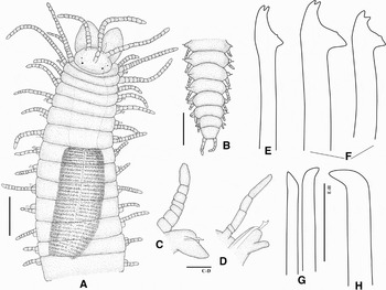
Haplosyllis nicoleae sp. nov. (A) Anterior end, dorsal view; (B) posterior end, dorsal view; (C) anterior parapodium, ventral view; (D) midbody parapodium, ventral view; (E) anterior chaeta; (F) midbody chaetae; (G) anterior aciculae; (H) midbody acicula. Scale bars: A–D = 200 µm; E–H = 20 µm.

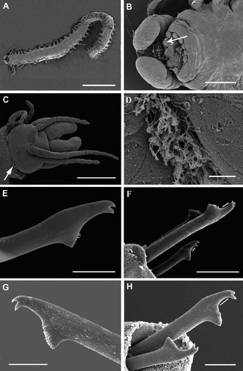
Haplosyllis nicoleae sp. nov. (A) Whole body, ventral view; (B) anterior end, ventral view, arrow pointing soft papillae; (C) anterior end, dorsal view, arrow pointing position of nuchal organs; (D) detail of nuchal organs; (E) anterior chaeta; (F) midbody chaetae; (G) posterior chaeta; (H) midbody chaeta. Scale bars: A = 1 mm; B–C = 100 µm; D, E, G, H = 5 µm; F = 10 µm. Bars of F and H indicating MJP.

TYPE MATERIAL
Holotype (RMNH 21112) and 35 paratypes (plus 2 for SEM) (RMNH 21111): Indonesia, Kudingareng Keke Island, 14 m, inside the sponge Clathria (Thalysias) reinwardti Vosmaer, 1880. 22 paratypes (RMNH 21114), same locality as holotype, 10 m. 8 paratypes (RMNH 21115): Spermonde Archipelago, Barang Lompo Island, inside C. (Thalysias) reinwardti, 3 m. Additional material: 1 specimen (RMNH 21109) and 55 specimens (RMNH 21108): Spermonde Archipelago, Kudingareng Keke Island: 11 m inside the sponge Biemna triraphis (Topsent, 1897). 18 specimens plus 1 for SEM (RMNH 21110): same locality, 16 m inside the sponge Melophlus sarassinorum Thiele, 1899.
DESCRIPTION
Body fragile, translucent. Small-sized, up to 2–7 mm long, 0.5 mm wide (excluding parapodia), for 30–41 segments, (20 in small individuals) (Figure 7A). Holotype length 5 mm, width 0.4 mm, with 40 segments. Preserved specimens pale-cream, without colour pattern. Prostomium oval, with two pairs of small eyes in trapezoidal arrangement (Figure 6A). Nuchal organs as a pair of lateral ciliated regions between peristomium and prostomium, only visible under SEM (Figure 7C–D). Antennae longer than palps; median antenna inserted on medium of prostomium, with 10–25 articles; lateral antennae inserted on anterior margin with 4–17 articles. Palps longer than prostomium, broadly triangular, fused at their bases, distally divergent. Sensory organs of ventral side of palps as rows of cilia. Pharynx orange, extending through 4–6 segments, with a long anterior tooth surrounded by a crown of 10–11 soft papillae and an inner ring of cilia (Figure 7B); ventral side of papillae also with cilia. Proventricle long, cylindrical, extending through 4–6 segments, with 27–39 muscular cell-rows. Peristomium well defined, shorter than subsequent ones. Dorsal tentacular cirri longer than ventral ones, with 10–22 articles. Dorsal cirri similar to antennae and tentacular cirri, short and slender; anterior six pairs of dorsal cirri with a distinct length pattern: first cirri longer than remaining ones (13–26 articles), second short (2–15 articles), third and fourth progressively longer (5–15 articles and 9–26 articles, respectively), fifth short (2–11 articles), sixth long (10–20 articles). Subsequent dorsal cirri gradually shorter to posterior end, alternating long (7–18 articles) and short (2–8 articles); longest midbody cirri do not exceed 1/2 of body width. Posterior dorsal cirri short, with 1–5 articles (Figure 6B). Anterior ventral cirri digitiform, broad, longer than parapodial lobes (Figure 6C), midbody and posterior ventral cirri, thinner, shorter than parapodial lobes (Figure 6D). Chaetae all bidentate, with spines on US of MF; anterior parapodia with 2–3 chaetae, small, with short MF (Figures 6E, 7E); midbody and posterior parapodia with two chaetae, with LMF similar to SW; MJP straight, long on largest chaeta of each parapodium (Figure 7F–H); apical teeth equal in size, well separated (Figure 7G). Aciculae with curved tips, more or less directed upwards; two aciculae on anterior and midbody parapodia (Figure 6G); one (sometimes two) on posterior segments (Figure 6H). Pygidium with two anal cirri.
REPRODUCTION
Haplosyllis nicoleae sp. nov. reproduces by acephalous stolons having one pair of blackish parapodial ocular spots per segment. The only specimen observed presented these ocular spots on the last three segments.
ECOLOGY
Haplosyllis nicoleae sp. nov. was found inside the sponges Clathria (Thalysias) reinwardti, Biemna triraphis and Melophlus sarassinorum between 3–18 m.
DISTRIBUTION
Indonesia, Spermonde Archipelago, Kudingareng Keke Island and Barang Lompo Island.
ETYMOLOGY
The name refers to Dr Nicole de Voogd, from the Institute for Biodiversity and Ecosystem Dynamics (Zoological Museum), University of Amsterdam, who kindly loaned us specimens of some of the species here described.
REMARKS
Haplosyllis nicoleae sp. nov. is characterized by its small, translucent body, short cirri, and a long MJP in largest chaetae of each parapodium. Haplosyllis nicoleae sp. nov. can be easily distinguished from H. spongicola, a more robust species having long whip-shaped cirri. Haplosyllis basticola, H. anthogorgicola and H. crassicirrata are all small-sized (about 4 mm long), with short cirri and occur in nearby geographical regions (see Table 1). Haplosyllis basticola may be distinguished by their wider chaetae, similar in shape all along the body, by lacking spines on US of MF, and by the short and curved MJP. Chaetae of Haplosyllis anthogorgicola clearly differs in chaetal shape from those of H. nicoleae sp. nov. (see Table 2); Haplosyllis crassicirrata differs in having dorsal cirri remarkably thick (Tables 1–2).
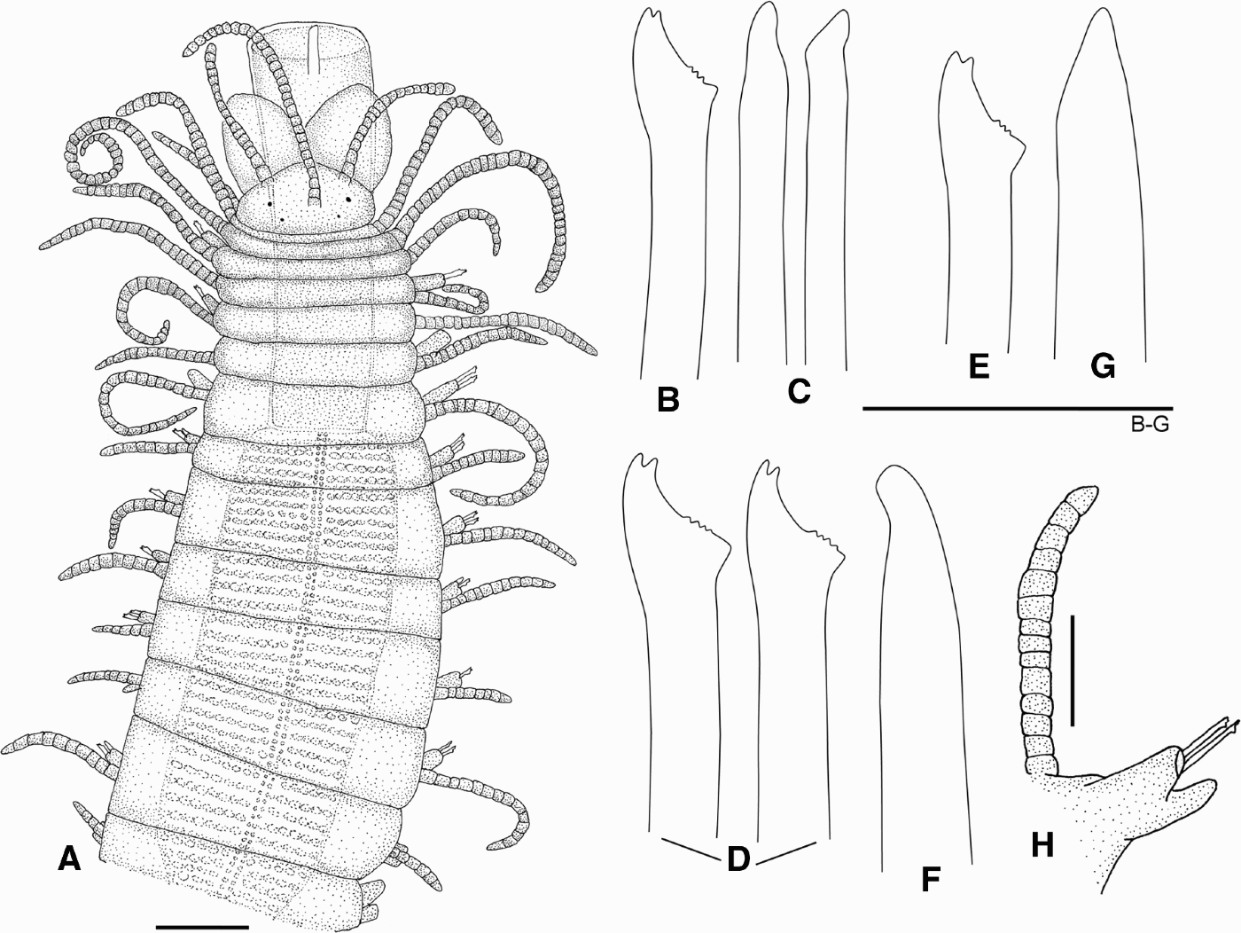
Haplosyllis tenhovei sp. nov. (A) Anterior end, dorsal view; (B) anterior chaeta; (C) anterior aciculae; (D) midbody chaetae; (E) midbody chaeta; (F) midbody acicula; (G) posterior acicula; (H) anterior parapodium. Scale bars: A = 200 µm; H = 48 µm; B–G = 20 µm.
TYPE MATERIAL
Holotype (RMNH 21099): Indonesia, Komodo, north-eastern Cape, 08°29′S 119°34.1′E. Snellius II, 30–34 m, Station 4.096, September 1984. Paratype (RMNH 21100): Indonesia, south-eastern of Karang Kaledupa, 05°57′S, 123°49′E. Snellius II, 350–500 m, Station 4.024, September 1984.
DESCRIPTION
Body broad, robust, wider at proventricular segments. Holotype 4.2 mm long, 0.7 mm wide excluding parapodia, with 31 segments. Paratype length 3.5 mm, width 0.5 mm, excluding parapodia with 29 segments. Pale-yellow: prostomium sub-pentagonal, with two pairs of small red eyes in trapezoidal arrangement (Figure 8A). Median antennae inserted on medium of prostomium, between posterior eyes, with 30 articles; lateral antennae inserted on anterior margin, with 19 articles. Palps long, slightly longer than prostomium, broad, triangular, fused at their bases, but clearly divergent all along their length. Cilia on palps and nuchal organs not seen. Pharynx orange, extending through 5–7 segments, with a large anterior tooth (Figure 8A) and a crown of about 10 soft papillae; pharyngeal ring of cilia not seen. Proventricle long, cylindrical, dark brown, 0.78 mm long, 0.21 mm wide, extending through 7–8 segments. Tentacular segment well defined, shorter than subsequent ones. Dorsal tentacular cirri longer than ventral ones, with 22–30 and 11–18 articles respectively. Antennae, tentacular cirri and dorsal cirri similar, gradually tapering from base to tips. Dorsal cirri long, slightly broad, usually coiled over dorsum (Figure 8A). Anterior six pairs of dorsal cirri with a characteristic length pattern: first cirri longer than remaining ones (37–42 articles), second short (16–19 articles), third and fourth intermediate (21–24 articles), fifth short (15–21 articles) and sixth intermediate (20–26). Dorsal midbody cirri alternating long (19–24 articles) and short (12–15 articles), decreasing in size to posterior end; longest cirri do not exceed body width. Ventral cirri digitiform, anterior ones longer (Figure 8H) than midbody and posterior cirri. Chaetae all bidentate, similar on all segments, 2–3 on each parapodium; anterior ones slightly smaller (Figure 8B); LMF similar to SW; MJP short and slightly curved; apical teeth equal in size, clearly separated (Figure 8E, D); spines on US of MF, not clearly seen. Aciculae straight, broad, with rounded tips (Figure 8C, G, F), 2–3 on anterior parapodia, two and one at midbody and posterior ones, respectively. Pygidium and stolons not seen.
ECOLOGY
Holotype collected from sandy bottoms with algae, sponges and tunicates, at 30–34 m deep. Paratype found in calcareous rocks, among dead corals and sponges at 350–500 m deep.
DISTRIBUTION
Komodo and Karang Kaledupa Islands (Indonesia).
ETYMOLOGY
The name refers to our colleague Dr Harry ten Hove from the Zoological Museum, University of Amsterdam, who has made important contributions to the knowledge of polychaetes and kindly loaned us the specimens of some of the species herein described.
REMARKS
Haplosyllis tenhovei sp. nov. can be distinguished by its small, broad body and the straight broad acicula. It differs from H. spongicola in the shape of aciculae and in having similar chaetae throughout (aciculae with hooked tips and marked differences between anterior and midbody and posterior chaetae in H. spongicola). Haplosyllis tenhovei sp. nov. clearly differs from H. uncinigera by shape of chaetae and aciculae by its small and fragile body, short cirri and non-serrated chaetae, and from H. crassicirrata in body shape and its characteristic broad cirri (see Tables 1–2).
ACKNOWLEDGEMENTS
This research was partly supported by the SYNTHESYS Project, financed by European Community Research Infrastructure Action under the FP6 ‘Structuring the European Research Area’ Programme for funding the visit by M.T.A. to the Zoological Museum of Amsterdam (NL–TAF). Many thanks are due to Dr Harry ten Hove (ZMA) for loaning material and for his warm hospitality. We are also very grateful to Dr Nicole de Voogd (RMNH, Leiden), the late Miguel Villena; and Javier Sánchez and Francisco Yagüe (MNCN), to Dr Dieter Fiege (ZMF) and Dr Birger Neuhaus (ZMB) for the loans of type material and other collection information. We also want to thank Dr Guillermo San Martín (UAM) for reading the manuscript and his important comments, to Dr Leslie Harris (NHM), for sharing her valuable observations on living worms in situ. Special thanks to Maria V. Plyuscheva (CEAB–CSIC) for helping us with the histological sections and to Jesus Sandi who kindly helped us with image editing. We also thank the two referees for their valuable comments. This paper is a contribution of the project ‘Taxonomía y Sistemática de la Familia Syllidae (Polychaeta)’, financed by ‘Ministerio de Educación y Ciencia’ of the Spanish Government, Project Number CGL2005–02442.