INTRODUCTION
Two myxozoans, Gadimyxa atlanticaKøie, Karlsbakk and Nylund, Reference Køie, Karlsbakk and Nylund2007 and Zschokkella hildae Auerbach, 1910, were detected in the excretory system of the kidney of Atlantic cod, Gadus morhua L. reared in a tank-based research facility on the Mull of Kintyre (Western Scotland) (Holzer et al. Reference Holzer, Wootten and Sommerville2010) and it was thought possible that they may be associated with mortalities in 0+ fry. Myxozoans are a large phylum of metazoan parasites with approximately 2200 known species (Lom and Dyková, Reference Lom and Dyková2006), occurring predominantly in fish but with only a small proportion reported to be pathogenic. However, several species that sporulate in the renal tubules cause severe pathologies resulting in mortalities of up to 100% of affected stocks e.g. Tetracapsuloides bryosalmonae Canning, Curry, Feist, Longshaw and Okamoura, 1999 (Feist et al. Reference Feist, Peeler, Gardiner, Smith and Longshaw2002; Wahli et al. Reference Wahli, Knuesel, Bernet, Segner, Pugovkin, Burkhardt-Holm, Escher and Schmidt-Posthaus2002), Sphaerospora renicola Dyková and Lom, 1982 (summarized in Lom and Dyková, Reference Lom, Dyková, Lom and Dyková1992) and Parvicapsula minibicornis Kent, Whitaker and Dawe, 1997 (St Hilaire et al. Reference St Hilaire, Boichuk, Barnes, Higgins, Devlin, Withler, Khattra, Jones and Kieser2002; Jones et al. Reference Jones, Prosperi-Porta, Dawe and Barnes2003). The kidney is a common site of infection for myxozoans where there may be more than 1 species present. However, little is known about the dynamics and interaction of mixed myxozoan infections. To gain a better understanding of such infections, particularly with regard to host age, G. morhua of different age classes ranging from 3 to 19 months were analysed with regard to prevalence, intensity of infection and location of G. atlantica and Z. hildae. At the facility on the Mull of Kintyre the growth performance of ongrowing fish is variable to the extent that they are divided into ‘good’ and ‘bad’ performers and, as growth performance has an economic impact, it was of interest to assess this in relation to infection. G. morhua at this site are derived from 3 hatcheries which differ in the age of initial exposure of fish to the parasites depending on the filtration system used at each site.
There are inherent problems in studying mixed myxozoan infections using the same organ owing to the difficulty of discriminating early life stages for which there are, as yet, no defining morphological characters. The methodologies developed for this study, therefore, included a rapid screening technique for fish tissues using a single-round PCR assay detecting both myxozoan species, and, for the first time in this parasite group, the use of a combined (double-label) in situ hybridization, which allows for the identification, differentiation and localization of all stages of development of the 2 myxozoan species on the same tissue section.
MATERIALS AND METHODS
Origin of G. morhua
A tank-based Atlantic cod research facility on the Mull of Kintyre (55°25′N, 5°44′W) Site D, is regularly supplied with cod fry from 3 different hatcheries i.e. Site A in Argyll (56°45′N, 5°51′W), Site B on the Isle of Man (54°12′N, 4°33′W) and Site C, on the Mull of Kintyre (55°25′N, 5°44′W), adjacent to Site D. On arrival at Site D, the different stocks from the hatcheries are mixed and on-grown. All hatcheries use local sea water and have a different water filtration system: at Site A, fry were maintained in 10 μm filtered water, at Site B fry were exposed to 1 μm filtered water for 58 days and then transferred to unfiltered water, and at Site C fry were cultured in 10 μm filtered water. At site D, the water was coarse filtered at approx. 60 μm.
Sampling regime and methodology
In July 2007, 20 fry, 90 days old, were obtained directly from each of the 3 hatcheries. For histology and in situ hybridization, 10 whole fry from each hatchery were fixed in 4% neutral buffered formalin after longitudinal incision of the abdominal cavity. Smears of skin, gills and kidneys of a further 10 fry from each hatchery were examined for the presence of parasites at ×400 magnification. The kidney smears were then transferred individually to Eppendorf tubes containing 300 μl TNES urea buffer (10 mm Tris-HCl (pH 8), 125 mm NaCl, 10 mm ethylenediamine tetraacetic acid, 0·5% sodium dodecyl sulphate, 4 m urea) for DNA extraction.
Between 2004 and 2007, sixty-four cod aged 6 to 19 months were obtained from Site D where fry from all 3 hatcheries are mixed and on-grown. At this site, mortalities and reduced growth had been observed amongst 0+ cod during summer months. In 2007, fish of the same lot had been graded according to their feeding and growth performance on site, and 15 representatives each of ‘good’ and ‘bad’ performers were sampled. From all fish, 4 μl of blood were collected from the caudal vein after removing the caudal fin. Subsequently the blood was mixed with TNES urea for DNA extraction to test for early myxozoan stages in the blood. Fresh smears of various organ pieces were viewed at ×400 magnification before they were used for DNA extraction and adjacent organ pieces were fixed for histology.
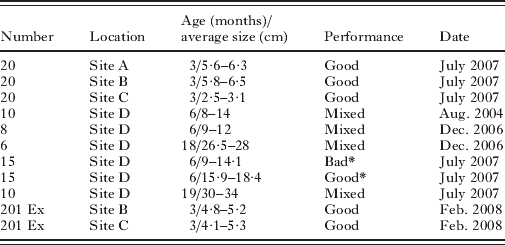
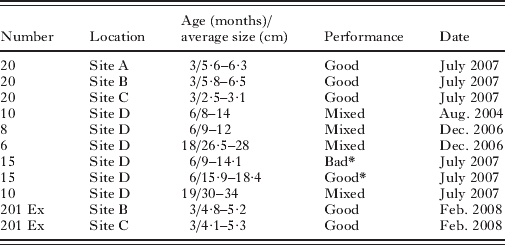
All fish were anaesthetized on arrival and processed immediately thereafter. The number, size and origin of Atlantic cod sampled at the different sites are summarized in Table 1.
Number, size and origin of Gadus morhua studied for myxozoan infections
(*Indicates fry from the same batch of fish which were separated into ‘good’ and ‘bad’ performers with regard to survival and growth; mo=months, Ex=experimental fry subsequently grown on for 29 days to study growth performance in relation to the development of myxozoan infections.)
DNA extraction and design of a specific PCR assay
All DNA extractions were performed according to a standardized phenol-chloroform protocol (Holzer et al. Reference Holzer, Sommerville and Wootten2004) after digestion of the samples with 100 μg/ml proteinase K overnight at 55°C. The extracted DNA was re-suspended in 50–10 μl of RNAse/DNAse-free water and left to dissolve overnight in a refrigerator.
Based on previously obtained 18S rDNA sequences of G. atlantica and Z. hildae from fish from Site D (Holzer et al. Reference Holzer, Wootten and Sommerville2010) two specific PCR primer pairs were designed in the variable regions of the 18S rDNA region of the 2 myxozoans: GadiF 5′-GTAATAGCATGGAACGAACAGT-3′ (current study) and GadiR 5′-AGGACCCGATTTAGTACATTATTA-3′ (current study) for G. atlantica (or related Gadimyxa species due to high sequence similarities), and ZHF 5′-GTTGCCACTTGTATGGTCAATTAT-3′ (current study) and ZHR 5′-GAGACAGATCCTGAGTCACAGAGCA-3′ (Holzer et al. Reference Holzer, Wootten and Sommerville2010) for Z. hildae. All primers were tested to exclude self-complementary binding or primer dimers, and they were then applied in a single-round PCR assay in a final volume of 10 μl containing 0·01 UμL−1 of Thermoprime Plus DNA polymerase and the related 10× buffer with 1·5 mm MgCl2 (ABgene, Epsom, UK), 0·2 mm of each dNTP, 0·5 mm of each primer and 50–100 ng of DNA. Cycling conditions were set to: 3 min initial denaturation at 95°C, 35 cycles of denaturation (94°C, 30 sec), annealing (60°C, 30 sec) and elongation (72°C, 30 sec), and a final elongation step of 3 min at 72°C. The PCR assay was used to test the DNA extractions of tissue and blood samples for the presence of the 2 myxozoans. Positive controls of fish containing morphologically identifiable spores of both myxozoans and negative controls containing DNA extractions of fins obtained from cod larvae hatched in 1 μm filtered sea water (site B) were included whenever PCR reactions were run.
Identification of Gadimyxa species
Due to very small differences in the 18S rDNA of G. atlantica and a second Gadimyxa species, i.e G. sphaerica, Køie, Karlsbakk and Nylund, Reference Køie, Karlsbakk and Nylund2007, described from cod, the PCR primers GadiF and GadiR designed in this study are not able to differentiate between the 2 species. However, the amplicons produced with the primers GadiF and GadiR include 2 areas with G. sphaerica-specific inserts of 6 and 4 bases, respectively, and an additional 4 individual base changes between G. atlantica and G. sphaerica. In order to determine whether different infection ratios of the 2 Gadimyxa species were responsible for the differences in the intensity of infection detected in fish from different hatcheries in 2007, PCR products obtained using primers GadiF and GadiR on DNA templates of 3 individual fish from Sites B and C (samples from 2007) were cloned into the pCR 2·1-TOPO vector (TOPO TA Cloning Kit, Invitrogen). Twelve clones from each fish (total of 72) were sequenced and analysed for species identity.
Histology and double-label in situ hybridization
For histological examination and in situ hybridization (ISH), organs or whole fry were fixed for 24–48 h in 4% neutral buffered formalin, subsequently dehydrated through an alcohol series, transferred into xylene and finally into paraffin. Longitudinal sections of whole fry were cut in a region where the maximum surface area of the kidney was present, whereas organ pieces of larger fish were combined in paraffin blocks and were cut arbitrarily. Six μm sections were fixed on 3% APES-coated slides for in situ hybridization and on uncoated slides for Giemsa staining.
For synchronous identification and localization of pre-sporogonic and early sporogonic stages of both, Z. hildae and G. atlantica in the same section, an in situ hybridization protocol, which uses 2 differently labelled probes targeting DNA of the 2 myxozoans in the sections, was designed and applied. The methodology of the double label protocol is based on the protocol of a single-label biotin-streptavidin-horseradish peroxidase detection system published earlier (Holzer et al. Reference Holzer, Sommerville and Wootten2003) which was combined with a digoxigenin (DIG)-anti-DIG-alkaline phosphatase system. A biotin-labelled probe GadiR designed for Gadimyxa sp. (Table 2) was applied simultaneously with a DIG-labelled probe, in this case ZHR, designed for Zschokkella (Table 2) for hybridization to the same section. After mixing the differently labelled probes with the hybridization buffer, denaturation and hybridization were conducted and the biotin label was detected as described by Holzer et al. (Reference Holzer, Sommerville and Wootten2003). Following the use of the Vector VIP substrate (Vectorlabs, Peterborough, UK), sections were washed in distilled water and transferred into TBS (pH 7·5). Subsequently, sections were incubated with 1:5000 anti-DIG-alkaline phosphatase Fab fragments (Roche Diagnostics, Mannheim, Germany) in TBS (pH 7·5) for 2 h at room temperature or overnight at 6°C. Following a wash in the TBS (pH 8·0), Vector Blue (Vectorlabs, Peterborough, UK) was used as a substrate to visualize the DIG-labelled probe according to the manufacturer's recommendations. After a rinse in distilled water, counterstaining was conducted in some cases using methyl green (Vectorlabs, Peterborough, UK) and the sections were dehydrated through an alcohol series and transferred through a xylene replacement, Citrosol (Panreac Quimica SAU, Spain), into a xylene-free mounting medium (modified acrylic resin, Panreac Quimica SAU, Spain).
Comparison of growth performance indicators of Atlantic cod fry originating from sites B and C, when cultured at 8°C and 12°C, respectively, for 29 days (mean±standard deviation)
(Data were pooled for replicate tanks of the same site maintained at 8°C where there were no statistical differences (3 tanks of site B and 2 tanks of site C); 12°C tanks were not replicated and therefore not analysed statistically. SGR=specific growth rate, FCR=food conversion ratio. * Statistically significant differences between sites.)

Comparison of infection and growth performance of fry from Site B and Site C over a 29-day period
Fry source
In February 2008, cod fry, 87 days post-hatch, were obtained from the hatchery at Site B and 84–90 days-post hatch fry from Site C. They were stocked into experimental tanks at Site D receiving only coarsely filtered (60 μm) water from a source close to Site C.
An initial sample of 15 fish was removed from each source prior to stocking in experimental tanks in order to obtain data on source infection status. These fish were individually weighed and measured and processed for histology.
Experimental design and fish maintenance
After removal of the initial subsample (n=15) of each stock, the remaining fish were randomly distributed in weighed batches of 10 into 8×73·5 L identical tanks. Six of the tanks were arranged in a randomized block design with 3 tanks×50 fish from Site B fry and 3 tanks×50 Site C fry. Ambient temperature was 8±0·5°C. A fourth tank of each stock was supplied with heated water to elevate the temperature to 12±0·5°C. These consisted of 1 tank×37 fish from Site B and 1 tank×36 fish from Site C. The flow rate in all tanks was 0·5±0·1 L min−1. The fry were fed to satiation for 29 days on Biomar Bio-optimal Start 1·1 mm at 6% biomass per day using an automatic feeding system. All tanks were siphoned twice a day and solids were dehydrated and weighed. Moribund fish were sacrificed, weighed and measured and processed for histology.
Sampling and data collection
After 29 days of maintenance all fish were sacrificed by an overdose of benzocaine. They were individually weighed and measured prior to fixation in 10% neutral buffered formalin and processed for histology. Fixed samples were trimmed prior to blocking to obtain a standardized longitudinal section of the kidney inclusive of its total length and giving the maximum surface area of kidney. After processing, 2 adjacent 5 μm sections of each fish were stained in Giemsa for parasitological analysis.
Parasitological analysis
Parasitological data were collected from a randomly selected subsample of 13 fish from each stock prior to the start of the trial and 10 fish from each of the 8 tanks after 29 days. Eighteen moribund fish were also examined. To estimate parasite intensity in each fish, sections were viewed under ×200 and ×400 magnification and all renal tubule sections were counted and recorded as either infected or uninfected. Although each tubule might have been sectioned more than once, the standardization of sections allowed for comparisons between fish. Two adjacent kidney sections per fish were counted using separate counters so as to remove any bias in the counts. Both counts were recorded and a mean taken for each fish. Any sections without a complete longitudinal section of the kidney were discarded resulting in a total of 28 fish of each stock at 8°C and 10 fish of each stock at 12°C being analysed at the end of the trial. Only parasitic stages with obvious, clear nuclear staining were counted and the number of parasite cells in the infected tubules was not taken into account. Thus, the relative percentage of infected tubules was calculated for each fish sampled. Individual sections of a subsample of the same paraffin blocks including 8 fish from each site and temperature regime were subjected to ISH to confirm the identity of the parasite stages.
Data analyses
Specific growth rates SGR=[(ln end weight in g−ln start weight in g)/experimental days]×100 for each fish in each tank were calculated and compared. Estimated food conversion ratio FCR=(amount of feed given−waste feed removed)/(increase in tank biomass+weight of mortalities) was calculated for each tank.
Parametric analysis was carried out using an independent value t-test or ANOVA on multiple groups. Where data sets did not conform to the assumptions of these analyses in terms of normality of distribution or homogeneity of variance, transformations using natural logarithm were performed and the data were again tested for conformity. Where data sets were unable to be transformed to a suitable form, non-parametric analysis was carried out using Mann Whitney or Kruskal-Wallis tests on multiple groups. Analysis was carried out on pooled replicates where appropriate for greater statistical robustness. The pre-determined alpha level for P-value significance in all tests was 0·05. Statistical tests were conducted using Minitab v13.32 (Minitab Inc.) or GraphPad InStat v3.01 (GraphPad Software, Inc). Only data from the replicated tanks held at 8°C were statistically analysed; the 12°C tanks were not replicated and data are reported as observations.
RESULTS
Age and sample site-related differences in the myxozoan infections
In fresh microscopic smears, spores (n=20) of G. atlantica measured 8·2–9·2 (8·8) μm in width, 6·8–8·2 (7·3) μm in length, 5·9–7·0 (6·5) μm in thickness, with a polar capsule diameter of 1·7–2·2 (1·9) μm and a polar filament length of 11–19 μm. The measurements obtained from Z. hildae have been reported by Holzer et al. (Reference Holzer, Wootten and Sommerville2010). Microscopically, G. atlantica was found to produce abundant disporous pseudoplasmodia containing maturing spores in fry and 6-month-old cod, whereas they were very scarce or absent in 1+ G. morhua. In contrast, Z. hildae plasmodia and maturing spores were not detected in fry ⩽6 months old, with small numbers present in some of the 6-month-old fish and larger numbers in all 1+ cod, where the infection was quite extensive in individual cases.
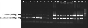
The specific single-round PCR assay designed in this study produced 2 PCR products, a 104 bp band for G. atlantica and a 336 bp band for Z. hildae (Fig. 1) and confirmed the microscopic observations. Fig. 1 shows the results of PCR reactions of DNA extracted from kidneys of some representative cod from different sites and of different ages (details in Table 1). The PCR results show the infection prevalence and indicate, semi-quantitatively, the intensity of infection. Fry became infected with both G. atlantica and Z. hildae at a very early age as bands for both species are present in the kidneys of the smallest, 3-month-old fry. These fry already presented a dominant 104 bp (G. atlantica) band, with fry from Site B showing a considerably larger amount of PCR product than those from Sites A and C. The highest amounts of G. atlantica PCR product were obtained from 6-month-old fry when compared with younger or older cod (Fig. 1). The oldest cod sampled (18 and 19 months old) showed a considerable decrease in the amount of G. atlantica amplicons. PCR bands of Z. hildae were very weak and sometimes absent in the smallest fry but PCR product increased in the 6-month-old fry and reached a maximum in the oldest (18 and 19 months old) cod. With regard to the presence or absence of PCR products of both species, no differences were detected between ‘good performers’ and ‘bad performers’ (Fig. 1). The quantities of Z. hildae PCR product were elevated in 7/30 fish (lane 10 of Fig. 1) when compared with other fish of the same age class, but this was independent of the performance status of the fish (3 ‘good performers’, 4 ‘bad performers’).
Simultaneous PCR detection of Zschokkella hildae and Gadimyxa atlantica in Gadus morhua kidney extractions from different sites in Scotland. Representative samples of: 1–9=3 month old G. morhua from 1–3: Site A, 4–6: Site B, 7–9: Site C; 10–15: 6 months old G. morhua from Site D with 10–12: ‘Bad’ performers and 13–15: ‘Good’ performers; 16–18: 19 months old G. morhua from Site D. M: size marker with bands every 100 bps.

The considerably larger amount of G. atlantica PCR product detected in fry from Site B when compared with the other hatcheries (Fig. 1) raised the question of whether the difference in intensity of infection is due to different infection ratios between more than 1 Gadimyxa species. Sequences obtained from the analysis of 72 clones of PCR amplicons produced with the primers GadiF and GadiR from 3 fry from each of Sites B and C showed that all but 1 clone were identical with G. atlantica (compared with GenBank Accession numbers EU163412-21), with interclonal differences of no more than 1–2 bases. The clone that differed considerably (4 bases and 2 inserts of 4 and 6 bases, respectively) was obtained from a fry from Site C and was identified as G. sphaerica, identical with previously obtained 18S rDNA sequences of this species (EU163424-5).
Parasite location
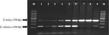
The PCR assay detected 18S rDNA of both G. atlantica and Z. hildae in kidney as well as in numerous other organs of infected fish, i.e. spleen, liver, heart, muscle, gills, brain and blood. PCR of DNA extractions from blood (Fig. 2) produced PCR products of G. atlantica in 56% (27/48) of the 6-month-old fry, whereas bands for G. atlantica were absent or, in exceptional cases, very weak in DNA extractions from blood of 1+ cod (lane 9 in Fig. 2). Blood samples were PCR-positive for Z. hildae in all the 6-month-old fry and in 62% (10/16) of 1+ cod (Fig. 2). Due to their small size and fixation on site, blood was not obtained from the 3-month-old fry and PCR data are thus not available. There was no difference in the presence or absence of blood stages of either myxozoan species when comparing ‘good performers’ and ‘bad performers’ but the amount of PCR product obtained from the blood samples was considerably higher in the cod sampled in the summer than in the winter (Fig. 2).
Simultaneous PCR detection of Zschokkella hildae and Gadimyxa atlantica in blood samples of Gadus morhua. Representative samples of: 1–3: 6 months old G. morhua (Site D) sampled in winter, 4–6: 6 months old G. morhua (Site D) sampled in summer, 7–9: 19 months old G. morhua (Site D) sampled in summer. M: size marker with bands every 100 bps.

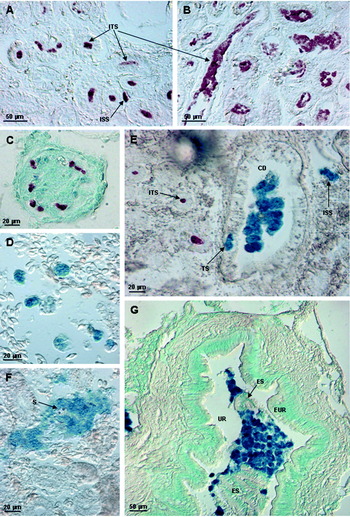
In situ hybridization (Fig. 3) confirmed the results obtained by PCR and added further information on the specific location of the 2 myxozoan species and the developmental stages present in fish of different ages and sites. Pre-sporogonic proliferative stages and spore-forming stages of G. atlantica were detected exclusively intratubularly, occurring in the proximal and distal parts of the renal tubules with the largest amount of parasite stages and numbers of tubules infected in 3-month-old fry. In some fry, pluricellular parasite stages of round to elongate shape and measuring 17·5–41·2 μm in diameter were present in the kidney interstitial tissue but blood stages of G. atlantica were not detected on the sections using ISH despite positive PCR reactions from blood DNA extractions. Frequently, numerous small parasite stages consisting of cell doublets and measuring 8–13·5 μm were detected in the glomeruli (Fig. 3C). In the oldest cod (18 and 19 months) the G. atlantica infection had almost disappeared and only isolated renal tubules were found infected with small numbers of this parasite. Despite the presence of weak PCR bands (Fig. 1), Z. hildae was not detected in sections of the smallest fry but multicellular stages measuring 16·4–35 μm were found in the blood (Fig. 3D) and interstitial kidney tissue in some of the 6-month-old cod and in larger, sometimes very high, numbers in 18- and 19-month-old cod (Fig. 3G). In some histological sections, interstitial stages of Z. hildae were found to invade the urinary collecting ducts by crossing the epithelium (Fig. 3E). Spore-forming stages of Z. hildae, first seen in 6-month-old cod, were mainly found in the large collecting ducts and the ureters, which, in 18- and 19-month-old cod, were sometimes completely filled with Z. hildae stages (Fig. 3G). Occasionally, large parasite stages and spore-formation of Z. hildae were observed in the kidney interstitial tissue (Fig. 3F). No pathological changes were evident in kidneys with large numbers of intratubular G. atlantica stages or blood and interstitial stages of Z. hildae. However, collecting ducts heavily infected by Z. hildae showed invasion of macrophages into the epithelium of the collecting ducts (not shown), and detached pieces of the epithelial lining were detected amongst masses of spore-forming parasites, indicating epithelial sloughing presumably caused by the parasites (Fig. 3G).
Simultaneous detection of Gadimyxa atlantica and Zschokkella hildae in histological sections of the kidney of Atlantic cod, using double-label in situ hybridization, with G. atlantica visualized in purple (Vector VIP substrate) and Z. hildae in dark blue (Vector Blue substrate); background staining in (C) and (G) is methyl green. (A) Intratubular sporogonic stages (ITS) and 1 interstitial, migratory stage (ISS) of G. atlantica in experimentally maintained (8°C) Site C fry with average level of infection. (B) Increased infection with larger numbers of tubules infected with G. atlantica and sporogonic stages filling the tubules, experimental growth trial (12°C) Site B fry. (C) Glomerulus infected with cell doublets of G. atlantica, 3-month-old fry (non-experimental, Site C). (D) Invasive blood stages of Z. hildae amongst erythrocytes, 6-month-old cod (Site D). (E) Interstitial migratory stage (ISS) of Z. hildae, a parasite traversing (TS) the epithelium of the urinary collecting duct (CD) and spore-forming stages of Z. hildae inside the duct, 18-month-old cod (Site D). (F) Mass of cells of Z. hildae forming spores (S) in the interstitial tissue of the kidney, 18-month-old cod (Site D). (G) Ureter (UR) with masses of spore-forming parasites and detached elements (ES) of the otherwise intact epithelium (EUR) of the ureter, 19-month-old cod (Site D).

G. atlantica infection dominated in histological sections of the 3-month-old fry, whereas in 1+ cod, Z. hildae infection was more prominent, with only a small number of tubules harbouring residual G. atlantica infection, confirming the PCR results. Thus, co-infection of the 2 myxozoans in the kidney was temporally restricted mainly to the 6-month-old G. morhua, where invasive blood and interstitial stages and, in some fish, also sporogonic stages of Z. hildae co-occurred with sporogonic stages of G. atlantica.
Comparison of infection and growth performance of fry from Site B and Site C after 29 days of growth
The comparison of the development of the myxozoan infection parallel to the performance of the host was studied in fry from 2 hatcheries with different parasite loads, Site B and Site C (compare Fig.1), before and after a 29-day period of maintenance at 2 different temperatures, 8°C and 12°C.
Fish growth and performance
The results of the parameters measured in the fish growth study are summarized in Table 2. The start weights of fry from Site B and C were not significantly different. The end weights of fry from the replicate tanks for each site maintained at 8°C were pooled for analysis except the results from 1 replicate tank of site B (tank 3) which was excluded from further analysis as the fry were significantly larger than the other replicates and this was attributed to an unknown tank effect. Fry from site B performed significantly better than those from Site C both in terms of specific growth rate and food conversion ratio. Furthermore, the numbers of moribund and dead fish were considerably higher for Site C fry than for Site B fry. At the higher temperature of 12°C a substantially higher final weight and SGR of fry in both stocks was observed although, since the data were not replicated, no statistical analysis was carried out.
Percentage of parasite-infected renal tubule sections
The results of the parasite analysis are summarized in Table 3. The percentage of infected tubule sections ranged from 3·17% to 45·38% (Table 3) in different individuals and varied greatly between the individuals from the same tank. In the pre-trial sample, cod fry from Site B had a significantly lower percentage of infected tubules (8·61%) than Site C fry (14·45%). However, after 29 days, Site B fish showed significant proliferation of the parasite in terms of the percentage of infected tubule sections, up to a mean of 19·04% but, in contrast, there was no significant change in the Site C fry, (12·78% tubule sections infected). The difference in the final parasite levels between Site B and C fry was highly significant.
Infection intensities=relative percentage of renal tubule sections infected with Gadimyxa atlantica per total number of tubule sections in 2 replicate histological sections per fish, before (start), during (moribund fish) and after (end) experimental 29 day growth trial of fry from Site B and Site C at 8°C and at 12°C
(Data were pooled for replicate tanks of the same site maintained at 8°C (3 tanks of site B and 2 tanks of site C). Statistically significant differences between a sites, b temperatures and c beginning and end of experimental period; moribund fish were not evaluated statistically due to low number of samples.)
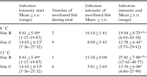
Despite the better growth performance of fry in replicate tank 3 from Site B when compared with fry from the other 2 Site B replicate tanks, the proportion of infected tubules in fish from this tank was not significantly different from the other 2 replicates. In the heated tanks, the proportion of infected tubules in Site B fry was markedly higher after 29 days with a mean of 27·81% tubules infected. In Site C fry-heated tanks there was little change from the levels recorded at the start of the experiment.
The number of moribund fish was considered to be too small for statistical analysis; however, examination of these fish demonstrated that the percentage of tubules infected was below the mean level for healthy fish originating from both sites at the end of the trial.
ISH showed that the myxozoan inside the tubules counted as ‘infected tubules’ in the experimental fish was exclusively G. atlantica. Individual pre-sporogonic stages of Z. hildae were found in the interstitial tissue and blood vessels only in a small number of sections.
DISCUSSION
Mixed infections of the 2 renal myxozoans G. atlantica and Z. hildae were detected in cultured G. morhua sampled at 3 different hatcheries and at a tank-based research facility located on the west coast of Scotland. Two specific assays based on the 18S rDNA sequence of the myxozoans, a single round PCR and a double-label ISH protocol were designed for G. atlantica and Z. hildae in this study. This permitted rapid, specific identification, visualization and localization of the 2 myxozoans in the same PCR reaction and on the same histological section, respectively. These methods proved to be invaluable tools to follow the development of the myxozoan infections in juvenile cod up to the age of 19 months. Possibly due to the lower number of cells and thus parasites in histological sections when compared with tissue samples taken for DNA extraction, and also due to the large copy number of 18S rDNA tandem repeats per cell, resulting in a strong PCR signal despite low numbers of parasites, PCR detection was more sensitive than ISH. However, only the combination of both techniques allowed detection, in situ localization and identification of different life-cycle stages of the myxozoans.
G. atlantica was first described by Køie et al. (Reference Køie, Karlsbakk and Nylund2007) who found 10% of Danish coastal cod and 50% of off-shore caught cod to be infected. However, in the present study, the infection prevalence of G. atlantica was 100%. G. atlantica was most numerous in fry and juvenile cod whereas parasite numbers had decreased considerably in 1+ G. morhua, despite their being reared in a continuously infectious environment. This decrease with age might explain the lower prevalences detected by Køie et al. (Reference Køie, Karlsbakk and Nylund2007) ; their fish measured 21–76 cm, equivalent to an age of 2–6 years in wild cod (Magnadóttir et al. Reference Magnadóttir, Jonsdottir, Helgason, Bjornsson, Jorgensen and Pilstrom1999). The current study suggests that cod acquire immunity to G. atlantica since invasive blood stages were detected by PCR in 56% of 6-month-old cod but had disappeared almost completely in fish older than 1 year although they were still in an infective environment. Acquired immunity to re-infection is mediated by lymphocytes and effected mainly through antibodies, a mechanism which takes 1–3 months depending predominantly on fish species and temperature (Magnadóttir, Reference Magnadóttir2006). The ontogenetic development of the immune system of cod is very slow when compared with other marine species (Magnadottir et al. Reference Magnadóttir, Lange, Gudmundsdottir, Bogwald and Dalmo2005) and is not fully competent until 2–3 months after hatching (Schrøder et al. Reference Schrøder, Villena and Jorgensen1998; Magnadóttir et al. Reference Magnadóttir, Lange, Steinarsson and Gudmundsdottir2004). Thus, the development of a specific immune response in cod would occur at the age of approximately 3–5 months. This coincides with our findings that only half of the 6-month-old cod presented G. atlantica blood stages and that blood stages had disappeared in 1+ cod. Resistance to re-infection, based on acquired immunity and the production of specific antibodies, has been reported for various other myxozoans, including T. bryosalmonae (Saulnier and de Kinkelin, Reference Saulnier and de Kinkelin1996), Enteromyxum spp. (Munoz et al. Reference Munoz, Sitja-Bobadilla and Alvarez-Pellitero2000, Reference Munoz, Cuesta, Athanassopoulou, Golomazou, Crespo, Padros, Sitja-Bobadilla, Albinana, Esteban, Alvarez-Pellitero and Meseguer2007; Sitja-Bobadilla et al. Reference Sitja-Bobadilla, Redondo, Macias, Ferreiro, Riaza and Alvarez-Pellitero2004, Reference Sitja-Bobadilla, Palenzuela, Riaza, Macias and Alvarez-Pellitero2007), Myxobolus cerebralis Hofer, 1903 (Hedrick et al. Reference Hedrick, El Matbouli, Adkison and MacConnell1998) and Myxobolus artus (Furuta et al. Reference Furuta, Ogawa and Wakabayashi1993). Intratubular sporogonic stages of G. atlantica in the renal tubules persisted in cod older than 2 years (Køie et al. Reference Køie, Karlsbakk and Nylund2007)). However, this does not contra-indicate acquired immunity to G. atlantica since persistence of sporogonic stages inside kidney tubules has also been observed in trout after acquiring immunity to T. bryosalmonae (Foott and Hedrick, Reference Foott and Hedrick1987; Holzer et al. Reference Holzer, Sommerville and Wootten2006). This might be due to spore production in an immunoprivileged coelozoic site (Sitja-Bobadilla, Reference Sitja-Bobadilla2008).
The second myxozoan found in the kidney of G. morhua was Z. hildae which, in contrast to G. atlantica, has been described as a myxozoan species with low host specificity, occurring in various gadoid fish and showing a wide geographical range in the northern Atlantic and Pacific (Gaevskaya and Kovaleva, Reference Gaevskaya and Kovaleva1979; MacKenzie, Reference MacKenzie1979; Bakay and Zubchenko, Reference Bakay and Zubchenko1981; Arthur, Reference Arthur1984; Aseeva, Reference Aseeva2002; Køie et al. Reference Køie, Steffensen, Moller and Christiansen2008).
Both Z. hildae and G. atlantica were found to produce blood stages in cod, indicating that both species use the blood as a medium of transport to the target organ, the kidney. The occurrence of blood stages of both G. atlantica and Z. hildae at all sites might indicate that they were continuously challenged by both species. Blood stages seem to be a common means of dissemination of myxosporean infections through the fish host. They were first described by Csaba (Reference Csaba1976) and have subsequently been reported from all main phylogenetic clades of the myxosporea, e.g. T. bryosalmonae (Kent and Lom, Reference Kent and Lom1986), Sphaerospora truttae Fischer-Scherl, El-Matbouli and Hoffmann, 1986 (McGeorge et al. Reference McGeorge, Sommerville and Wootten1994), Ceratomyxa shasta Noble, 1950 (Bartholomew et al. Reference Bartholomew, Smith, Rohover and Fryer1989) and S. renicola (reviewed by Lom and Dyková, Reference Lom, Dyková, Lom and Dyková1992). It is therefore not surprising that more than 1 myxozoan occurs in the blood of an individual fish host at the same time. In brown trout, 4 myxozoans, i.e. Chloromyxum shurovi Shul'man & Ieshko, 2003, T. bryosalmonae, S. truttae and Chloromyxum truttae Léger, 1906, occurred co-temporally in the blood (Holzer et al. Reference Holzer, Sommerville and Wootten2006). In the current study, Z. hildae and G. atlantica blood stages were present at the same time in cod aged 6 months and they possibly co-occur in younger fry, for which no blood samples were analysed. All the 6-month-old cod and 62% of the 1+ cod tested PCR-positive for blood stages of Z. hildae. This indicates that, in contrast to G. atlantica, which was not found in the blood of 1+ fish, Z. hildae is able to continuously invade and establish in cod at least up to the age of 19 months (oldest cod studied), and that specific immunity is not acquired or is only acquired at a later date.
Z. hildae showed massive proliferation in the blood of 1+ cod, i.e. once G. atlantica had disappeared from the bloodstream. Similarly, the maximum G. atlantica spore-formation was detected in 3-month-old fry and spore numbers were still prominent in 6-month-old cod, whereas spore-formation of Z. hildae was only initiated in some of the 6-month-old cod but spores were numerous in all 1+ cod. This suggests that the presence of maximum numbers of blood stages and the proliferation and spore-formation of the 2 myxozoans in the kidney is separated in time, despite the fact that invasion by both G. atlantica and Z. hildae takes place in the smallest fry. Reciprocally varying prevalences with one species increasing in prevalence while another decreases, despite parallel transmission rates of both, strongly suggests within-host competition of the parasites (reviewed by Mideo, Reference Mideo2009). In the present case, there are 2 possible mechanisms for this: (1) a competitive advantage in resource acquisition may cause G. atlantica to proliferate and develop more rapidly and thus be more virulent than Z. hildae which, in contrast, takes more time to establish itself (exploitation competition), or (2) G. atlantica might directly suppress the development of Z. hildae using some type of allelopathy or even cross-reactive antibodies (interference competition). A number of studies have reported that different species of the malaria agent Plasmodium in human blood suppress each other in mixed-infected individuals (e.g. Bruce et al. Reference Bruce, Donnelly, Alpers, Galinski, Barnwell, Walliker and Day2000; Mayxay et al. Reference Mayxay, Pukrittayakamee, Chotivanich, Imwong, Looareesuwan and White2001), which may be mediated by cross-reactive IgM (Nagao et al. Reference Nagao, Kimura-Sato, Chavalitshewinkoon-Petmitr, Thongrungkiat, Wilairatana, Ishida, Tan-Ariya, de Souza, Krudsood and Looareesuwan2008). Usually, the suppressed species rebounds after the other species has declined, and may appear as a prolonged infection (Richie, Reference Richie1988). This general scheme has parallels with the progression of the infection with G. atlantica and Z. hildae observed in cod and might explain the chain of events. Competition between the parasites could also explain the spatial separation of the 2 myxozoans in the excretory system of the kidney with G. atlantica sporulating in the renal tubules and Z. hildae in the collecting ducts and ureters and, less frequently, in the interstitial tissue, as antagonistic interactions between parasitoids can be mediated by niche separation (Pedersen and Mills, Reference Pedersen and Mills2004). For example sympatric, congeneric trematode species of Diplostomum have been shown to inhabit specific microhabitats in the avian intestine (Karvonen, Reference Karvonen2006 a) as well as different eye tissues of the fish host (Karvonen, Reference Karvonen2006 b).
The development of G. atlantica and Z. hildae might be strongly influenced by the maturation of the host's immune system parallel to the expansion of the infections and independently of the influence of the suggested interspecific competition on the development of the myxozoan infections. The ontogenetic development of the immune system of cod is not fully competent until 2–3 months after hatching (Schrøder et al. Reference Schrøder, Villena and Jorgensen1998; Magnadóttir et al. Reference Magnadóttir, Lange, Steinarsson and Gudmundsdottir2004) so that, prior to that, cod are wholly reliant on non-specific mechanisms for their defence against myxozoan infections.
The results of the growth study of fry originating from 2 different hatcheries are interesting because, during the experimental period, G. atlantica showed significant proliferation in fry from Site B but not in those from Site C. It would appear that the fry from Site C were able to mount an effective immune response against the parasite, thus preventing its proliferation. This may be related to the differences regarding the initial time and the duration of exposure to infective actinosporean stages of G. atlantica at the 2 sites. The alternate host of G. atlantica is an abundant spirorchid polychaete belonging to the genus Spirorbis (Køie et al. Reference Køie, Karlsbakk and Nylund2007). The actinosporean spores liberated by this invertebrate measure 7–8 μm (Køie, Reference Køie2002) and thus Site B fry had established the first contact with G. atlantica infective stages at a later stage than Site C fry that had been maintained in 10 μm filtered water and thus exposed to G. atlantica actinosporeans from the time of hatching onwards. Site B fry were on a 1 μm filter for 58 days and only 29 days on unfiltered water prior to the experiment and may not have had time to develop an effective immune response to keep parasite numbers down during the experimental period.
It has been shown that increased temperatures allow myxozoans to proliferate more rapidly in both vertebrate and invertebrate hosts (Ferguson, Reference Ferguson1981; Redondo et al. Reference Redondo, Palenzuela, Riaza, Macias and Alvarez-Pellitero2002; Yasuda et al. Reference Yasuda, Ooyama, Iwata, Tun, Yokoyama and Ogawa2002; Foott et al. Reference Foott, Harmon and Stone2004; Sitja-Bobadilla et al. Reference Sitja-Bobadilla, Redondo, Bermudez, Palenzuela, Ferreiro, Riaza, Quiroga, Nieto and Alvarez-Pellitero2006; Tops et al. Reference Tops, Lockwood and Okamura2006). This might explain the higher infection load observed in site B fry sampled in summer (2007) and in winter (2008) (see Table 1). The elevated temperature resulted in greater proliferation only in the Site B fry and this might also reflect differences in the immune response, with site C fry being better able to control the parasite because of a greater immune competency.
Despite the heavy expansion of the G. atlantica infection in Site B fry, they showed a significantly better growth and lower mortality and, whilst this might be explained by their genetic differences, or better conditions during the earlier rearing period, it might also reflect the bioenergetic cost of maintaining immunity. The innate immune reactions as well as the adaptive responses have been shown to be energetically costly in a wide range of species from invertebrates, which rely exclusively on non-specific defence mechanisms, e.g. insects (Freitak et al. Reference Freitak, Ots, Vanatoa and Horak2003) to vertebrates, e.g. fish (Beamish et al. Reference Beamish, Sitja-Bobadilla, Jebbink and Woo1996) or birds (Martin et al. Reference Martin, Scheuerlein and Wikelski2003). The significantly lower growth rate in Site C fish may support the finding that infection generally leads to a hypermetabolic state to support up-regulation of the immune system (see review by Lochmiller and Deerenberg, Reference Lochmiller and Deerenberg2000), here resulting in a greater proportion of the energy intake in the food being partitioned to immunoregulatory processes and repair than to growth. This is supported by the poorer food conversion ratio in the Site C fry.
A further explanation may be that more than 1 Gadimyxa species is present, and that the different species may be differentially pathogenic and the stocks have a predominance of one species. G. atlantica had been shown to occur in mixed infections with G. sphaerica in cod (Køie et al. Reference Køie, Karlsbakk and Nylund2007); however, in the current study, only 1 out of 72 clones was identified as G. sphaerica, indicating that G. atlantica is the dominant species at both sites and that species composition is not responsible for the differences observed between sites.
Overall, G. atlantica infection did not have a negative effect on the performance of Atlantic cod; there were no obvious pathological changes observed in the affected kidney tubules and it did not appear to contribute towards mortality in the growth study. Supporting this is the observation that the percentage of infected tubules in moribund fish was lower than in the healthy fish which was site independent. In contrast, Z. hildae was found to cause damage to the epithelium of the collecting ducts, induce localized macrophage invasion, and connective tissue proliferation in the walls of the collecting ducts of the kidney. Furthermore, a higher number of blood stages were present in the summer months than in the winter months indicating that host invasion and/or pre-sporogonic proliferation was greater in summer, coinciding with the summer-time mortalities that occurred at Site D. However, the mild pathological changes observed, make it doubtful that the proliferation of the myxozoans was severe enough to be the exclusive cause of the observed mortalities but may have contributed amongst other factors, and thus further studies are needed.
According to the present knowledge, this is the first report that investigates the dynamics of a mixed myxozoan infection during the ontological development of the fish host. The results of the study strongly indicate the presence of within-host competition resulting in both, temporal and spatial separation of the infections during the host's development. As a future aim, it would be especially interesting to investigate the course of the 2 myxozoan infections independently and to try to isolate blood stages in order to investigate interactions in vitro. This could contribute considerably to our understanding of myxozoan interactions in their hosts, with the potential of using non-pathogenic species as biological control for pathogenic species.
FINANCIAL SUPPORT
This work was supported by the External Facilities of the University of Stirling (A. S. H. July-August 2007); by the Council of Education of the Valencian Autonomous Government (A. S. H. grant number GVPRE/2008/185) and by the Spanish Ministry of Science and Innovation (A. S. H. grant number JDCI2007-1054-1215).