Case description
This is a case of a 9-year-old girl with the diagnosis of tricuspid atresia and dextro-transposition of the great arteries with associated hypoplastic right ventricle. She underwent staged palliation initially with pulmonary artery banding and subsequently bidirectional cavopulmonary shunt. As part of her staged palliation, she finally underwent an extracardiac Fontan procedure utilising an 18-millimetre stretched Gortex graft (W. L. Gore and Associates, Flagstaff, Arizona, United States of America) and concurrent ligation of her main pulmonary artery. At the time of her Fontan operation, she was found to have a moderately restrictive ventricular septal defect with moderate left ventricular outflow tract obstruction, which was not addressed. She was followed up closely for her restrictive ventricular septal defect and related left ventricular outflow tract obstruction and ultimately became progressively fatigued and short of breath upon exertion with pedal oedema, which was medically managed by optimising her diuretic and afterload reducing agents.
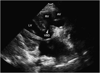
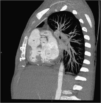
Her recent transthoracic echocardiogram, 5 years after her Fontan procedure, demonstrated preserved left ventricular function with increased circumferential hypertrophy. The ventricular septal defect was significantly restrictive (peak gradient of 78 mmHg with mean gradient of 44 mmHg) with severe dynamic subaortic obstruction (peak instantaneous gradient of 130 mmHg and a mean gradient of 77 mmHg; Fig 1). The bidirectional cavopulmonary shunt and Fontan pathway were widely patent with good calibre pulmonary artery branches. A spiral cardiac computed tomography was obtained as a pre-operative imaging tool to explore the relationship of the great vessels and the size of the pulmonary artery stump for the planned creation of a Damus–Kaye–Stansel connection (Fig 2).
Echocardiographic imaging of the restrictive VSD. AV = aortic valve; LV = left ventricle; RV = right ventricle; VSD = ventricular septal defect.

Cardiac computed tomography revealing the presence of a small main pulmonary stump and its relationship to the aorta. AV = aortic valve; LAA = left atrial appendage; LV = left ventricle; MPA = main pulmonary artery; PV = pulmonary valve; RV = right ventricle; VSD = ventricular septal defect.

The decision was made to surgically relieve her systemic outflow obstruction in the form of a Damus–Kaye–Stansel connection. Through a midline sternotomy, she was placed on cardiopulmonary bypass via ascending aortic and right femoral venous cannulation using a 17-French Bio-Medicus (Medtronic Incorporation, Minneapolis, Minnesota, United States of America) venous cannula, which was advanced into the Fontan conduit under transesophageal echocardiographic guidance. Moderate hypothermic cardiopulmonary bypass support was initiated. The pulmonary venous drainage was managed by placing a left ventricular vent through the left atrial appendage. After achieving a diastolic arrest with antegrade cardioplegia administration, the aorta was transected above the sinotubular junction and the distal aorta was reflected to dissect and mobilise the main pulmonary artery stump. The stump was densely adherent to the Fontan anastomosis and was only a few millimetres long. The stump was opened and enlarged by extending the arteriotomy into the left and right pulmonary sinuses. The pulmonary valve appeared tricuspid and pliable with good coaptation of the leaflets. There was also no evidence of thrombus in the stump or presence of subpulmonic narrowing. The facing sinuses of the aortic and pulmonary valves were sutured with polypropylene suture. Using the modified Damus–Kaye–Stansel double-barrel technique, the ascending aorta was anastomosed in an end-to-end manner to the amalgamation of aortic and pulmonary sinuses without a need for a patch. Post-operative transesophageal echocardiogram demonstrated a widely patent Damus–Kaye–Stansel and mild aortic and neoaortic insufficiency with an estimated peak gradient of 10 mmHg across the ventricular septal defect and no left ventricular outflow tract obstruction. She was extubated in the operating room and was discharged home 3 days post-operatively. At the 3- and 6-month follow-up, she seemed very active with no limitation in her exercise activity and stable echocardiographic parameters.
Discussion
In patients with tricuspid atresia with transposed great arteries and right ventricular hypoplasia, the natural trend is to undergo the single-ventricle pathway with completion of the Fontan procedure. The patient may undergo pulmonary artery banding or division with a systemic-to-pulmonary shunt to control high pulmonary blood flow early in infancy.Reference Fontan and Baudet 1 The majority of the patients will have a subsequent bidirectional cavopulmonary shunt or Fontan operation without developing systemic outflow obstruction, either from subaortic infundibular narrowing, restrictive bulboventricular foramen, or a combination of both.Reference Freedom, Sondheimer, Dische and Rowe 2 Surgical management of left ventricular outflow tract obstruction is addressed early at the time of bidirectional cavopulmonary shunt or Fontan operation in the form of subaortic muscle resection, enlargement of the restrictive ventricular septal defect, or creation of the Damus–Kaye–Stansel connection.Reference Trusler and Freedom 3
Advantages of the Damus–Kaye–Stansel procedure include better outflow gradient relief, lower incidences of post-operative heart block, and reoperation. Disadvantages of this approach include increased semilunar valvar insufficiency, lack of feasibility when attempting septation-type operations for univentricular hearts, and a technically more difficult operation.Reference Pass, Solowiejczyk and Quaegebeur 4 , Reference Cerillo, Murzi, Giusti, Crucean, Redaelli and Vanini 5 However, direct bulboventricular foramen muscle resection is less reliable for long-term relief, and it also carries a significant risk of heart block and ventricular dysfunction. An unobstructed competent pulmonary outflow allows the Damus–Kaye–Stansel procedure to be performed with complete relief of left ventricular outflow tract obstruction, even in patients with a transected main pulmonary artery or in the presence of a thrombus.Reference Broekhuis, Brizard, Mee, Cochrane and Karl 6 , Reference Fiore, Rodefeld and ViJay 7
We present a case of tricuspid atresia and dextro-transposition of the great arteries with associated left ventricular outflow tract obstruction and restrictive ventricular septal defect, which had worsened several years after the Fontan procedure. The relive of left ventricular outflow tract and restrictive ventricular septal defect, in most part, are performed at the time of the bidirectional cavopulmonary shunt or Fontan procedure in the form of Damus–Kaye–Stansel or direct ventricular septal muscle resection to relieve pressure load on the systemic ventricle while there is minimal scarring from previous operation. This reoperation was further complicated because of the dense adhesions created by the Fontan conduit, making dissection for venous cannulation difficult. Therefore, venous drainage was accomplished via single femoral venous cannula advanced into the Fontan conduit under transesophageal echocardiographic guidance. The pulmonary venous and coronary sinus drainage were accomplished by placing a vent into the left atrial appendage. The aorta was cannulated directly underneath the innominate artery and was transected above the sinotubular junction. This allowed exposure and dissection of the posteriorly located main pulmonary artery stump, which was short and densely adhered to the Fontan anastomosis. Mobilisation of the ascending aorta and aortic and pulmonary roots are critical for a tension-free anastomosis, avoiding posterior compression of the Fontan and branch pulmonary artery confluence, as well as neoaortic insufficiency. The stump of the main pulmonary artery required enlargement by extending the arteriotomy into the facing sinuses of the pulmonary valve. The facing sinuses of the aortic and pulmonary valves were sutured together and the amalgamation was anastamosed in an end-to-end, double-barrel manner to the ascending aorta without patch augmentation.
Although echocardiography and cardiac catheterisation are the mainstay modalities in determining intracardiac anatomy and haemodynamic measurements, spiral cardiac computed tomography was instrumental in delineating extracardiac anatomy and status of the pulmonary artery stump. The electrocardiographic gated cardiac computed tomography provides three-dimensional representations of the heart and the great vessels images, which are reconstructed at a given cardiac cycle. The cardiac computed tomography aided in surgical planning by providing a clear anatomical picture of the relationship of the great vessels and the Fontan confluence, while avoiding the risk of cardiac catheterisation and femoral vessel injury.
Conclusion
A case of tricuspid atresia with dextro-transposition of the great arteries and associated hypoplastic right ventricle underwent a successful repair of systemic outflow obstruction by creation of a modified double-barrel Damus–Kaye–Stansel connection several years after the Fontan procedure. Despite the fact that the main pulmonary artery was ligated and the pulmonary valve was non-functional for many years, the pulmonary valve could still be utilised for this procedure with minimal insufficiency or stenosis. Pre-operative spiral cardiac computed tomography scan was beneficial in determining the feasibility of the Damus–Kaye–Stansel connection and details of the relationships of the great arteries, pulmonary artery stump, and the Fontan complex.