Introduction
In the recent era of paediatric heart transplant, improvements in donor organ selection, post-operative care, and immunosuppressive regimens have resulted in 1-year survival of approximately 90%.Reference Rossano, Dipchand and Edwards1 Concomitantly, while there has been a decrease in the incidence of early rejection from 60% to 20–40%, there has not been a decrease in haemodynamically compromising rejection, the most severe form of rejection with significant long-term sequelae.Reference Gossett, Canter and Zheng2 Timely detection of an acute rejection event remains critical as outcomes are worse when rejection leads to haemodynamic compromise,Reference Mills, Naftel and Kirklin3 and rejection still accounts for 20% of overall long-term mortality.Reference Rossano, Dipchand and Edwards1
Endomyocardial biopsy is the currently accepted standard for diagnosing rejection after heart transplant.Reference Kobashigawa4 National guidelines and consensus statements have recommended frequent scheduled surveillance endomyocardial biopsies in the first year after transplantReference Costanzo, Dipchand and Starling5–Reference Rosenthal, Chin and Nishimura8 with widespread adoption.Reference Godown, Harris, Burger and Dodd9 This biopsy-reliant surveillance, however, remains costly and invasive. Biopsy-related complications occur at a rate of 0.2–3%Reference Bhat, Burwig and Walsh10, Reference Baraldi-Junkins, Levin, Kasper, Rayburn, Herskowitz and Baughman11 and in the paediatric population there is a 0.6% rate of serious complications.Reference Pophal, Sigfusson and Booth12 Concomitantly there is the recognition that biopsy-related costs are significant drivers of surveillance expense: 52% in the first post-operative year.Reference Ye, Parker, Brink, Weintraub and Konstantinov13 Yet despite efforts to decrease the number of endomyocardial biopsies, there remains significant inter-centre variability reported in the intensity of biopsy frequency for rejection detection following paediatric heart transplantation.Reference Godown, Harris, Burger and Dodd9, Reference Dodd, Brady, Carden, Frist, Boucek and Boucek14, Reference Valantine, Yeoh and Gibbons15 While many centres incorporate echocardiography in rejection surveillance/screening, published single centre reports evaluating the adequacy of various imaging strategies for rejection detection have had mixed results.Reference Rosenthal, Chin and Nishimura8, Reference Moran, Lipshultz and Rifai16 The integration of endomyocardial biopsies with echocardiography for rejection surveillance in the first critical year has not been systematically evaluated. Yet, as the population of post-transplant patient increases, optimising rejection surveillance protocols—in terms of both medical and cost efficacy—grows in importance.
Given the wide variability of current practices for rejection detection in paediatric recipientsReference Godown, Harris, Burger and Dodd9 and the limited evidence on which to initiate a multi-centre clinical trial to establish standard practices for the intensity of endomyocardial biopsies, we retrospectively assessed the effectiveness of a low-intensity biopsy surveillance protocol with less frequent scheduled surveillance endomyocardial biopsies integrated with clinical and non-invasive monitoring including standardised serial echocardiography. The intent of this study is not to demonstrate the ability of our specific echocardiographic methodology to detect rejection; our aim is to describe the outcomes of recipients monitored with a low-intensity biopsy surveillance protocol in relation to contemporary outcomes reported in paediatric registries.
Methods
Seattle Children’s Hospital Institutional Review Board approved the study protocol. A retrospective chart review identified patients who received cardiac transplant at our institution from January 2008 through November 2014. Re-transplant or multi-organ transplant patients were excluded. In addition, patients who died during the initial post-operative hospitalisation or were not followed post-operatively at our institution were excluded. We extended the first annual date to 13 months as some patients received their annual surveillance biopsy slightly after 12 months. For clinical information, an investigator reviewed each patient’s medical record from the time of transplant until January 2017.
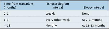
Standard immunosuppression at our institution consists of induction with rabbit anti-thymocyte globulin (thymoglobulin), steroid coverage for the first 10 weeks, and mycophenolate mofetil with either cyclosporine (less than 2 years of age) or tacrolimus during the maintenance phase. Graft surveillance is scheduled according to Table 1. Serial protocolised multi-parametric echocardiographic analyses were employed both for scheduled surveillance and for symptom-initiated rejection screening that included computer-assisted measurements of 2D-guided M-mode tracingsReference Boucek, Mathis and Boucek17–Reference Boucek, Boucek and Asante-Korang19 as well as pulse wave and tissue Doppler imagingReference Asante-Korang, Fickey, Boucek and Boucek20, Reference Pauliks, Pietra and DeGroff21 to detect acute changes in the following: left ventricular systolic and diastolic function, left ventricle chamber size and wall thicknesses, pericardial effusion, and/or atrioventricular valve incompetence.Reference Putzer, Cooper, Keehn, Asante-Korang, Boucek and Boucek22 This echocardiographic protocol, used by a small number of United States paediatric transplant centres, has both retrospective and prospective concordance with biopsy evidence for treatable cellular rejection.Reference Boucek, Mathis and Boucek17, Reference Tantengco, Dodd, Frist, Boucek and Boucek18, Reference Putzer, Cooper, Keehn, Asante-Korang, Boucek and Boucek22 Endomyocardial biopsies were performed in the cardiac catheterisation laboratory where right heart haemodynamics and endomyocardial biopsies were evaluated for histopathologic evidence of cellular or antibody-mediated rejection. Cellular rejection was graded according to the 2005 criteria whereby any patient with acute cellular rejection grade 2R or higher was treated for rejection.Reference Stewart, Winters and Fishbein23 Antibody-mediated rejection was based, in part, on C4D staining with any patient with pAMR2 or higher treated.Reference Berry, Angelini and Burke24
Low-intensity biopsy schedule used at Seattle Children’s Hospital between 2008 and 2014.

The frequency of echocardiograms, endomyocardial biopsies, and cardiology clinic visits was individually reviewed to determine if these were performed as part of routine surveillance or because of a new concern for rejection during the first 13 months after transplant. Assessments performed for another reason, such as evaluation for residual anatomic defect alone, were not included. For the same reason, echocardiograms were tabulated starting 2 weeks after transplant so as not to include studies mainly evaluating for post-operative surgical and procurement complications.
Consistent with the Pediatric Heart Transplant Study registry, a rejection event was defined as escalation in immunosuppression to treat rejection.Reference Everitt, Donaldson and Casper25 In this report, we further defined treated rejection as the escalation of immunosuppression and admission to the hospital to clearly differentiate rejection from adjustment of immunosuppression similar to Rossano et al who classified treated rejection conditional on being discharged from the hospital.Reference Rossano, Dipchand and Edwards1 Clinical evidence of a possible rejection event consisted of new arrhythmias or electrocardiogram changes, signs and symptoms of heart failure, and/or changing laboratory markers such as rising B-type natriuretic peptide concentrations. Patients with clinical and/or echocardiographic changes consistent with rejection were either scheduled for a confirmatory endomyocardial biopsy and/or admitted for augmentation of immunosuppressive therapy, typically with high-dose corticosteroids. Haemodynamically compromised rejection was defined as the need for inotropic support in the first 24 hours of a rejection-related hospitalisation. Clinical outcome measures, which would be implicated in both over- as well as under-immunosuppression, included cardiac allograft vasculopathy, post-transplant lymphoproliferative disorder, and all-cause graft loss. To facilitate comparisons, these outcome measures were published both conditional and non-conditional on survival to discharge.
To quantify the financial impact of surveillance strategies, Seattle Children’s cost information was extracted for two representative types of patient encounters: an outpatient endomyocardial biopsy with an associated echocardiogram without overnight hospitalisation as well as an outpatient clinic visit with an echocardiogram. Costs were analysed from the perspective of hospital costs, inclusive of overhead allocation, in 2015 United States dollars. Costs associated with laboratory testing were not included as the utilisation of laboratory measures during these illustrative outpatients’ visits would vary between practices.
Statistical analysis
Patient demographics and clinical characteristics are summarised using counts and percentages for categorical data and means and standard deviations for continuous data. Survival, both conditional and non-conditional on survival to discharge, was calculated, the latter to permit using registry results as reference standards. Comparisons between those with rejection and those without rejection were made using t-tests for continuous variables and chi-square tests for categorical variables. Kaplan-Meier time-to-event curves were created for time to first rejection in the first 13 months after transplant, as well as time to coronary artery vasculopathy, post-transplant lymphoproliferative disorder, and graft loss censored to the latest follow-up in this study. Analyses and summaries were performed using StataCorp. 2017 (Stata Statistical Software: Release 15. College Station, TX: StataCorp LLC).
Results
Study cohort
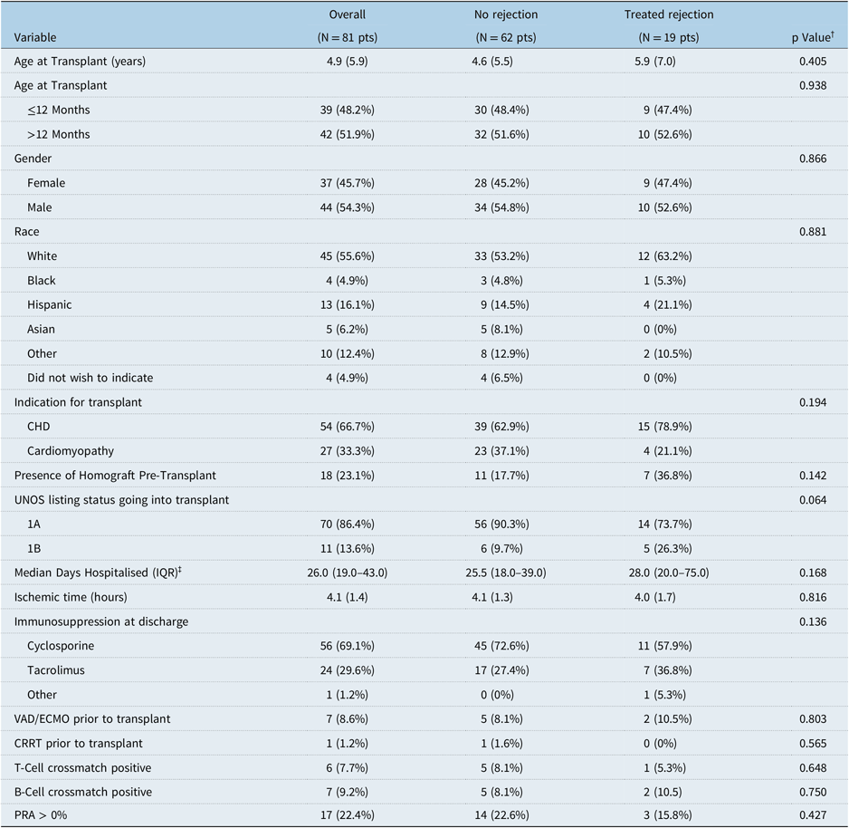
A total of 91 patients underwent cardiac transplantation during the study period, 10 of whom were excluded for the following indications: 4 died prior to discharge (1–137 days post-transplant), 2 had received a prior transplant, and 4 were transferred to another centre within 13 months after transplant. Clinical and demographic characteristics of the 81 remaining discharged study patients are outlined in Table 2. Although some differences were observed between those who suffered a rejection event and those who did not, none of these characteristics were statistically significant except for the length of hospital stay. Of note, age, presence of a homograft, positive panel reactive antibodies, and positive direct cross match were not associated with rejection in the first-year post-transplant.
Recipient characteristics and by rejection status in the first 13 months after heart transplant. All continuous variables stated as mean (standard deviation). All categorical variables stated as count (percentage).

CHD = Congenital Heart Disease; CRRT = Continuous Renal Replacement Therapy; ECMO = Extracorporeal Membranous Oxygenation; VAD = Ventricular Assist Device; UNOS = United Network for Organ Sharing.
† p-Value from t-test for continuous variables chi-squared for categorical comparing rejection to no rejection groups.
‡ p-Value from Wilcoxon rank sum.
Early rejection and surveillance studies
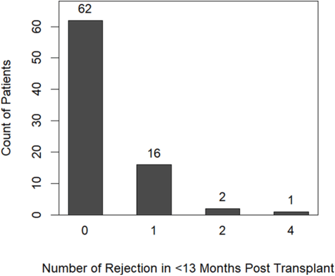
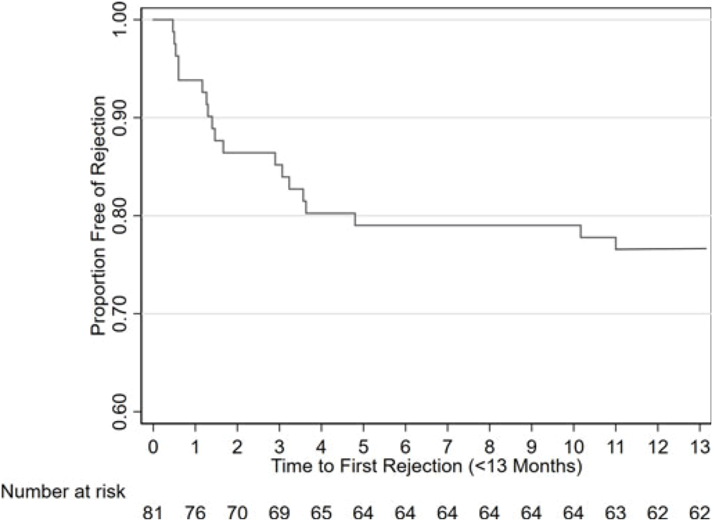
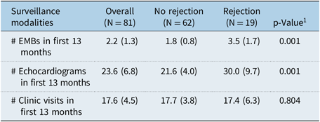
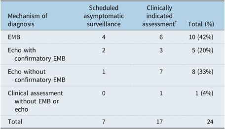
In the first 13 months after transplant, 19 recipients (23.4%) had 24 treated rejection events (Fig 1). As expected, these recipients had a higher number of endomyocardial biopsies and echocardiographic investigations compared to those without rejection in the first year (p = 0.001, Table 3). Overall, there was a mean of 1.9 (SD ± 1.0) months between an episode of treated rejection and subsequent endomyocardial biopsy. For recipients without treated rejection, the mean interval between endomyocardial biopsies was 7.6 ± 4.1 months while for recipients with rejection, the interval decreased to 1.5 ± 1.2 months. Among the 19 patients with early rejection, the majority were diagnosed within 4 months of transplant, with an average time to first rejection of 2.8 months (Fig 2). Of the 24 treated rejection events, 4 were identified at scheduled surveillance biopsies, 3 were identified by asymptomatic surveillance echocardiogram, and 17 were identified based on new clinical suspicion for rejection (Table 4). Of the 4 discovered by asymptomatic surveillance biopsy in the 13 months following transplant, 3 were early (average 2.5 months post-transplant) and 1 was late in the first year (10 months post-transplant). Of the 24 rejection events, 9 were diagnosed and treated without an endomyocardial biopsy. Of the remaining 15 treated rejection events supported by an endomyocardial biopsy, 11 also had antecedent clinical or echocardiographic evidence of rejection. Of the 24 rejection events, 4 (16%) were haemodynamically significant (4.9% of the total cohort).
Distribution of rejection events in the first 13 months after transplant.

Kaplan-Meier curve illustrating freedom from rejection in first 13 months.

Method of diagnosis for rejection utilised in the first 13 months after transplant. All continuous variables presented as mean (standard deviation).

EMB = Endomyocardial Biopsy.
1 p-Value from Student t-test for comparing rejection to no rejection groups.
Initial modality for detection of treated rejection.

EMB = Endomyocardial Biopsy.
† Patients with symptoms, laboratory or ECG changes.
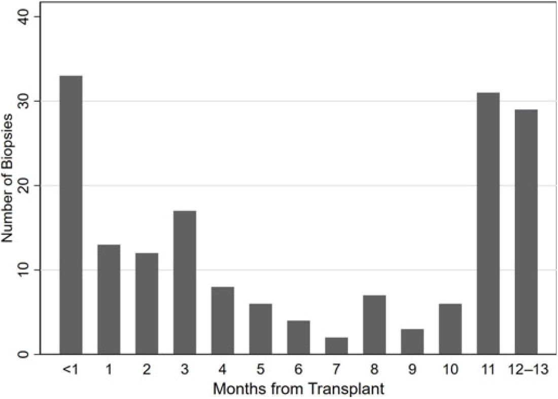
Overall using low-intensity biopsy surveillance resulted in an average of 2 ± 1.3 endomyocardial biopsies, 24 ± 6.8 echocardiograms, and 17 ± 4.4 clinic visits per patient in the first 13 months after transplant (Table 3). Reflecting the low-intensity biopsy surveillance schedule outlined in Table 1, 28 biopsies (16%) were performed at 3 ± 1 month and 60 biopsies (34%) were performed at 12 ± 1 month (Fig 3). The remainder (50%) were performed outside of the surveillance endomyocardial biopsy protocol at an average of 4 months post-transplant.
Biopsy count by month after transplant.

Intermediate-term follow-up and adverse events
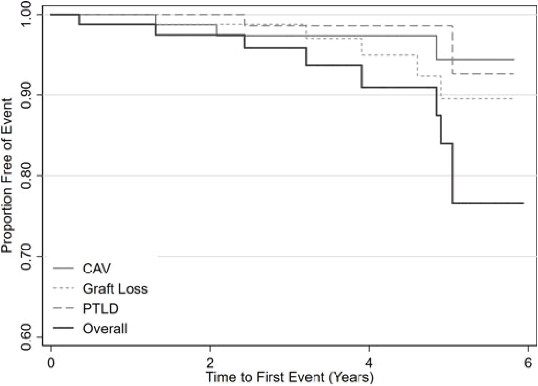
At an average latest follow-up of 4.7 years + 2.12 the outcome measures were as follows: four patients (4.9%) had coronary artery vasculopathy at an average 3.6 years post-transplant, three patients (3.7%) were diagnosed with post-transplant lymphoproliferative disorder at an average of 4.2 years post-transplant, and seven patients (8.6%) suffered graft loss at an average of 4.2 years post-transplant with four deaths at an average of 3.4 years post-transplant (Fig 4). Of these adverse outcomes, there was a rejection history in one of four patients who went on to develop cardiac allograft vasculopathy, three of seven graft loss, and none of the three post-transplant lymphoproliferative disorder patients. The 3-, 12-, 36-, and 60-month graft survival rates conditional on survival to hospital discharge were 100%, 98.8%, 98.8%, and 90.4%, respectively. The non-conditional 3-, 12-, 36-, and 60-month graft survival rates were 97.7%, 94.1%, 94.1%, and 85.3%, respectively, which included the four patients who died prior to discharge. Patients who transferred care to other institutions were not included in either set of survival statistics.
Kaplan-Meier curves illustrating time-course (years) of adverse events for those discharged after transplant. CAV = Cardiac Allograft Vasculopathy; PTLD = Post-Transplant Lymphoproliferative disorder.

Institutional costs
Regarding the costs implicated in either surveillance strategies, the hospital costs for an outpatient surveillance clinic encounter was $1264 dollars, of which $513 was for the echocardiogram, $178 was for the electrocardiogram, and $573 was for the professional fees. On the other hand, costs for a same-day stay with right heart catheterisation and endomyocardial biopsy was $8975 dollars which includes a post-procedural echocardiogram.
Discussion
This study describes the incidence of early rejection as well as major adverse events in the intermediate term follow-up of paediatric cardiac transplant recipients who underwent rejection surveillance by a low-intensity biopsy surveillance protocol in the first year. When surveilled with such a protocol, our patients demonstrated similar short- and intermediate-term outcomes when referenced to aggregate results from registry data where in general the biopsy frequency is higher.Reference Godown, Harris, Burger and Dodd9, Reference Zinn, Wallendorf, Simpson, Osborne, Kirklin and Canter26 For example, 23.4% of our patients discharged from the hospital had early rejection within the first year post-transplant. By comparison, Gossett et al reported a decreasing incidence of early rejection from 60% in 1993 to 40% in 2005,Reference Gossett, Canter and Zheng2 while Rossano et al reported in 2016 a more contemporary cohort, less than 20% incidence of early rejection also conditional on being discharged from the hospital.Reference Rossano, Dipchand and Edwards1 In our study, the incidence rate for rejection within the first 13 months was 0.27 rejection episodes per patient-year (95% CI: 0.17–0.42) compared to incident rates in an earlier era of between 0.5 and 1.0 rejection episodes per patient-year depending on era, as reported by Gossett.Reference Gossett, Canter and Zheng2 The rejection incidence rate for the study by Rossano et al is not reported but would likely be lower. Lastly, 4.9% of our cohort experienced haemodynamically compromised rejection which is similar to the 5% previously reported in the recent era.Reference Everitt, Pahl and Schechtman27
As shown in Table 2, there was no difference in the distribution of rejection by age in the first year post-transplant, which is dissimilar from registry reports where rejection in infants is expected to be less common compared to adolescents.Reference Zinn, Wallendorf, Simpson, Osborne, Kirklin and Canter28 However, while a relatively high percentage of our heart transplant recipients are less than 12 months (48%), there is a large proportion (67%) with congenital heart disease which could have imposed a higher rejection risk in these younger patients. If the rejection rate was lower in our infant population, then one can conjecture that it is because of fewer endomyocardial biopsies in our protocol, but the opposite was found. In an international survey of 47 sites, the median and mode of programs perform between five and seven biopsies in the first year after transplant across all age groups.Reference Zinn, Wallendorf, Simpson, Osborne, Kirklin and Canter26 The dependence on performing clinically indicated endomyocardial biopsies, as opposed to surveillance biopsies, to diagnose rejection is also consistent with the recent report by Zinn et al that of all treated rejection events in the first year after transplant reported to Pediatric Heart Transplant Study, an accompanying surveillance biopsy accounted for only 25.5% of such events in infants and 51.7% in adolescents.Reference Zinn, Wallendorf, Simpson, Osborne, Kirklin and Canter28
A theoretical concern regarding a low-intensity biopsy surveillance protocol is that the diagnosis of rejection could be delayed until the patient is “symptomatic”. Alternatively, a low-intensity biopsy surveillance protocol could also lead to over-treating rejection based on echocardiographic or clinical findings (false positives) that are not always confirmed by endomyocardial biopsy. Because this study was not designed to assess the accuracy of echocardiographic or clinical indicators of rejection, we addressed these concerns by examining intermediate-term adverse outcomes as surrogates for the misdiagnosis of rejection with low-intensity biopsy surveillance, resulting in either under- or over-immunosuppression. Importantly, none of the 62 patients who were considered rejection-free during the first year post-transplant using the low-intensity biopsy protocol were found to have rejection at their annual surveillance endomyocardial biopsies. In addition, the cumulative incidences of coronary artery vasculopathy (4.9%), post-transplant lymphoproliferative disorder (3.7%), and graft loss (8.6%) up to 6 years post-transplant using the low-intensity biopsy protocol were also non-inferior in reference to what is reported by large registry studies. For example, Dipchand et al reported an average 5-year freedom from coronary artery vasculopathy of ~83%, post-transplant lymphoproliferative disorder of 94%, and graft loss of 72%.Reference Dipchand, Rossano and Edwards29 If indeed the rate of rejection has continued to decrease since 2005 in paediatric recipients, as reported by Gossett et alReference Gossett, Canter and Zheng2 and Rossano et al,Reference Rossano, Dipchand and Edwards1 then our findings using low-intensity biopsy surveillance may be even more compelling in lessening reliance on invasive procedures. Support for a less intensive surveillance biopsy protocol can also be found in adult literature, with a recent similar single centre study of 282 adult patients followed using a similar combination of scheduled and “symptom-triggered” endomyocardial biopsies.Reference Weckbach, Maurer and Schramm30 This group reported non-inferior outcomes using an average of seven endomyocardial biopsies in the first year—which is up to 50% fewer biopsies than comparable reporting centres.
Of note, there is precedent for asymptomatic, mild–moderate grade rejection resolving without intensification of immunosuppression or having wide variation of interpretation by pathologists.Reference Winters, Loh and Schoen31–Reference Brunner-La Rocca, Sutsch, Schneider, Follath and Kiowski34 There are also data suggesting that the number of biopsies in the first year is not associated with survival and undergoing surveillance endomyocardial biopsies beyond the first 6 months illustrate no survival advantage in adults.Reference Sethi, Kosaraju, Arabia, Roasdo, McCarthy and Copeland35 Furthermore, it is still unclear if treatment of low–moderate grade rejection in children will produce better clinical outcomes. In our cohort, the majority (87.5%) of rejection diagnoses were made in the context of echocardiographic and/or clinical change, rather than with scheduled surveillance biopsies. Accordingly, half of the biopsies in our sample were performed outside of our surveillance biopsy schedule, many of which in the first month after transplant. This is contrary to other studies in which the majority of rejection episodes assigned the International Society for Heart and Lung grade 2R and above were asymptomatic at the time of diagnosis.Reference Zinn, Wallendorf, Simpson, Osborne, Kirklin and Canter28, Reference Chin, Akhtar, Rosenthal and Bernstein36 Importantly, the definition of “symptomatic” and the threshold for clinically diagnosing rejection are both practitioner and centre specific, and this subjectivity likely contributes to the discrepancy in the frequency of reported “asymptomatic” rejection.Reference Sethi, Kosaraju, Arabia, Roasdo, McCarthy and Copeland35, Reference Chin, Akhtar, Rosenthal and Bernstein36 It is possible that we under-diagnosed subclinical or “asymptomatic” rejection events using our low-intensity biopsy surveillance protocol. However, our study aim examined clinical outcomes which illustrated that we did not have a higher rate of adverse events in the short or intermediate term, nor did we observe a higher than expected rejection rate that would have accrued by under-diagnosing rejection at the first annual surveillance endomyocardial biopsy.
Potential cost implications
While the patient outcomes at our institution support both the safety and effectiveness of this low-intensity biopsy surveillance protocol, an increasingly relevant consideration is cost in resource utilisation decisions. Considering averages from our population of 2 endomyocardial biopsies, 24 echocardiograms, and 17 clinic visits in the first 13 months after transplant, this equates to an average cost of $43,037 per patient, $17,950 of which is assigned to the biopsy cost. Recently published survey data from multiple paediatric heart transplant centres indicate an average of six scheduled surveillance biopsies are performed in the first year which would result in an estimated $53,851 per patient in biopsy costs alone at Seattle Children’s Hospital.Reference Godown, Harris, Burger and Dodd9, Reference Zinn, Simpson, Wallendorf, Osborne, Kirklin and Canter37 If we applied the frequency of six scheduled surveillance endomyocardial biopsies in the first year to our approach at Seattle Children’s Hospital—substituting the four additional endomyocardial biopsies for clinic visits and echocardiograms—the cost would be $73,881 per patient in the first 13 months after transplant.
Study limitations
Limitations to this report include those intrinsic in a single-centre retrospective study such as smaller sample size and style of practice and demographics not fully representative of transplant care at other centres, respectively. When fewer endomyocardial biopsies are performed, it is certainly possible that mild-to-moderate grade rejection could have been undiagnosed by clinical or non-invasive testing. However, the design of this study presents intermediate-term outcome measures and demonstrated that these are equivalent to representative registry outcomes. Although it is of interest to assess the accuracy of echocardiography + clinical/laboratory findings versus endomyocardial biopsies to detect rejection and coronary artery vasculopathy, an adequately powered multi-centre prospective study with a sufficiently robust echocardiographic diagnostic and surveillance protocol would be required.Reference Kobashigawa4, Reference Alonzo and Pepe38 Similarly, we cannot conclude on what is the best timing or frequency of endomyocardial biopsies, only that similar outcomes can be achieved with a low-intensity biopsy surveillance protocol. Regarding cost analysis, the lack of comparable data from a more intense endomyocardial biopsy surveillance program on ancillary direct costs (such as chest imaging and laboratory tests) limits our ability to perform a true cost comparison; we assumed large registry data represent predominantly a more intensive biopsy surveillance practice.Reference Zinn, Wallendorf, Simpson, Osborne, Kirklin and Canter28, Reference Zinn, Simpson, Wallendorf, Osborne, Kirklin and Canter37 Furthermore, while we are able to model possible cost-reductions in the setting of similar clinical outcomes, a formal cost-utility analysis of low-intensity biopsy surveillance across a patient’s life-expectancy horizon is beyond the scope of this manuscript and would be an avenue of future inquiry. Finally, since the follow-up in this study is short to intermediate term we cannot predict how the low-intensity biopsy surveillance protocol would affect longer term outcomes.
Conclusions
The standard reference of using endomyocardial biopsies to detect rejection has costs and risks that must drive careful consideration of surveillance strategies for determining its proper use. Much has been done in minimising immunosuppression in children such as lowering targeted calcineurin inhibitor levels and steroid use, but not enough has been done to study lessening invasive monitoring by endomyocardial biopsy.Reference Auerbach, Gralla, Campbell, Miyamoto and Pietra39, Reference Singh, Faber and Blume40 This study provides the first description of outcomes of a low-intensity biopsy surveillance protocol integrated with standardised clinical/echocardiographic monitoring across paediatric age groups in a contemporary era. In this study, our patient population experienced similar aggregate outcomes across both a short and intermediate term in relation to what is reported in registries. Of increasing importance, these medically equivalent results were achieved with significant anticipated cost savings compared to a more intensive surveillance biopsy schedules. Future studies are now warranted to compare a low-intensity biopsy-based protocol integrated with standardised echocardiography to a more intensive endomyocardial biopsy-based surveillance protocol in a prospectively designed multi-centre study.
Author ORCIDs
Patrick D. Evers 0000-0002-9299-1926
Acknowledgements
None
Financial Support
This research received no specific grant from any funding agency, commercial or not-for-profit sectors.
Conflicts of Interest
None.