Achiasma is a rare congenital abnormality of the visual pathway, characterized by the complete or partial (hypochiasma) absence of decussation of nasal retinal fibers at the optic chiasm. Since its first description by Apkarian et al. in 1994, Reference Apkarian, Bour and Barth1 only a small number of cases have been reported in the literature. This malformation can occur as part of broader malformative syndromes, such as midline brain defects and microphthalmia spectrum, or as an isolated anomaly. Reference Sami, Saunders and Thompson2
Clinically, achiasma manifests with a wide spectrum of findings, including congenital nystagmus, particularly of the “see-saw” type, Reference Apkarian and Bour3 visual field defects, Reference Pilat, McLean, Vanina, Dineen and Gottlob4 and characteristic ipsilateral asymmetry of visual-evoked potentials (VEPs). Reference Jansonius, van der Vliet, Cornelissen, Pott and Kooijman5 Therefore, diagnosis is usually done in childhood, and identification in adulthood is exceedingly uncommon.
We report the case of an 86-year-old patient with incidentally discovered isolated achiasma.
An 86-year-old male patient was referred for a progressive decrease in visual acuity (VA) of his left eye. His main antecedents were a congenital nystagmus and a right exotropia with right amblyopia. He also presented an arterial hypertension and a left sylvian ischemic stroke few years ago.
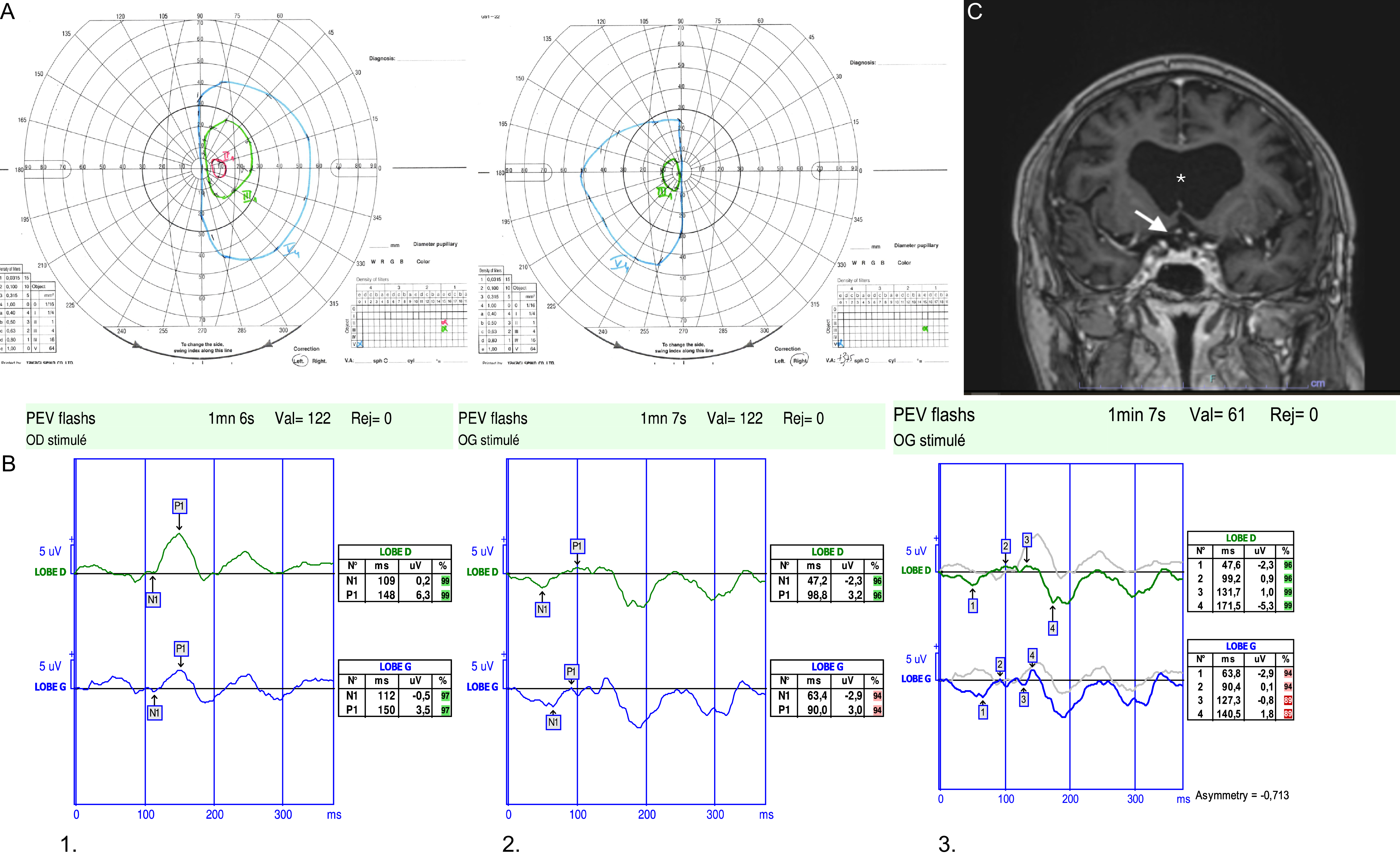
Best corrected VA was 2/10 right eye and 5/10 left eye. Slit lamp examination found a bilateral nuclear cataracts. Fundus examination showed central areolar atrophy due to age-related macular degeneration and optic disc pallor. Goldman visual field revealed bitemporal hemianopia (Figure 1A); the visual field of the right amblyopic eye did not demonstrate a pan-field loss, likely reflecting the predominantly macular origin of its reduced VA rather than a diffuse perimetric deficit. A brain MRI was performed and first interpreted as normal. Specifically, no sella lesion was found.
Multimodal imaging. (A) Goldmann visual field showing bitemporal hemianopia. (B) Monocular flash visual-evoked potential (VEP)responses. 1. Right eye responses showed ipsilateral asymmetry with better amplitudes on the right occipital lobe. 2. Left eye responses were unclonclusive. 3: Chiasm coefficient showing a value of – 0.713 of asymmetry. (C) Coronal MRI showing absence of optic chasm (arrow), note absence of septum pellucid (asterisk).

Figure 1. Long description
Panel A: Two Goldmann visual field charts showing bitemporal hemianopia. The charts display radial grid patterns with plotted points indicating visual field defects. Panel B: Three sets of monocular flash visual-evoked potential (VEP) responses. Each set includes two line graphs representing responses from the right and left occipital lobes. The graphs show time in milliseconds on the x-axis and microvolts on the y-axis. The first set shows better amplitudes on the right occipital lobe for the right eye. The second set shows inconclusive responses for the left eye. The third set shows a chiasm coefficient value of -0.713 of asymmetry. Panel C: A coronal MRI scan showing the absence of the optic chiasm and the septum pellucidum. The MRI image highlights specific areas with an asterisk and an arrow.
Patient was reassessed one year later. VA and the bitemporal visual field defect were stable. A close look at his ocular movements highlighted a pendular see-saw nystagmus in which one eye moves upward and intorts when the other eye moves downward and extorts in the first half cycle, followed by inversion of eye movement direction during the next half cycle. Flash and pattern-reversal VEPs were performed and showed ipsilateral asymmetry, more prominent on the right eye stimulation (Figure 1B). We also calculated the chiasm coefficient Reference Jansonius, van der Vliet, Cornelissen, Pott and Kooijman5 and found a value of −0.713 (Figure 1B), indicative of strong interlobar asymmetry. These new elements prompted us to re-examine the MRI: an almost complete absence of the chiasm was detected (Figure 1C).
Achiasma/hypochiasma results from an embryological defect in the guidance of retinal ganglion cell axons, leading to failure of nasal fibers to cross at the optic chiasm. Reference Sami, Saunders and Thompson2 The resulting abnormal organization of visual pathways produces distinctive clinical and electrophysiological patterns. Reference Apkarian, Bour and Barth1
In our patient, a bitemporal hemianopia was initially suggestive of a compressive lesion of the optic chiasm. However, the absence of any structural abnormality on MRI, combined with the presence of a see-saw nystagmus and the ipsilateral asymmetry of VEP responses, led to the suspicion of achiasma, subsequently highlighted by a careful review of the brain MRI.
Even if MRI scans provide objective anatomical proof of achiasma, their analysis remains subjective. VEP features may then play a key role in helping physicians to adjust their diagnostic process. Indeed, VEP may demonstrate ipsilateral asymmetry, characterized by ipsilateral response (i.e., recorded in front of the same occipital lobe) of the stimulated eye exceeding contralateral (i.e., recorded in front of the contralateral occipital lobe) response Reference Brecelj6 as it was shown for our patient. Unfortunately, nystagmus may also suppress VEP responses to pattern-reversal stimulation and make the results inconclusive as it was most probably for the left eye of our patient.
Chiasmal coefficient calculation may enhance the sensitivity of VEP for chiasmal crossing abnormalities. The chiasmal coefficient is a quantitative index derived from monocular flash VEP recordings, calculated as the ratio of the difference between ipsilateral and contralateral occipital responses to their sum; it provides a measure of the proportion of crossing fibers at the optic chiasm, with values near zero reflecting normal decussation and negative values indicating a reduced or absent crossing (as in albinism or in achiasma).
See-saw nystagmus is a rare ocular motor instability where a half cycle consists of the rise and intorsion of one eye and the depression and extorsion of the other eye. This sequence is then followed by an inversion of the ocular movements in the other half cycle. While the mechanism of this pattern is still unclear, it is mostly observed in congenital or acquired chiasmatic dysfunction, such as lesion to the optic chiasm or mesencephalon, parasellar masses, cone-rod dystrophy, or acquired severe visual loss. Reference Sami, Saunders and Thompson2
Interestingly, bitemporal hemianopia has been rarely described in association with achiasma. Indeed, Apkarian et al. described non-decussating retinal fugal fiber syndrome as the absence of optic chiasm, accompanied by specific criteria including normal visual field. Reference Apkarian, Bour and Barth1 More recently, Sami et al. proposed three types of achiasma: Type A in patients with reduced decussation with optic nerves of normal appearance on clinical examination, type B in those with reduced decussation, chiasmal hypoplasia and midline brain defects reminiscent of septo-optic dysplasia (absent septum pellucidum, optic nerve and olfactive bulbs hypoplasia and hypopituitarism), and type C in those with reduced decussation, chiasmal hypoplasia, clefting disorders and encephaloceles. Our patient appears to fall in both type B. Reference Sami, Saunders and Thompson2 We found only two other reports describing this visual field defect associated with achiasma. Reference Little7,Reference Rudich and Lesser8 This interpretation supports the notion that achiasma can reproduce visual field patterns traditionally attributed to acquired chiasmal lesions, despite an entirely different underlying mechanism.
Achiasma is a rare congenital anomaly that may go unrecognized in adults due to its subtle or compensated manifestations. This case emphasizes the importance of thorough evaluation in patients presenting with bitemporal hemianopia without tumoral sella lesions on imaging. The combination of a see-saw nystagmus and VEP ipsilateral asymmetry should alert clinicians about chiasmal crossing disorders and prompt detailed MRI investigation of the optic chiasm. This case highlights both the rarity of achiasma in the elderly and the key clinical and electrophysiological clues for this diagnosis.
Author contributions
MS: corresponding author, doctor in charge of the case from the date of the follow-up visit.
VP: first doctor in charge of the patient.
VS: assistance and advice given based on the file.
Funding statement
We declare having no funding.
Competing interests
We declare no competing interests for any authors.