INTRODUCTION
Apicomplexan parasites (Plasmodium, Toxoplasma, Cryptosporidium, Neospora, Eimeria, Theileria, etc.) are responsible for major loss of human life and livestock worldwide. Coccidiosis is an important disease caused by several different species of the genus Eimeria, and is responsible for significant losses of revenue in the chicken industry of about 2 billion dollars per annum. Like other apicomplexans, the Eimeria parasite undergoes a complex life cycle (i.e. sporogony, merogony, gamogony) in the epithelial cells lining the intestinal mucosa of an infected host. The parasite is transmitted by environmentally stable oocysts, which develop as a result of sexual reproduction, whereby male gametes (micro-) fertilize the female (macro-) gametocytes.
We and others have studied the antibody response to Eimeria sexual stage parasites and showed that chickens exposed to a live Eimeria infection secrete highly protective anti-gametocyte antibodies that can be transferred via passive or maternal immunization to young offspring chicks (Wallach et al. Reference Wallach, Mencher, Yarus, Pillemer, Halabi and Pugatsch1989, Reference Wallach, Pillemer, Yarus, Halabi, Pugatsch and Mencher1990, Reference Wallach, Halabi, Pillemer, Sarshalom, Mencher, Gilad, Bendheim, Danforth and Augustine1992, Reference Wallach, Smith, Braun and Eckert1995a , Reference Wallach, Smith, Petracca, Miller, Eckert and Braun1995b , Reference Wallach, Ashash, Michael and Smith2008; Smith et al. Reference Smith, Wallach, Miller, Braun and Eckert1994a , Reference Smith, Wallach, Miller, Morgenstern, Braun and Eckert b , Reference Smith, Wallach, Petracca, Braun and Eckert c ; Wallach, Reference Wallach1997, Reference Wallach2010; Ferguson et al. Reference Ferguson, Belli, Smith and Wallach2003). These antibodies act to markedly reduce parasite development and oocyst shedding thus preventing the transmission of the disease (i.e. transmission blocking immunity). Indeed, this principle has been used in the development of a subunit vaccine against coccidiosis, CoxAbic®. Over the years research has been carried out on the molecular basis for the immunity induced by this vaccine, however, no direct evidence was provided showing that the induced anti-gametocyte antibodies are capable of reaching the parasites developing in the intestinal epithelial cells in vivo.
In order to evaluate the ability of anti-gametocyte IgG to reach Eimeria maxima macrogametocytes in vivo, chickens were immunized with E. maxima extracted wall forming body (WFB) antigens and 1 week later were challenged with E. maxima oocysts. After 6 days, sexual stage parasites were isolated from the infected chicken and were incubated with secondary antibody conjugated to FITC in order to localize the serum antibodies by 3D confocal microscopy. Results presented here show that WFB antigens induced the production of high-titre serum IgG, which has the ability to reach in situ gametocytes in the intestinal lumen and permeate the enterocyte/parasite membranes in order to bind to the cytoplasmic Type 1 and Type 2 WFBs.
MATERIALS AND METHODS
Animals and parasites
The Houghton strain of E. maxima used throughout these experiments was originally provided by Martin Shirley (Institute for Animal Health, Compton, Newbury, Berkshire, UK). The oocysts were periodically propagated in 3–7 week-old light breed Australorp chickens (Barter and Sons Hatchery, Luddenham, Australia) using established methods (Wagenbach et al. 1966). Oocysts were harvested from the faeces, sporulated in vitro at 30 °C in an incubator and isolated by salt flotation and bleach treatment (2%, Milton solution). Gametocytes were isolated at 134 h post infection (p.i.) and purified from infected chicken intestines by techniques published previously (Wallach et al. Reference Wallach, Mencher, Yarus, Pillemer, Halabi and Pugatsch1989), and stored as aliquots at −80 °C until required.
Preparation of wall-forming body antigens
To prepare antigen for immunization experiments, wall-forming bodies (WFBs) of E. maxima macrogametocytes were extracted essentially as described previously (Frölich et al. Reference Frölich, Johnson, Robinson, Entzeroth and Wallach2013) with a minor modification. Briefly, the clean freeze–thawed gametocytes (6×10⁶ cells) were extracted with 0·1% Saponin in TNEP (10 mm Tris–HCl, pH 7·4, 50 mm NaCl, 2 mm EDTA) for 20 min at room temperature and centrifuged at 1000 g for 5 min. The resulting pellet was washed three times in TNEP by centrifugation at 3000 g for 2 min, and the supernatant discarded. The pellet was re-suspended in TNEP and mechanically dispersed by ultrasonication (Vibra Cell Sonicator) using a 2-mm microtip and a wave amplitude of 2·5 W, 3 times for 10-s with intervals with cooling on ice between each burst. The sexual stage parasites were checked for complete lysis based on microscopy. The sonicate was then centrifuged at 15 000 g for 10 min and the pellet was resuspended in 1% sodium-dodecyl-sulphate (SDS) (w/v) and the mixture was centrifuged at 15 000 g for 10 min. The resulting WFB rich extract was then concentrated on a 1000 kDa cut-off filter (MWCO, VivaSpin) and washed three times in TNEP by centrifugation (4000 g for 20 min). Protein concentration was measured using the bicinchoninic protein assay kit (Pierce) with bovine serum albumin (BSA) as the standard prior to storing at −20 °C for use in immunization experiments (see below).
Immunization, challenge with oocysts and collection of mucosal scrapes
The flow chart describing steps involved in the immunization regime is shown in Fig. 1. Chickens kept coccidia free were allowed unlimited time to feed. Immunization of chickens with purified WFBs antigens was carried out as follows. At 3 weeks of age, the chickens were immunized intramuscularly with either 50 μg of WFB-rich fraction (WFB-immunized group), or sham-immunized (adjuvant-immunized group) with PBS in Freund's adjuvant (Sigma), or were left unimmunized (un-immunized group). The chickens were injected intramuscularly with 0·5 mL emulsion into their breast muscle. The first boost was administered at 4 weeks of age and the final boost was administered 2 weeks prior to challenge. Reactivity of sera from all groups was screened by Western blotting and indirect immunofluorescence analysis.
Flow chart of the experimental protocol for immunization.

At 7 weeks of age, the chickens were challenged with 50 000 sporulated E. maxima oocysts per os and infected small intestines were removed 134 h post infection. Following extensive washing with ice-cold PBS, the intestines were slit open lengthwise and a 1 cm2 piece was cut from around the diverticulum. The exposed mucosa was then gently scraped using a glass cover slip and scrapings collected into an Eppendorf tube. The scrapings were re-suspended in TNEP buffer (10 mm Tris–HCl, pH 7·4, 50 mm NaCl, 2 mm EDTA) and mixed by brief vortexing. The quality of harvested parasites was tested by Evans blue staining using methods published previously (Ferguson et al. Reference Ferguson, Belli, Smith and Wallach2003). Secreted IgG was analysed by incubating suspended scrapings with FITC conjugated rabbit anti-chicken IgG (Fab`)2 antibody (Jacksons Research Laboratories) diluted to 1:500. Following 1-hour incubation at 4 °C in the dark, samples were washed three times in PBS by centrifugation at 800 g for 5 min, pipetted onto a slide coated with 0·1% poly-L-Lysine and mounted using Vectashield® (Vector Laboratories, Burlingame, CA, USA) prior to confocal microscopy.
Indirect immunofluorescence analysis
To evaluate specificity of the extracted WFB antigens, isolated gametocytes were fixed for 5 min at room temperature with either ice-cold methanol (95% v/v), or ice-cold acetone (95% v/v), permeabilized in 0·1% Tween20/PBS and non-specific binding was blocked with either normal mouse or chicken serum (1:500 dilution) for 1 h, at room temperature. The harvested gametocytes were then incubated with a panel of antibodies, including: mouse anti-affinity purified gametocyte antigen (APGA) antibody (1:200 dilution) generated to a fraction of macrogametocyte proteins enriched for gam56 and gam82, amongst other molecules (Fried et al. Reference Fried, Mencher, Sarshalom and Wallach1992); mouse monoclonal anti-WFB2 1E11-11 antibody (1:200 dilution) raised against E. maxima gametocyte glycoprotein gam56, or chicken anti-WFB polyclonal antibodies (pAbs) generated to a fraction enriched for E. maxima macrogametocyte WFBs (1:500 dilution). Incubations were carried out for 1 h at room temperature in the dark. Following washes with PBS, primary antibodies were detected with Alexa Fluor 549–conjugated goat anti-mouse IgM (1:500 dilution, Invitrogen, Australia), or with FITC conjugated rabbit anti-chicken IgG (Fab`)2 antibody (1:500 dilution, Jacksons Research Laboratories). As a control for non-specific binding, primary antibody was either omitted or substituted with the normal mouse or chicken sera (pre-bleed, 1:500) or the sera collected from birds immunized with adjuvant (1:500).
Confocal microscopy
Localization of bound antibodies was visualized with an inverted Nikon A1 laser scanning confocal microscope equipped with 60× and 100× oil objective lens (Plan Apo NA 1.4 aperture) MadCity ultra-fast piezo Z-drive, galvano scanners and Perfect Focus System™ for continuous maintenance of focus. Stained cells were examined with a green (488/500–550 nm), red (561/570–620 nm) and far red (637/662–737 nm) lasers. To acquire three-dimensional volumes, ranging from 25 to 91-z serial optical sections were collected with a step size of 0·25 μm, using NIS Elements software (Nikon). The images were processed in NIS-Elements Viewer software, Advanced Version (Nikon, Melville, NY, USA). The laser intensity, photomultiplier tube gains (PMT-HV), and photomultiplier offset (PMT-offset) levels were kept constant during the image acquisition. Raw images were reconstructed and rendered in 3D using IMARIS v.7 software (Bitplane Scientific, Zurich, Switzerland).
SDS-PAGE and Western blotting
For identification of immunogenic WFB antigens, proteins from gametocyte lysate were separated by size as described previously (Frölich et al. Reference Frölich, Johnson, Robinson, Entzeroth and Wallach2013). Laemmli sample buffer containing β-mercaptoethanol and 10% SDS was added prior to boiling for 10 min at 95 °C. Samples were then centrifuged at 12 000 g for 5 min, at room temperature to remove insoluble and aggregated matter prior to SDS–PAGE on NuPAGE® Novex 4–12% Bis–Tris gradient gels (Invitrogen). Total protein (4 μg) loaded per lane was determined by bicinchoninic protein assay (Pierce). The proteins were then visualized by staining with Coomassie or, alternatively, transferred to polyvinylidene difluoride (PVDF, Pall Corporation) membranes and incubated with mouse anti-1E11–11 monoclonal antibody (1:500), mouse anti-APGA (1:500), chicken anti-WFB antibody (1:2000) or normal mouse or chicken serum (1:500). PVDF membranes were then incubated with alkaline-phosphatase conjugated rabbit anti-mouse IgM or anti-chicken IgG (1:1000; Sigma) and developed with 5-bromo-4-chloro-3-indolyl-phosphate/nitro blue tetrazolium (SIGMA FAST ™ BCI/NBT, Sigma) according to the manufacturer's instructions.
RESULTS
Polyclonal anti-WFB antibody localizes to the intracellular WFBs and the oocyst wall
We purified gametocyte WFBs as described in Materials and Methods, emulsified them in Freund's adjuvant and used 50 μg of protein to immunize naïve chickens for the production of pAbs. The resulting sera were tested by IFA on E. maxima purified gametocytes to study the sub-cellular localization of the WFBs. In addition, the sera were tested by Western blotting using purified gametocyte lysate to analyse the molecular weights and band intensity of the antigens that were recognized in comparison with control anti-APGA serum. A monoclonal antibody to the 56 kDa gametocyte protein (1E11-11) was used as a positive control for detection of the Type 2 WFBs, while sham immunized and untreated chicken sera were used as negative controls.
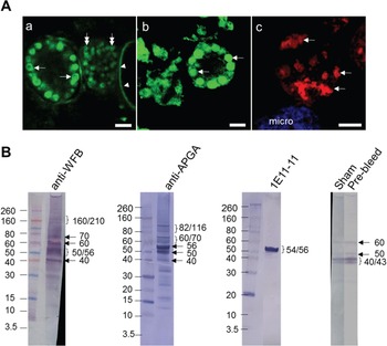
As can be seen in Fig. 2Aa, the anti-WFB pAb was found to intensely stain Type 1 (arrows) and Type 2 (double arrows) WFBs as well as the oocyst wall (arrowheads). Antisera raised against APGA and a monoclonal antibody to the 56 kDa WFB1 protein (1E11-11) used for comparison stained Type 1 and Type 2 WFBs of E. maxima macrogametocytes (Fig 2Ab and Ac). Gametocytes double-labelled to reveal both Type 1 and Type 2 WFBs simultaneously are shown in the Appendix Fig. A1. Negative control sera gave very low levels of staining and there was no reactivity detected in enterocytes collected from the intestinal mucosa of uninfected birds (not shown).
Immuno-labelling (A) and Western blot analysis (B) of sexual stage parasites probed with anti-WFB sera. (Aa) Confocal micrographs of harvested macrogametocytes probed with chicken anti-WFB polyclonal antibodies generated to a fraction enriched for E. maxima macrogametocyte WFBs (1:2000), mouse anti-APGA antibody (1:200 dilution) generated to a fraction of macrogametocyte proteins enriched for gam56 and gam82, amongst other molecules (Wallach et al. Reference Wallach, Mencher, Yarus, Pillemer, Halabi and Pugatsch1989), or mouse monoclonal 1E11-11 antibody (1:200 dilution) raised against E. maxima gametocyte glycoprotein gam56 (Fried et al. Reference Fried, Mencher, Sarshalom and Wallach1992). Primary antibodies were detected with Alexa Fluor 549–conjugated goat anti-mouse IgM (1:500 dilution), or with FITC conjugated rabbit anti-chicken IgG (Fab`)2 antibody (1:500 dilution). (Aa) Isolated macrogametocytes at various stages of maturation with stained large Type 1 WFBs (arrows), Type 2 WFBs (double arrows) and oocyst wall (arrowheads). Note the labelling of the large Type 1 WFBs vesicles, which appear as large rings easily discerned from the very small ‘doughnut-shaped’ Type 2 WFBs aggregated in the cytoplasm of an early macrogametocyte. (Ab) Mature macrogametocyte labelled with anti-APGA antibody. Note the staining of the luminal content of the WFB1s (arrows). (Ac) Early macrogametocyte with doughnut-shaped Type 2 WFBs clustered in the cytoplasm (arrows). Microgametocyte is highlighted in blue stained with DAPI. Scale bar, 5 μm. (B) An immunoblot probed with anti-WFB polyclonal antibody (anti-WFB lane), anti-APGA antibody (anti-APGA lane) and 1E11-11 monoclonal antibody (1E11-11 lane). Note the presence of a prominent 54/56 protein band when all three antibodies are used. Note the presence of 40/45, 50 and 60 kDa band detected non-specifically in negative controls (sham and pre-bleed lanes). A total of 5 μg of protein was loaded per lane. The position and molecular weight (in kDa) of protein standards are indicated on the left.

In comparing the antisera raised against the WFB preparation to anti-APGA, we observed some differences in the labelling of the parasites. While anti-APGA strongly detected antigens found within the lumen of the Type 1 WFBs (Fig. 2Ab), in several cases the anti-WFB predominantly stained the membrane surrounding the WFB1s producing a large ring with a central luminal hole (Fig. 2Aa, arrows). As expected, the control anti-56 kDa monoclonal antibody exclusively stained ‘doughnut-shaped’ WFB2s clustered within the cytoplasm of an early macrogametocyte (Fig. 2Ac, arrows).
Western blot analysis of E. maxima gametocyte lysates showed that all three antibodies detected a prominent band of 54/56 kDa (Fig. 2A–C), while only the polyclonal anti-APGA and WFB showed an additional band at 50 kDa. Multiple smaller weak bands migrating between 40 and 10 kDa were also detected with both anti-APGA and anti-WFB pAbs . Using anti-APGA, bands were also detected at 60, 70, 82 and 116 kDa with some weaker bands in between. The 60 and 70 kDa bands were also seen with the anti-WFB antibody, although not very distinctly. The polyclonal anti-WFB detected additional high molecular weight bands at 160 and 210 kDa, which were difficult to detect with anti-APGA, indicating that there are substantial differences in the recognition pattern of these two types of sera. As expected the monoclonal 1E11-11 antibody strongly reacted with a doublet protein with a molecular weight of 54/56 kDa. Finally, there were two bands migrating at approximately 40/45 kDa along with two weak bands at 50 and 60 kDa when control sera were used (Fig. 2B, pre-bleed and sham sera lanes).
Determining the quality of gametocytes used in in situ experiments
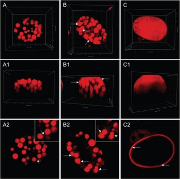
As a control for our in situ experiment, immunized and control chickens (used to produce the sera described in the experiments above) were challenged with 20 000 sporulated E. maxima oocysts. They were euthanized at 134 h post-infection and the intestines were removed, carefully washed and very gently scraped to release gametocytes without damaging the microvasculature, which were fixed and stained with Evans blue. The gametocytes were then analysed by 3D confocal microscopy to observe the cell geometry and three-dimensional shape of the WFB1s at various developmental stages. The perspectives chosen for analysis were from the top (x, y, z-axis, Fig. 3A–C), the side view (x or y-axis, Fig. 3A1, B1, B2) and corresponding single 2D focal slices (Fig. 3A2–C2). Figure 3A shows a mature macrogametocyte with a total of 37 concentrically arranged Type 1 WFBs, which varied in size from 0·3 to 3 μm in diameter (Fig. 3A and B). The focal slice of the macrogametocyte showed the WFB1s linked to one another (Fig. 3A2, arrows). These membranous projections interconnected WFB1s into structures resembling beads on a chain.
Three-dimensional confocal microscopy images of macrogametocytes harvested 134 h post infection. Harvested sexual stage parasites stained with Evans blue dye (red), highlighting the large Type 1 WFBs and the outer oocyst wall. (A-A1) 3D reconstruction of a mature macrogametocyte showing numerous Type 1 WFBs of different size occupying the entire cytoplasm of the cell. (A2) A single focal slice of a cell shown in A1. Note the membrane projections connecting WFB1s making them appear as beads on a chain (arrows). (B–B1) 3D reconstruction of an ovoid macrogametocyte at early stages of oocyst wall formation with large Type 1 WFBs fusing at the cell surface (arrows). (B2) A single focal slice of a cell shown in B1. Note the fused WFB1s (arrows). (C–C1) 3D reconstruction of a fully formed oocyst showing Evans blue positive outer oocyst wall. (C2) A single focal slice of a cell shown in C. Note the presence of a single layer around the oocyst. A, A1, B, B1, C, C1 represent 3D volumetric view with blending effect. A1, B1, C1 show side views for each cell (x and y planes are indicated). Quadrants in each box represent 5 μm.

The 3D confocal image in Fig. 3B shows an example of an early oocyst in which WFB1 fusion and wall formation has commenced. The top view enabled visualization of the WFB1s fusion taking place at the surface of the parasite (Fig. 3B and B1, arrows). The focal slice enabled visualization of the luminal content of the organelle which appeared uniformly distributed throughout (Fig. 3B2, arrows). Using 3D confocal microscopy to view all planes, the oocyst outer wall exhibited homogeneous staining (Fig. 3C-C1), consistent with its appearance in 2D (Fig. 3C2, arrows). The 3D digital reconstructions enabled visualization of the entire outer oocyst wall matrix.
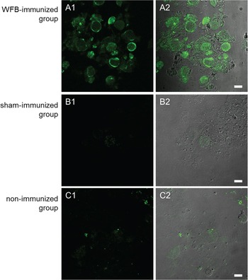
Distribution of fluorescence signals in macrogametocytes harvested from immunized (A1-3) and sham- (B1-3) or non-immunized (C1-3) birds. Low magnification (40×) of sexual stage parasites extracted from all groups were washed and immediately incubated with the FITC conjugated anti-chicken IgG (Fab) secondary antibody. Fluorescence signals indicative of secondary antibody binding to the target antigen were analysed using confocal microscopy keeping fluorescence intensities and gains constant. (A1–2) Macrogametocytes isolated from the chickens immunized with the purified WFB antigens in which strongly fluorescing parasites can be observed. Note the weak fluorescence signals in sham-immunized (B1–3) and non-immunized (C1–2) birds. Scale bar, 20 μm.

Spatial distribution of endogenous IgG antibodies at different stages of oocyst biogenesis
We used the WFB immunized chickens to determine if the resulting serum antibodies have the ability to reach E. maxima gametocytes in vivo to bind to the intracellular antigens. Immediately after euthanizing the chickens, the intestines were very gently scraped and the bound IgG was visualized by incubating the gametocytes with FITC-conjugated anti-chicken (Fab’)2 IgG secondary antibody. Figure 4 shows low magnification confocal micrographs of gametocytes from WFB immunized, sham-immunized and non-immunized control birds. While strong labelling could be observed in gametocytes harvested from birds immunized with WFB antigens (Fig. 4A1–2), very weak staining was detected in gametocytes collected from non- and sham-immunized birds (Fig. 4B1-2 and C1-2). This strong difference in fluorescence intensity shows that the serum IgG antibodies were able to reach the gametocyte, permeate the cellular membranes and bind to the cytoplasmic Type 1 and Type 2 WFBs.
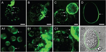
2D and 3D high-resolution confocal microscopy were used to observe the subcellular localization of the antibodies. Analysing single focal planes of the cells, we were able to visualize binding of the IgG to both the parasite's surface and the intracellular compartments. Figure 5A-A1 shows that the large Type 1 WFBs were strongly stained giving a crescent-shaped appearance (Fig. 5A-A1, arrows). Also strongly stained were small granules approximately 0·7–1·5 μm in diameter (WFB2s) which condensed near the surface of the parasite (Fig. 5B-B1, arrows). In parasites which were at the start of oocyst wall formation, the IgG was found bound to the exocytosed WFB1s (Fig. 5C-C1, arrows). The IgG was also found to bind to the inner oocyst wall of formed oocysts (Fig. 5D-D1, arrows).
Localization of secreted IgG in macrogametocytes harvested from birds immunized with purified WFB antigens. Sexual stage parasites extracted from WFB-immunized birds were washed and immediately incubated with the FITC conjugated anti-chicken IgG (Fab) secondary antibody. Fluorescence signals indicative of secondary antibody binding to the target antigen were analysed using confocal microscopy. (A) A late stage macrogametocytes in which fluorescing Type 1 WFBs can be observed inside the parasites. (A1) Insert shows a close-up of the crescent-shaped Type 1 WFB organelles (arrows). (B-B1) A macrogametocyte with fluorescing Type 2 WFBs clustered at the parasite's surface (arrows). (C) Zygotes at different points of oocyst wall secretion. (D-D1) An oocyst with fluorescing oocyst wall (arrows). Scale bar, 5 μm.

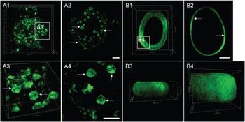
We next processed the z-stack images of gametocytes in order to obtain a 3D reconstruction as shown in Fig. 6. Digital 3D reconstruction of macrogametocytes (Fig. 6, A1) showed that the anti WFB pAbs were not uniformly distributed inside the WFB1s and instead showed a punctate staining of the organelle lumen (Fig. 6A2).
Spatial distribution of endogenous IgG at different stage of oocyst biogenesis. Following harvest from WFB-immunized birds, sexual stage parasites were washed extensively and incubated immediately with the FITC conjugated anti-chicken IgG (Fab) secondary antibody. The localization and distribution of fluorescence was analysed by confocal microscopy. (A1) 3D reconstruction of a mature macrogametocyte showing positive labelling of the cell surface and the large Type 1 WFBs occupying the cytoplasm of the cell. (A2) A single focal slice of a cell shown in A1. (A3) A close-up of a region depicted in A1 with numerous positively stained large Type 1 WFBs (arrows). (A4) A focal slice of vesicles shown in A3. Note the punctate staining of the WFB1s (arrows). (B1) 3D reconstruction of an oocyst showing positive labelling of the inner oocyst wall. (B2) A focal slice of the oocyst shown in B1. (B3) Side view of the inner oocyst wall scaffold. (B4) A close-up of a region depicted in B1 with clearly visible inner oocyst wall ‘scaffolding’ pattern. Images in A1, A3, B1, B3, B4 represent 3D volumetric view with blending effect. Quadrants in A1, B1, B3 and B4 represent 5 μm, quadrants in A3 show 1 μm.

Furthermore, the close-up of the region depicted in A1 showed fluorescently labelled IgG embedded into the membrane coating the Type 1 WFBs (Fig. 6A3 and A4). In digital reconstructions of the fully formed oocyst (Fig. 6B1-4), the IgG is associated with the surface of the oocysts, which now appeared embedded in the surface matrix forming a scaffold (Fig. 6B4).
DISCUSSION
The results of the present study have shown that wall-forming bodies extracted from E. maxima gametocytes were highly immunogenic in chickens. Antibodies produced against these organelles were found to localize specifically to the Type 1 and 2 WFBs as well as the oocyst walls in isolated macrogametocytes and early-stage oocysts as expected. Chickens immunized with WFBs were challenged with E. maxima oocysts and then euthanized at 134 h post-infection to localize the anti WFB antibodies produced in situ. Using 2D confocal fluorescence microscopy and 3D computer reconstruction, we found that the anti-WFB antibodies were able to reach the gametocytes located in the intestinal mucosa and bind to the internal Type 1 and 2 WFB organelles. These results help elucidate the means by which antibodies produced against gametocyte antigens can provide protection against challenge Eimeria infections.
The geometry and three-dimensional shape of the WFB1s was studied at different stages of oocyst biogenesis using a 3D confocal microscope to visualize oocyst wall secretion in both lateral and axial planes. Micrographs showed the expected concentrically arranged WFB1s of different sizes in the mature macrogametocytes. During the early stages of oocyst wall development, we observed the secretion of the contents of the WFB1s which was characteristic of vesicles undergoing exocytosis. Evans blue showed that the WFB1s fused prior to liberating their luminal content at the surface of the developing oocyst.
We incubated gametocytes extracted from immunized and control chickens with FITC-conjugated secondary antibody only to detect anti-WFB antibodies produced in situ. Both low magnification and high-resolution confocal micrographs, showed strong labelling of the surface as well as the internal WFB Type 1 and 2 organelles. These results show that the serum IgG antibodies were able to reach the gametocyte in the intestinal mucosa, permeate the cellular membranes and bind to the cytoplasmic Type 1 and Type 2 WFBs. In a recent study we had also shown that even relatively large 100 nm nanoparticles are able to transverse membranes and reach the intracellular WFB1s where they accumulate (unpublished). Thus, we postulate that via endocytosis, antibodies to the WFBs are taken into the gametocytes and are able to bind to the internal organelles.
Interestingly, we found by IFA that WFB1s labelled with the anti-WFB antibodies in situ gave a crescent-shaped appearance (Fig. 5A-A1). There are a few possible explanations for this result. Firstly, the specific antigens recognized by these antibodies may have a polar distribution over the surface of the organelle in situ. Secondly, antibody uptake into the gametocytes may be occurring from only one side of the parasite and they thereby accumulate in one portion of the organelle. Further work is required to elucidate this phenomenon.
In our previous studies, we used antibodies raised against APGA to study the subcellular localization of these antigens (Frölich et al. Reference Frölich, Johnson, Robinson, Entzeroth and Wallach2013). In the present study, we compared antibodies raised against purified WFBs to those produced against APGA. We found that although the two types of antisera gave similar results there were some noted differences. By IFA we found that the anti WFBs predominantly stained the surface of the E. maxima WFB1s whereas the anti APGA stained the whole organelle. This result is similar to that we found previously using a monoclonal antibody raised against a WFB1 protein (Frölich et al. Reference Frölich, Johnson, Robinson, Entzeroth and Wallach2013). We therefore concluded that the antibodies produced by vaccination with WFBs are mainly directed against the proteins found on the surface of the WFB1s and not in the lipid core. By Western blot analysis we found a difference in the banding pattern whereby the anti WFB sera produced more prominent bands in the 160–210 kDa range as compared with the anti APGA antisera.
The results described above may be explained by the differences in the extraction methods. While preparation of APGA relies on the use of non-ionic, non-denaturing detergent (Pugatsch et al. Reference Pugatsch, Mencher and Wallach1989; Wallach et al. Reference Wallach, Mencher, Yarus, Pillemer, Halabi and Pugatsch1989), the extraction of WFBs relies heavily on the use of high concentrations (1% w/v) of strong anionic surfactant SDS (Frölich et al. Reference Frölich, Johnson, Robinson, Entzeroth and Wallach2013). SDS surfactant is a strong protein denaturant able to disrupt non-covalent bonds within and between proteins. In addition it is commonly used to remove lipids from experimental samples (Samso et al. Reference Samso, Daban, Hansen and Jones1995). Given that the Type 1 WFBs are a lipid-rich organelle with a protein-rich membrane coat (Frölich et al. Reference Frölich, Johnson, Robinson, Entzeroth and Wallach2013), it is very likely that the SDS denatured and/or removed some of the antigens recognized by the anti-APGA, hence, the difference in IFA and in the recognition pattern of proteins between these two types of anti-sera.
Finally, digital reconstructions of the fully formed oocysts showed an intriguing ‘scaffolding’ pattern, indicating the incorporation of the processed WFB proteins into the inner oocyst wall. A similar scaffolding pattern was reported previously in the inner layer of the oocyst wall of Eimeria tenella (Bushkin et al. Reference Bushkin, Motari, Magnelli, Gubbels, Dubey, Miska, Bullitt, Costello, Robbins and Samuelson2012), which, in addition to tyrosine rich proteins, was found to be composed of fibrils of beta-1, 3-glucan that form a trabecular scaffold. Therefore, it is plausible that the WFB antigens were also incorporated into the wall in a similar pattern to that of the glucan scaffold.
Taken together, we conclude that immunization with WFB-rich extract induces a large production of antibodies which have the ability to reach the parasite in situ, and bind to the WFBs. We postulate that via this process antibodies block the development of E. maxima in vivo. These findings broaden our understanding of avian intestinal immunity and the importance of the humoral immune response.
ACKNOWLEDGEMENTS
The authors are grateful to the Ernst Animal Facility at the University of Technology, Sydney for maintaining the chickens. Confocal microscopy and data analyses were performed at the Microbial Imaging Facility of the University of Technology, Sydney.
FINANCIAL SUPPORT
The authors wish to thank the Faculty of Science, University of Technology, Sydney and PhiBro Animal Health Corporation, Israel [Linkage grant, FR110089] for financial support. The funders had no role in study design, data collection and analysis, decision to publish, or preparation of the manuscript.
Appendix
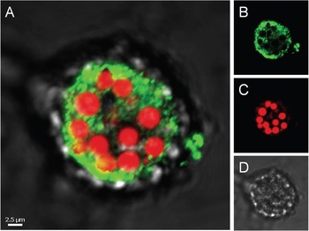
Localization of Type 1 and 2 WFBs in macrogametocytes harvested from chickens 134 h post infection. Confocal micrographs of harvested macrogametocytes probed with mouse monoclonal 1E11-11 antibody (1:200 dilution) raised against E. maxima gametocyte glycoprotein gam56 (Fried et al. Reference Fried, Mencher, Sarshalom and Wallach1992) and counter stained with Evans blue. Primary antibodies were detected with Alexa Fluor 488–conjugated goat anti-mouse IgM antibody (1:500 dilution). (A) Merged image of transmission, green and red signals; (B) green fluorescing 1E11-11 positive Type 2 WFBs; (C) Evans blue stained Type 1 WFBs; (D) transmission detection. Scale bar indicated.