Introduction
Congenital nasal obstructions are generally classified according to their anatomical location as anterior, midnasal and posterior.Reference Sohal and Schoem1 The most common posterior type (1 in 5000 to 8000 births) and the most severe congenital nasal obstruction is choanal atresia, followed by pyriform aperture stenosis anteriorly (1 in 25 000 births).Reference Sohal and Schoem1–Reference Yang, Orta, Renk and Inman3 Congenital midnasal stenosis has been previously described as a component of many syndromes, but it was first reported precisely in 2004 by Raghavan et al.Reference Raghavan, Fuad and Gibbin4 Case series have subsequently been presented, and diagnosis and treatment modalities have been discussed in the literature.Reference Levi, Kornreich, Hilly, Raveh and Gilony5–Reference Sahin Onder, Sahin-Yilmaz, Gergin Tinay and Unsal8
Choanal atresia, pyriform aperture stenosis and midnasal stenosis are clinically similar entities. Endoscopic evaluation plays an important role in diagnosis. Congenital midnasal stenosis should be considered in an infant if the middle turbinate is not visible with a fibre-optic endoscope after passing the anterior end of the inferior turbinate, despite no severe septal deviation.Reference Syed, Raja, Kolethekkat, Varghese, Al Abri and Kurien6 Levi et al. have established diagnostic principles of clinical examination and computed tomography (CT) findings in newborns with midnasal stenosis. These authors have determined comprehensively the normative measurements of neonatal endonasal anatomical distances related to midnasal stenosis.Reference Levi, Kornreich, Hilly, Raveh and Gilony5
The aetiology of nasal obstruction in adults is different from that of newborns. Nasal obstruction in adults may be due to mucosal causes such as rhinosinusitis and turbinate hypertrophy, or have structural causes such as septal deviation and nasal valve collapse. Polyps and neoplasms obstructing the nasal cavity are less common. While pyriform aperture stenosis in adults has been reported by Erdem et al.,Reference Erdem, Ozturan, Erdem, Akarcay and Miman9 midnasal stenosis in adults has not been previously described in the literature. This study aimed to reveal midnasal normative radiological measurements in adults, and to present representative patients who were diagnosed with midnasal stenosis and treated surgically.
Materials and methods
The study commenced after ethics committee approval. A total of 161 individuals aged 18–60 years were studied retrospectively. The paranasal CT scans of 161 cases, conducted for various reasons other than nasal obstruction, were studied to obtain the endonasal anatomical distance measurements. Axial and coronal sections of the paranasal CT scans were grouped according to gender. Those CT scans belonging to patients with nasal polyposis, concha bullosa, chronic sinusitis, and severe and moderate septal deviation were not included. Patients with a history of nasal surgery, facial syndromic findings, suboptimal CT measurement parameters, or cases whose CT image did not cover the entire nasal cavity, were excluded.
Three patients (two females and one male, with a mean age of 26 years) with complaints of nasal obstruction were diagnosed with midnasal stenosis in the last three years. Midnasal stenosis should be suspected in adult patients without well-known common causes of nasal obstruction, in whom a 3 mm rigid endoscope cannot be inserted posterior to the anterior end of the inferior turbinate, and in whom, following decongestion, the middle turbinate and uncinate process are visualised as more medially located than usual on endoscopic nasal examination.
Nasal Obstruction Septoplasty Effectiveness (‘NOSE’) scoring was applied in these patients.Reference Stewart, Smith, Weaver, Witsell, Yueh and Hannley10 Paranasal CT was performed for diagnostic purposes and to rule out other causes following clinical examination. Definitive midnasal stenosis was diagnosed utilising measurements made in axial and coronal CT planes. Additionally, acoustic rhinometry and rhinomanometry evaluations were performed.
As these patients did not benefit from conservative medical treatments, the stenotic midnasal area was treated surgically. They underwent endoscopic sinus surgery performed by the first author (OO), whereby middle turbinectomy, complete uncinectomy, mega middle meatal antrostomy and partial anterior ethmoidectomy were performed in both nasal cavities to relieve midnasal stenosis. If indicated, accompanying minor septal and conchal pathologies were also remedied during the same surgical session.
These patients were followed up for at least one year. One patient underwent post-operative CT for another reason and the findings are presented for the purpose of comparison with the pre-operative CT findings (Figure 1).
(a) Pre- and (b) post-operative coronal, and (c) pre- and (d) post-operative axial, computed tomography sections. P = posterior

Radiological measurements
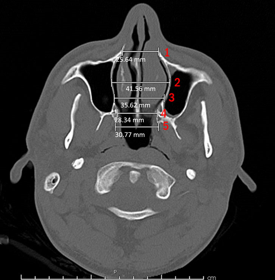
The CT measurements of endonasal dimensions were made as reported previously in the literature.Reference Reeves, Discolo and White11,Reference Belden, Mancuso and Schmalfuss12 Measurements were carried out by two otolaryngologists on CT sections using the landmarks described above with the help of radiological software. Evaluations in axial sections were performed by measuring the horizontal distances, as follows: (1) at the widest part of the pyriform aperture; (2) at the point between the pyriform aperture and the choana; (3) at the point between measurement ‘(2)’ and the choana; (4) at the widest part of the choana; and (5) between the medial pterygoid protrusions (Figure 2).
Evaluations in axial sections were performed by measuring horizontal distances as follows: (1) at the widest part of the pyriform aperture (‘A.Prif’); (2) at the point between the pyriform aperture and the choana (‘A.50’); (3) at the point between ‘A.50’ (measurement 2) and the choana (‘A.75’); (4) at the widest part of the choana (‘A.Cho’); and (5) between the medial pterygoid protrusions (‘A.Mp’). P = posterior
Four landmarks were used for the coronal section measurements. Pyriform aperture width was measured horizontally in the coronal section where the anterior head of the inferior turbinate is visible (Figure 3a). The distance between the nasolacrimal duct was measured at the last section where both nasolacrimal ducts were visible (Figure 3b). The width of the posterior nasal cavity was measured in the section where the last molar teeth were seen (Figure 3c). The distance between the line drawn vertically, passing tangentially from the medial wall of the orbit, and the vertical line over the inferior turbinate attaching to the medial wall of the maxillary sinus, was measured horizontally in the first section where the maxillary sinus ostium was seen in coronal views on both sides (Figure 3d). It was recorded whether the ostium was medial or lateral to this vertical line. The location of the ostium as medial or lateral to the vertical orbital line determined whether the measurements were negative or positive, respectively (Figure 3d).
Evaluations in coronal sections were performed by measuring distances as follows: (a) horizontal width at the widest part of the pyriform aperture (‘C.Prif’); (b) horizontal distance between the nasolacrimal ducts (‘C.Nld’); (c) horizontal distance between two lateral nasal walls where the molar teeth in the upper jaw are visible (‘C.Md’); and (d) distance between the line drawn vertically passing the medial wall of the orbit and the part where the inferior turbinate attaches to the medial wall of the maxillary sinus, measured horizontally in the first section where the maxillary sinus ostium is visible on both sides (‘C.Omw’).

Statistics
Statistical analysis was carried out using SPSS software for Windows, version 24.0 (SPSS, Chicago, Illinois, USA). All quantitative variables were estimated using measures of central location (i.e. mean and median) and measures of dispersion (i.e. standard deviation (SD)). Data normality was checked using the Kolmogorov–Smirnov tests of normality. The independent t-test was used to compare the two groups according to the pathological diagnoses of the tissues. A p-value of less than 0.05 was considered statistically significant.
Results
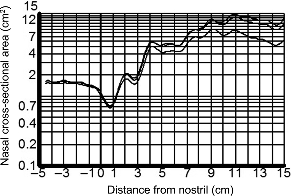
Three patients were diagnosed with adult midnasal stenosis in our clinic between 2018 and 2020. Endoscopic nasal examination before and after decongestion clearly showed a narrow passage at the midsection of the nasal cavity. When acoustic rhinometry and rhinomanometry were performed after nasal decongestion, it was observed that the obstruction was in the midnasal area (Figure 4); there was no other remarkable obstruction. Their pre-operative Nasal Obstruction Septoplasty Effectiveness scores were 9, 12 and 15, respectively (mean of 12). Nasal CT findings substantiated the diagnosis of midnasal stenosis. These patients did not have sufficient benefit from the conservative medical treatment.
Acoustic rhinometric measurements following decongestion denote midnasal stenosis.

Paranasal sinus CT measurements of the 3 patients and the 161 control individuals whose radio-anatomical measurements were taken for normative data are presented in Figure 5.
Mean values for paranasal sinus computed tomography (CT) measurements for our 3 patients and 161 control individuals (except for the coronal measurement defined in Figure 3d (‘C.Omw’)); these are the axial section measurements 1–5, as defined in Figure 2 (i.e. ‘A.Prif’, ‘A.50’, ‘A.75’, ‘A.Cho’ and ‘A.Mp’), and the coronal section measurements defined in the Figure 3a–c images (i.e. ‘C.Prif’, ‘C.Nld’ and ‘C.Md’).

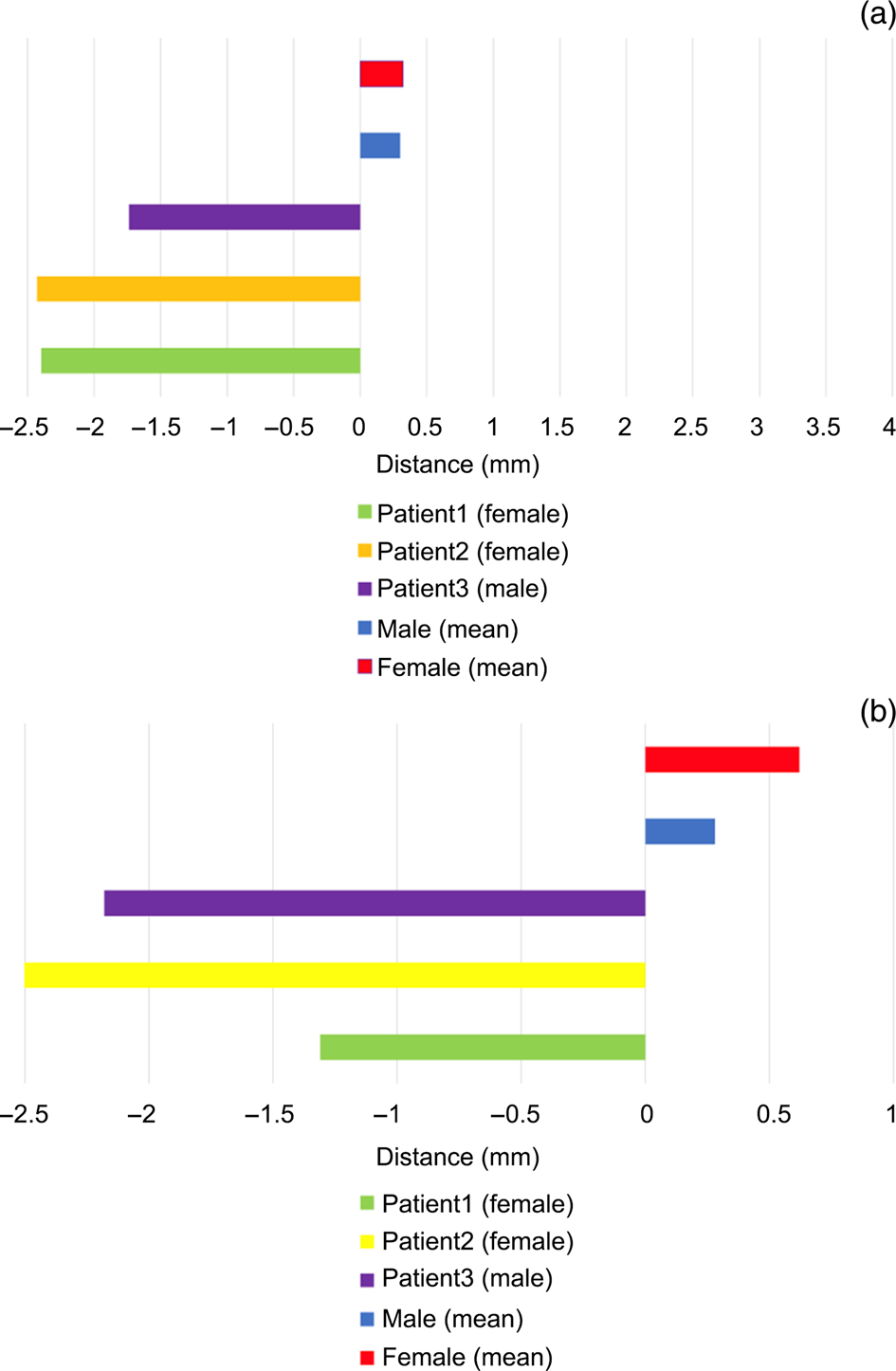
For the normative values, significant differences between males and females were found for all parameters. All measurements were larger in males than in females, except for the distance between the line drawn vertically passing the medial wall of the orbit and the part where the inferior turbinate attaches to the medial wall of the maxillary sinus, measured horizontally in the first section where the maxillary sinus ostium is visible on both sides (Figure 6). The maxillary sinus medial wall was slightly more lateralised on the right side in females, and significantly more lateralised on the left side in females compared to males.
Mean values for the coronal measurement defined in Figure 3d (i.e. ‘C.Omw’), for our 3 patients and 161 individuals, on the (a) right and (b) left sides.

In our patients, the maxillary sinus medial walls were in an overtly medialised position, causing midnasal stenosis. The values for other parameters were similar to the normative group, demonstrating that while the pyriform aperture and choana are identical, only the midnasal region is constricted. As can be observed in Figure 5, the following measurements were significantly lower in our three patients with midnasal stenosis compared to the normative measurements: the horizontal distance at the point between the pyriform aperture and the choana in the axial section; the horizontal distance at the point between the previous measurement and the choana in the axial section; the horizontal distance between the nasolacrimal ducts in the coronal section; and the horizontal distance between two lateral nasal walls where the molar teeth in the upper jaw are visible in the coronal section.
Our patients underwent functional endoscopic sinus surgery. Middle turbinectomy, complete uncinectomy, mega maxillary antrostomy and partial anterior ethmoidectomy were performed to enlarge the midnasal stenotic site. The patients were followed up for at least 12 months post-operatively. The post-operative Nasal Obstruction Septoplasty Effectiveness scores were 3, 2 and 3 respectively (mean score of 2.6). These patients reported that their nasal obstruction symptoms significantly improved after surgery.
Discussion
Newborns are dependent on nasal breathing. Therefore, atresia, stenosis or severe nasal congestion in the newborn can be life-threatening. Structural nasal obstructions of the newborn are respectively classified as pyriform aperture stenosis, midnasal stenosis and choanal atresia in the anterior, middle and posterior anatomical locations.Reference Gnagi and Schraff2 These three conditions clinically present as similar disturbances.
Congenital midnasal stenosis may result from medial overgrowth of the lateral nasal wall or thickening of the septum. A preliminary diagnosis is made by fibre-endoscopic evaluation in the out-patient setting. Midnasal stenosis should be suspected in newborns when the anterior end of the inferior turbinate cannot be seen on endoscopy and where no other cause of nasal obstruction can be detected. Following an endoscopic nasal examination, CT is the ‘gold standard’ means of evaluating the location and size of obstruction, and eliminating other causes. Levi et al. recently presented the principle CT measurements for diagnosing midnasal stenosis in newborns.Reference Levi, Kornreich, Hilly, Raveh and Gilony5 In their study, the measurements between the bone of the inferior turbinate and the septum in the midnasal area were significantly different between the newborns diagnosed with midnasal stenosis and the healthy group.Reference Levi, Kornreich, Hilly, Raveh and Gilony5 However, midnasal stenosis has not been described in adults before. In addition, normal measurements of the midnasal area in adults have not been previously reported in the literature. The current investigation highlights consideration of midnasal stenosis as a cause for nasal obstruction in adults.
Isolated midnasal stenosis is a diagnosis that can be easily missed in adults given its rareness and the lack of clinician awareness, which hinders its detection. This condition should be considered in the differential diagnosis during endoscopic nasal examination. Clinical suspicion can be corroborated by deliberate CT evaluations. Furthermore, diagnosis can be supported with acoustic rhinometry and rhinomanometry.
In CT scans, special attention should also be paid to images of the midnasal region. The measurements showing the midnasal region were accepted in previous studies to be: the horizontal distance at the point between the pyriform aperture and the choana in the axial section; the horizontal distance at the point between the previous measurement and the choana in the axial section; the horizontal distance between the nasolacrimal ducts in the coronal section; and the horizontal distance between two lateral nasal walls where the molar teeth in the upper jaw are visible in the coronal section.Reference Reeves, Discolo and White11,Reference Belden, Mancuso and Schmalfuss12 These anatomical and radiological evaluation criteria were followed in this study as well. In our three patients, the midnasal passage was overtly narrow as seen in CT examinations.
Measurement of the distance between the line drawn vertically passing the medial wall of the orbit and the part where the inferior turbinate attaches to the medial wall of the maxillary sinus, determined horizontally in the first section where the maxillary sinus ostium is visible on both sides, was introduced for the first time in this study. In our patients, the maxillary wall was quite medial compared to the control group. Utilisation of this specific measurement can be considered for diagnosing adult midnasal stenosis in future studies.
There are different approaches to the treatment of neonatal midnasal stenosis. Conservative treatments such as nasal steroids and topical antibiotics can be applied initially.Reference Levi, Kornreich, Hilly, Raveh and Gilony5 Topical decongestants are generally not recommended in infants.Reference Scadding13 Surgical treatment may be considered in more severe cases, and in cases unresponsive to medical treatments administered for 10–15 days.Reference Gnagi and Schraff2 Surgical treatment options include balloon dilatation, lateralisation of the inferior turbinates, and nasal stent placement for a few days to weeks.Reference Raghavan, Fuad and Gibbin4,Reference Syed, Raja, Kolethekkat, Varghese, Al Abri and Kurien6 The key point is not to damage the mucosa during surgery.
The aetiology and treatment of adult midnasal stenosis differ from that of newborns. Midnasal stenosis in adults is caused by the medialised position of the medial wall of the maxillary sinus and the uncinate process bilaterally, as found on the CT scans of our three patients. Therefore, middle turbinectomy, complete uncinectomy, mega middle meatal antrostomy and partial anterior ethmoidectomy were utilised as functional endoscopic surgical techniques. Concomitant septal and inferior turbinate interventions can also be performed during surgery, if required. Patients were relieved of the nasal obstruction symptoms, and their nasal passages were favourably large following the operative interventions.
• Midnasal stenosis should be considered in the differential diagnoses in patients with nasal obstruction, in addition to the well-known causes
• Midnasal stenosis is suspected based on endoscopic visualisation of medially located middle turbinates and uncinate processes in nasal cavities
• Definite diagnosis of midnasal stenosis can be made by intentional examination of paranasal sinus computed tomography scans
This is the first study to focus on midnasal stenosis in adults, and the first to define normative anatomical measurements in adults. It is unknown whether midnasal stenosis in adults is congenital or developmental. Our study is limited by the small number of patients. The diagnosis and treatment of midnasal stenosis in more patients, and the sharing and reporting of colleagues’ experiences, are certainly needed.
Conclusion
Midnasal stenosis should be considered in the differential diagnoses of patients presenting with nasal obstruction, in addition to the well-known, common causes. Midnasal stenosis is suspected based on endoscopic visualisation of medially located middle turbinates and uncinate processes in the nasal cavities. Definite diagnosis of midnasal stenosis can be clearly made by intentional examination of the paranasal sinus CT scans. As the surgical treatment should be based on aetiology, the authors recommend endoscopic middle turbinectomy, complete uncinectomy, mega maxillary antrostomy and partial anterior ethmoidectomy. However, larger case series are needed to further expand our knowledge on the diagnosis and treatment of midnasal stenosis in adults.
Acknowledgement
The authors would like to thank Edward F Wright, for his critical review and editing of the manuscript.
Competing interests
None declared