Individuals with Down syndrome have increased risks of developing pulmonary hypertension including left-to-right shunt CHD, an abnormal growth of pulmonary vasculature such as thinning of the media, hypoxia due to upper airway obstruction, and an imbalance in pulmonary vasoconstriction and relaxation due to abnormal synthesis of nitric oxygen or prostanoids.Reference King and Tulloh 1 – Reference Fukushima, Kosaki and Sato 6 An appropriate diagnosis and a combined multidisciplinary approach are critical to the management of pulmonary hypertension in individuals with Down syndrome. Pulmonary veno-occlusive disease is an extremely rare cause of pulmonary hypertension, which is pathologically characterised by extensive and diffuse fibrous obliteration of small pulmonary veins.Reference Montani, Lau and Dorfmüller 7 In this article, we present the case of an infant with Down syndrome who unexpectedly developed rapidly progressive pulmonary veno-occlusive disease and fatal pulmonary hypertension.
Case report
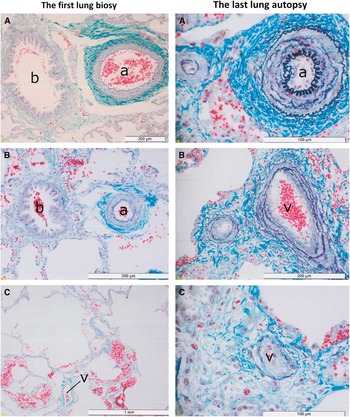
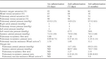
A 15-day-old girl with Down syndrome was referred to our hospital because of respiratory distress and feeding difficulty. She was born at 35 weeks of gestation with a birth weight of 2.3 kg. The patient had neither a history of neonatal respiratory distress syndrome nor an infection. Physical examination revealed an accentuated pulmonary second heart sound and systolic murmur on the left upper sternal border. Chest X-ray showed a cardiothoracic ratio of 0.55. Echocardiography showed partial atrioventricular septal defect with trivial atrioventricular valve regurgitation, and patent ductus arteriosus with a diameter of 3.5 mm. The flow direction in the ductus arteriosus was left to right. Preoperative cardiac catheterisation at 53 days of age revealed that the pulmonary-to-systemic blood flow ratio, the mean pulmonary arterial pressure, and pulmonary vascular resistance were 1.88, 35 mmHg, and 3.59 Wood units·m2, respectively (Table 1). As we speculated that the patent ductus arteriosus mainly contributed to the increase in pulmonary blood flow and pulmonary hypertension, the patient underwent closure of the ductus arteriosus and lung biopsy via left thoracotomy at 60 days of age. In our institution, lung biopsy is indicated when pulmonary vascular resistance abnormally increases over 3–4 Wood units·m2, especially in patients with Down syndrome. The postoperative course was uneventful. Histopathological examination revealed almost normal intimal and medial structures of small pulmonary arteries, but pre-acinar small pulmonary arteries with diameter <100 μm were hypoplastic. The pulmonary veins were normal (Fig 1). Follow-up cardiac catheterisation was performed to assess haemodynamics at 72 days of age. It revealed that the pulmonary-to-systemic blood flow ratio and the pulmonary-to-systemic arterial pressure ratio were decreased (Table 1), although pulmonary vascular resistance remained unchanged. The patient was scheduled for intracardiac repair of the partial atrioventricular septal defect and was followed-up regularly at the outpatient clinic. Neither oxygen supplement nor pulmonary vasodilator agents were administrated because of the associated risk of an increased pulmonary blood flow. At 4 months of age, however, the patient unexpectedly developed feeding difficulty, dyspnoea, oliguria, and pallor. Her oxygen saturation levels decreased to 82%. A physical examination revealed an accentuated pulmonary second heart sound and hepatomegaly. Her chest X-ray showed an increased cardiothoracic ratio of 0.65. Parasternal short-axis view on echocardiography showed dilation of the right ventricle and a semilunar shape of the left ventricle, suggesting deterioration of pulmonary hypertension. Subsequent urgent cardiac catheterisation revealed that the pulmonary-to-systemic flow ratio was decreased and pulmonary vascular resistance was extremely increased up to 13.3 Wood units·m2 (Table 1). The pulmonary venous pressure was normal, and pulmonary arterial angiography showed no pulmonary venous stenosis. Oxygen and nitric oxide inhalation test revealed that the pulmonary-to-systemic arterial pressure ratio and pulmonary vascular resistance were decreased and the pulmonary-to-systemic blood flow ratio was increased, which suggested a possibility of reversible pulmonary hypertension. The patient was treated with inhalation oxygen therapy, oral administration of bosentan, and intravenous administration of prostacyclin. Her clinical manifestations, however, deteriorated despite these aggressive pulmonary vasodilation therapies. Her chest X-ray showed increased cardiomegaly and pulmonary vascular congestion. We should have ideally discontinued these therapies, because increased pulmonary congestion after the introduction of pulmonary vasodilation therapies suggested a suspicion of pulmonary veno-occlusive disease. On the basis of previous histopathological findings of normal architecture of the pulmonary vein, however, we assumed rapid development of pulmonary “arterial” disease. Unfortunately, the infant died from heart failure at 5 months of age. Lung autopsy was performed after obtaining consent from parents. Histopathological examination revealed narrowing pulmonary veins with intimal fibrous thickness and arterialised media with an increase in elastic fibres. Small pulmonary veins with diameter <100 μm were completely obliterated with intimal fibrous hypertrophy. The pulmonary arteries were hypoplastic, with medial thickness (Fig 1). These findings are consistent with pulmonary veno-occlusive disease.
Left side: histopathological findings of the first lung biopsy at 60 days of age (Elastica-Goldner stain): pulmonary artery (a), pulmonary vein (v), and bronchioles (b). ( A ) A small pulmonary artery with a diameter of 500 μm has almost normal architecture and size. ( B ) A small pulmonary artery with a diameter of 100 μm is hypoplastic. Note that the diameter of the pulmonary artery is almost 60% of the diameter of the terminal bronchiole. ( C ) A pulmonary vein appears normal. The alveolar ducts are dilated. Right side: histopathological findings of the last lung autopsy at 5 months of age (Elastica-Goldner stain): pulmonary artery (a) and pulmonary vein (v). ( A ) A pulmonary artery with a diameter of 100 μm is shown with medial thickness. ( B ) A pulmonary vein with a diameter of 250 μm is arterialised and narrowing, with an increase in elastic fibres. ( C ) The lumen of a small pulmonary vein with a diameter of 200 μm is completely obliterated with intimal fibrous hypertrophy.

Summary of cardiac catheterisation data.

Pulmonary and systemic arterial pressures are described as the systolic pressure/the diastolic pressure (the mean pressure). Right and left atrial pressures and pulmonary venous pressures are described as the mean pressure. Left ventricular pressure was described as the systolic pressure/the end-diastolic pressure. An oxygen test was not performed at the first catheterisation
Discussion
Pulmonary veno-occlusive disease can be misdiagnosed because its clinical manifestations are similar to those of idiopathic pulmonary arterial hypertension, despite the histopathological differences. Pulmonary veno-occlusive disease, however, is characterised by a worse prognosis and the possibility of developing severe pulmonary oedema with specific pulmonary vasodilator therapy, which justifies the importance of diagnosing this disease. The actual incidence of pulmonary veno-occlusive disease is probably underestimated, because many cases may be classified as idiopathic pulmonary arterial hypertension. Pulmonary veno-occlusive disease is extremely rare with a frequency of 0.1–0.2/1,000,000.Reference Montani, Lau and Dorfmüller 7 Among individuals with Down syndrome, the leading cause of death is CHD associated with pulmonary hypertension.Reference Yang, Rasmussen and Friedman 8 We assume that pulmonary veno-occlusive disease may also be misdiagnosed in patients with Down syndrome and pulmonary hypertension. Although the underlying cause as well as the pathophysiology of pulmonary veno-occlusive disease remain unclear, pulmonary veno-occlusive disease is occasionally associated with bone marrow transplantation, autoimmune connective tissue disease, human immunodeficiency virus infection, sarcoidosis, and Langerhans cell histiocytosis.Reference Woerner, Cutz, Yoo, Grasemann and Humpl 9 However, pulmonary veno-occlusive disease in childhood seems to be unrelated to such underlying diseases. Woerner et al.Reference Woerner, Cutz, Yoo, Grasemann and Humpl 9 described a case series consisting of nine patients, in whom pulmonary veno-occlusive disease was diagnosed at the mean age of 14 years; the mean survival time of these patients was 14 months. This study emphasises the importance of lung biopsy and differential diagnosis of pulmonary veno-occlusive disease from pulmonary arterial hypertension, because of the worse prognosis of pulmonary veno-occlusive disease. The present case demonstrated that pulmonary veno-occlusive disease was only proven after the second lung autopsy and it rapidly developed within only 4 months. Therefore, the possibility of pulmonary veno-occlusive disease should be considered among patients with Down syndrome who show unexpected deterioration of pulmonary hypertension. In the present case, overall, the approach of performing a postoperative cardiac catheterisation at 72 days of age might not be common practice, and the patient with pulmonary vascular resistance of 3.59 Wood units·m2 would have been eligible for corrective cardiac surgery without lung biopsies; however, a lung biopsy may be warranted to provide an appropriate management in patients with Down syndrome and atypical pulmonary hypertension.
High-resolution CT is a non-invasive diagnostic tool for the screening of pulmonary veno-occlusive disease. The presence of lymph node enlargement, centrilobular ground-glass opacity, and thickening of septal lines is the radiographic triad of pulmonary veno-occlusive disease.Reference Montani, Lau and Dorfmüller 7 , Reference Günther, Jaïs and Maitre 10 Biopsy may be reserved for patients in whom the diagnosis of pulmonary arterial hypertension remains in the differential despite the less invasive testing such as high-resolution CT; however, we assume that it is difficult to distinguish with certainty between pulmonary veno-occlusive disease and an increase in the pulmonary blood flow in left-to-right shunt CHD.
In conclusion, this is the first case report of development of pulmonary veno-occlusive disease in an infant with Down syndrome and pulmonary hypertension. Pulmonary veno-occlusive disease as a cause of pulmonary hypertension should be considered in the management of individuals with Down syndrome.
Acknowledgements
The authors thank Dr Shigeo Yamaki from the Japanese Research Institute of Pulmonary Vasculature, Miyagi, Japan, for performing histopathological examinations.
Financial Support
This study received no specific grant from any funding agency or from commercial or not-for-profit sectors.
Conflicts of Interest
There are no conflicts of interest.