Introduction
A high occurrence has been reported for Taenia taeniaeformis (Batsch, 1976), a cestode of cosmopolitan distribution that parasitizes the small intestine of cats (Felis catus) (Morgan & Hawkins, Reference Morgan and Hawkins1949). Its infective form Cysticercus fasciolaris, also named Strobilocercus fasciolaris, is commonly found in rodents. According to Lagapa et al. (Reference Lagapa, Konno, Oku, Nonaka, Mamoru and Kamiva2002) and Kumar et al. (Reference Kumar, Reddy, Aparna, Srinivas, Nagarajan, Venkatesan, Sreekumar and Sesikaran2006), in intermediate hosts, such as rats and mice, the occurrence of T. taeniaeformis larvae can vary by locality and, in these animals, has a close relationship with the occurrence of fibrosarcomas and immunodeficiency. Infections by T. taeniaeformis and other adult cestodes cause little damage to cats. Nevertheless, treatment and control is recommended to prevent new gastrointestinal helminth infections (Bowman et al., Reference Bowman, Lynn, Eberhard and Alacaraz2006). Taenia taeniaeformis larvae are transmitted to intermediate hosts by the ingestion of eggs present in faeces or in contaminated environments (Urquhart et al., Reference Urquhart, Armour, Duncan, Dunn and Jennings1998). Morgan & Hawkins (Reference Morgan and Hawkins1949) affirm that it would be almost impossible to control T. taeniaeformis, thus the need for alternative control methods to prevent new infections.
Fungal egg parasitism is an important biological phenomenon that may be used as an alternative method of control of helminthiasis in domestic animals (Braga et al., Reference Braga, Ferreira, Araújo, Araujo, Silva, Carvalho, Campos and Freitas2010), and nematophagous fungi of the genus Pochonia stand out among potential biological control agents (Braga et al., Reference Braga, Araújo, Campos, Carvalho, Silva, Tavela and Maciel2007). Pochonia chlamydosporia is a facultative deuteromycete which parasitizes helminth eggs. With a wide distribution, it has been used successfully in laboratory conditions in the biological control of eggs of gastrointestinal helminth parasites (Braga et al., Reference Braga, Araújo, Campos, Carvalho, Silva, Tavela and Maciel2007; Araújo et al., Reference Araújo, Braga, Araujo, Silva and Tavela2008). It is harmless to animals and humans and produces large amounts of chlamydospores, resistant structures that allow passage of the fungus through the gastrointestinal tract of domestic animals and enhances its survival in the soil for long periods (García et al., Reference García, Bulnes, Melchor, Veja, Ileana, de Oca, Hidalgo and Marrero2004; Lopes et al., Reference Lopes, Ferraz, Ferreira, Freistas, Dhingra, Gardiano and Carvalho2007; Braga et al., Reference Braga, Araújo, Carvalho, Silva, Araujo, Tavela, Campos and Costa2009).
The objective of this study was to evaluate, in vitro, increasing concentrations of P. chlamydosporia chlamydospores (1000, 10,000 and 20,000 per Petri dish) on T. taeniaeformis eggs.
Materials and methods
Fungal cultures and chlamydospore production
An isolate of P. chlamydosporia (VC4) was used in the assays. This isolate was obtained from soil located in Viçosa, Minas Gerais, Brazil, 20°45′20″S; 42°52′40″W, 649 m altitude.
The P. chlamydosporia isolate was kept in test tubes containing 2% corn-meal-agar (2%CMA) and subcultured in 9-cm Petri dishes containing 2% water-agar (2%WA). Plates were incubated at 25°C in the dark, for 10 days. Agar discs, 4 mm in diameter, were removed from the periphery of uncontaminated fungal colonies with a platinum loop and inoculated in Petri dishes containing 20 ml of YPSSA (yeast extract, 4 g; K2HPO4, 1 g; MgSO4, 0.5 g; soluble starch, 20 g; agar, 20 g; water to complete 1 litre of solution).
The plates were incubated at 25°C, in the dark, for 28 days, and surfaces were washed (10 ml of distilled water) with a paint brush. The suspension obtained was sieved into a plastic container (50 μm) to remove mycelium fragments. Chlamydospores were identified according to Gams & Zare (Reference Gams and Zare2001). Recovered spores were quantified using a Neubauer chamber (10 counts) and classified (chlamydospores) using three 10 μl aliquots. Finally, dilutions for the assessed concentrations (1000, 10,000 and 20,000) were prepared with distilled water.
Collection and assays of eggs of Taenia taeniaeformis
Eggs of T. taeniaeformis were dissected from proglottids of an adult specimen expelled from a cat and morphologically analysed for integrity under a light microscope (10 × objective lens) according to Urquhart et al. (Reference Urquhart, Armour, Duncan, Dunn and Jennings1998). The eggs were washed ten times in distilled water and centrifuged at 1000 rpm for 5 min each time. The supernatant was discarded at the end of each centrifugation cycle. The eggs were then incubated at 25°C for 14 days with a solution containing 0.05% formalin, 0.005% streptomycin sulphate and 0.01% chloramphenicol as described by Araújo et al. (Reference Araújo, Santos and Ferraz1995).
Eggs of T. taeniaeformis were plated on 9-cm Petri dishes containing 2%WA or 2%CMA containing the different chlamydospore concentrations (1000, 10,000 and 20,000 chlamydospores/Petri dish), with 12 repetitions for each group. The treatments consisted of plates containing 1000 T. taeniaeformis eggs in each chlamydospore concentration and control plates without chlamydospores also containing 1000 eggs. One hundred eggs were removed from each plate at intervals of 7, 14 and 21 days, using the technique described by Araújo et al. (Reference Araújo, Santos and Ferraz1995), and examined under a light microscope at 40 × following parameters described by Lysek et al. (Reference Lysek, Fassatiová, Pineda and Hernández1982): type 1 effect, biochemical and physiological effect without morphological damage to the eggshell; type 2 effect, lytic effect with morphological alteration of the eggshell and embryo; and type 3 effect, lytic effect with morphological alteration of eggshell and embryo with hyphal penetration and internal egg colonization. Data of each studied interval were examined by Friedman's non-parametric test at 1% probability level and linear regression analysis. The analyses were performed using BioEstat 3.0 software (Ayres et al., Reference Ayres, Ayres, Ayres and Santos2003).
Results and discussion
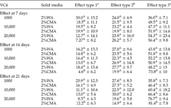
Table 1 shows the results for type 1, 2 and 3 effects of P. chlamydosporia (VC4) at concentrations of 1000, 10,000 and 20,000 chlamydospores/Petri dish in the studied intervals. In the control group (without fungus) all results were negative (zero infection).
Percentages of ovicidal activity and standard deviations for type 1, 2 and 3 effects at 7, 14 and 21 days of interaction with Pochonia chlamydosporia (VC4) chlamydospores at concentrations of 1000, 10,000 and 20,000 per Petri dish on Taenia taeniaeformis eggs in 2% water-agar (2%WA) and 2% corn-meal-agar (2%CMA).

Percentages followed by same letter (A, B) in the same column are not significantly different (P>0.01) – Friedman test.
a Physiological, biochemical effect without morphological damage to eggshell, with hyphae adhered to the shell.
b Lytic effect with morphological alteration of embryo and eggshell, without hyphal penetration through the eggshell.
c Lytic effect with morphological alteration of embryo and eggshell, besides hyphal penetration and internal colonization.
At the end of the experiment, examinations under light microscopy (40 × ) showed eggs of T. taeniaeformis destroyed by P. chlamydosporia. Significant differences (P < 0.01) were found for egg destruction (type 3 effect) at the tested concentrations compared with the control. Differences (P < 0.01) between the culture media 2%WA and 2%CMA were observed at day 7 only for the concentration of 1000 chlamydospores. Differences (P < 0.01) were also observed for effects type 1 and type 3 between the culture media 2%WA and 2%CMA at 14 and 21 days, respectively, at the concentration of 20,000 chlamydospores. The highest percentages for type 3 effect were observed in 2%WA and 2% CMA at the concentration of 20,000 chlamydospores: 76.7 and 81.4%, respectively, at 21 days.
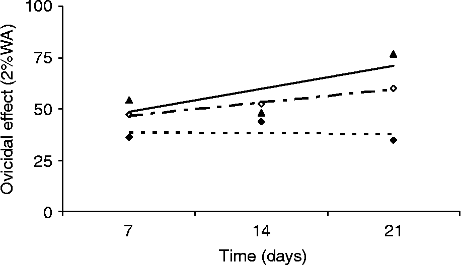
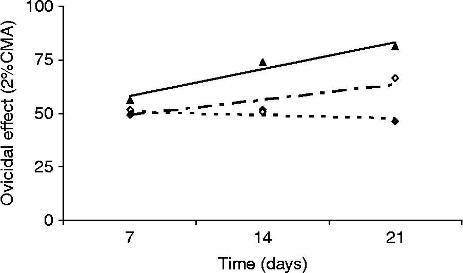
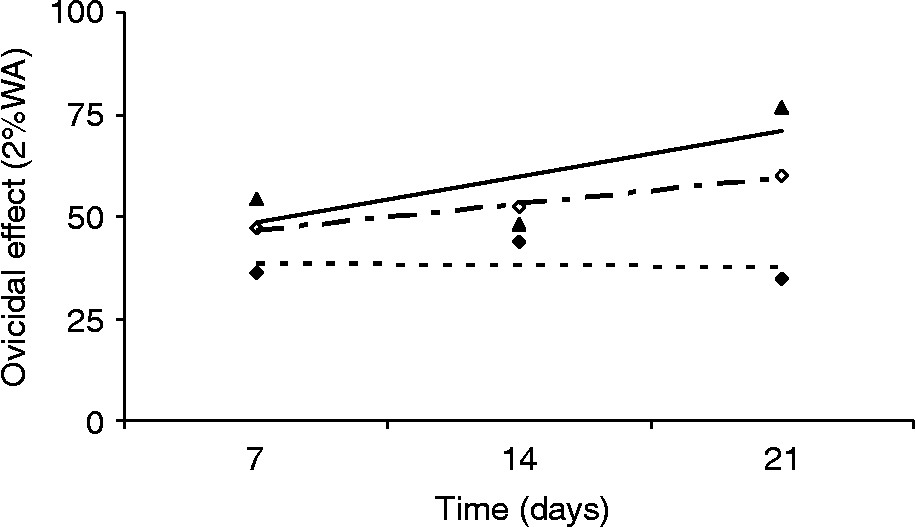
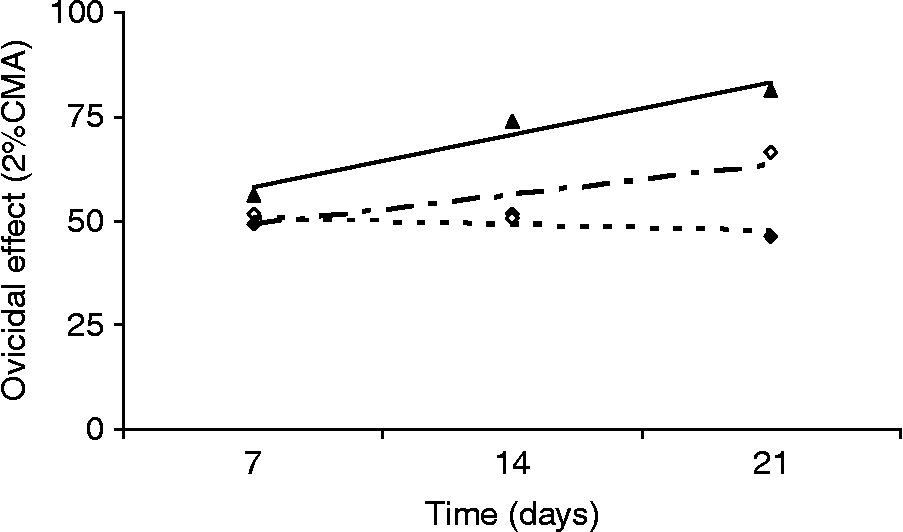
Linear regression and correlation coefficients (R 2) for type 3 effect in 2% WA at the three chlamydospore concentrations (1000, 10,000 and 20,000 per Petri dish) at 7, 14 and 21 days for the treated groups were − 0.5, R 2 = 0.0108; 6.35, R 2 = 0.9829 and 11.2, R 2 = 0.557 respectively. Linear regression and correlation coefficients (R 2) for 2%CMA at the same concentrations were − 1.55, R 2 = 0.3272; 7.25, R 2 = 0.6985 and 12.5, R 2 = 0.9513, respectively. Regression curves for effect type 3 in 2%WA and 2%CMA at the tested concentrations showed higher ovicidal activity with increasing chlamydospore concentrations (figs 1 and 2).
Linear regression curves for effect type 3 in 2% water-agar (2%WA) at chlamydospore concentrations of (1000, 10,000 and 20,000 per Petri dish), at 7, 14 and 21 days of interaction. ♦ 1000 chlamydospores/Petri dish, linear regression (- - -): y = − 0.5x+39.267, R 2 = 0.0108; ⋄ 10,000 chlamydospores/Petri dish, linear regression (— - —): y = 6.35x+40.467, R 2 = 0.9829; ▲ 20,000 chlamydospores/Petri dish, linear regression (—): y = 11.2x+37.333, R 2 = 0.55.
Linear regression curves for effect type 3 in corn-meal-agar (2%CMA) at chlamydospore concentrations of (1000, 10,000 and 20,000 per Petri dish), at 7, 14 and 21 days of interaction. ♦ 1000 chlamydospores/Petri dish, linear regression (- - -): y = − 1.55x+52.333, R 2 = 0.3272; ⋄ 10,000 chlamydospores/Petri dish, linear regression (— - —): y = 7.25x+41.9, R 2 = 0.6985; ▲ 20,000 chlamydospores/Petri dish, linear regression (—): y = 12.5x+45.533, R 2 = 0.9513.
There are reports in the literature that P. chlamydosporia has proven action on eggs of parasitic nematodes of animals and plants (Lysek, Reference Lysek1978; Mizobutsi et al., Reference Mizobutsi, Ferraz and Ribeiro2000). Recent studies have shown that this fungus is also able to destroy, under in vitro conditions, eggs of trematodes, cestodes and nematodes (Braga et al., Reference Braga, Araújo, Campos, Carvalho, Silva and Tavela2008a, Reference Braga, Araújo, Campos, Araujo, Silva, Carvalho, Correa and Pereirab; Araújo et al., Reference Araújo, Braga, Araujo, Silva and Tavela2008). These studies were carried out with grown fungal isolates plated on 2% WA with no distinction between vegetative structures (conidia and chlamydospores). In this study, each chlamydospore concentration effectively destroyed T. taeniaeformis eggs in vitro after 21 days.
This is an important finding, since P. chlamydosporia produces large amounts of chlamydospores, resistant survival structures that enable the passage of this fungus through the gastrointestinal tract of domestic animals (Araújo et al., Reference Araújo, Mota and Campos2004; Lopes et al., Reference Lopes, Ferraz, Ferreira, Freistas, Dhingra, Gardiano and Carvalho2007).
Using a different P. chlamydosporia isolate (VC1) against T. taeniaeformis eggs, Braga et al. (Reference Braga, Araújo, Carvalho, Silva, Araujo, Tavela, Campos and Costa2009) reported results of 32.2% and 54.0% for type 3 effect at 7 and 14 days, respectively. In this study, the isolate VC4 showed 49.25% and 53.4% of type 3 effect at the same intervals. Comparing the results obtained for type 3 effect, it was observed that although at 7 days of interaction the isolate VC4 showed higher activity against the eggs, overall, the ovicidal percentages were very similar. However, it is discussed that the action of fungal isolates (VC1 and VC4) against intestinal helminth eggs has not shown differences (P>0.01) (Braga et al., Reference Braga, Ferreira, Araújo, Araujo, Silva, Carvalho, Campos and Freitas2010). Therefore, the use of both P. chlamydosporia isolates in the biological control of gastrointestinal helminth eggs can be suggested.
In the present study, the highest percentages for type 3 effect were observed in both culture media (2%WA and 2%CMA), 76.7 and 81.4% respectively, at the concentration of 20,000 chlamydospores/Petri dish, at 21 days. Stirling & West (Reference Stirling and West1991) discussed that nematode eggs consist of proteins and lipids that serve as energy sources for the fungus.
Although significant differences (P < 0.01) were found at day 7 for type 1 effect between 2%CMA and 2%WA at the concentration of 1000 chlamydospores/Petri dish and at 14 and 21 days of observation, at the concentration of 20,000 chlamydospores/Petri dish, these results did not interfere with the fungal predatory capacity, since according to Lysek et al. (Reference Lysek, Fassatiová, Pineda and Hernández1982), the ovicidal activity is characterized by egg destruction (type 3 effect).
Ascending regression curves were showed for concentrations of 10,000 and 20,000 chlamydospores/Petri dish (figs 1 and 2). This may be explained by the higher ovicidal activity on T. taeniaeformis eggs with increasing interaction time. Braga et al. (Reference Braga, Araújo, Campos, Carvalho, Silva and Tavela2008a) also showed that a long exposure time of eggs to fungi enhances ovicidal activity.
In this study, there was a difference (P < 0.01) in egg destruction between poor (2%WA) and enriched (2%CMA) culture media, at a concentration of 20,000 chlamydospores/Petri dish at intervals of 14 and 21 days. A higher ovicidal activity (81.4% type 3 effect) was found for 2%CMA. De et al. (Reference De, Sanyal, Sarkar, Patel, Pal and Mandal2008) verified that P. chlamydosporia effectively destroyed eggs of Fasciola gigantica and Gigantocotyle explanatum under laboratory conditions, after 7 days of interaction. In the same work the authors demonstrated that 2%CMA was the best culture medium for fungal growth. These results are in accordance with the percentages found in the present work for the type 3 effect. This is an interesting result, since Eren & Pramer (Reference Eren and Pramer1965) found that the periodic provision of nematode-trapping fungi in poor culture media reduced their saprophytic growth, increasing their activity as a natural antagonist.
The penetration mechanism of ovicidal fungi on parasitized eggs is still not fully elucidated. However, several authors admit that enzymatic activity is a major component in the process of attack and penetration through eggshells (Lysek & Sterba, Reference Lysek and Sterba1991).
The results indicated that chlamydospores of P. chlamydosporia at concentrations of 1000, 10,000 and 20,000 chlamydospores/Petri dish effectively destroyed T. taeniaeformis eggs. Therefore, P. chlamydosporia can be used as a biological control agent for this cestode.
Acknowledgements
J.V.A. received a scholarship from CNPq, Rio de Janeiro, Brazil. The authors would like to thank CAPES, FINEP-Pró-equipamentos, Fapemig and CNPq for financial support and grants.