Implications
North-eastern Sicily is an area with iodine deficiency disorders occurring both in humans and in domestic animals. However, limited studies have attempted to evaluate the thyroid function in horses. This is largely due to the inadequacies of current routine diagnostic capabilities.
Our study showed that endemic areas for humans are not necessarily so for horses, which seem to be able to adopt an adaptive strategy without presenting any clinical signs of thyroid disorders. However, goitre areas in Sicily still remain a serious environmental and health problem for humans and represent a potential economic threat for the animal industry. Recently, thyroid disease in the horse was ranked second as a research priority in Italy.
Introduction
Thyroid hormones are important physiological indices commonly used in studies concerning metabolic rate (Chen and Riley, Reference Chen and Riley1981), animal thermogenesis (McGuire et al., Reference McGuire, Beede, Collier, Buonomo, DeLorenzo, Wilcox, Huntington and Reynolds1991; Bird et al., Reference Bird, Clarke and Symonds1998), central nervous system development and changes in cognitive function in animals and humans (Schantz and Widholm, Reference Schantz and Widholm2001; Helmreich et al., Reference Helmreich, Crouch, Dorr and Parfitt2006; Long and Voice, Reference Long and Voice2007). Their supplementation increases the metabolic rate and promotes significant weight loss with dietary restriction (Graves and Schott, Reference Graves and Schott2002). In the horse, a deficiency in thyroid hormones results in severe growth retardation (Irvine and Evans, Reference Irvine and Evans1975; Allen et al., Reference Allen, Fretz, Card and Doige1998). Iodine deficiency occurs in pregnancy with retardation of foetus development resulting in foetal death or the birth of weak neonates affected by hyperplastic goitre (Baker et al., Reference Baker, Wyn-Jones and Eley1983; Osame and Ichijo, Reference Osame and Ichijo1994; Durham, Reference Durham1995). The effects of iodine deficiency in thyroid disorders, especially in endemic goitre areas, have been reported both in humans (Squatrito et al., Reference Squatrito, Delange, Trimarchi, Lisi and Vigneri1981; Vigneri, Reference Vigneri1988; Vermiglio et al., Reference Vermiglio, Sidoti, Finocchiaro, Battiato, Lo Presti, Benvenga and Trimarchi1990; Vermiglio et al., Reference Vermiglio, Lo Presti, Scaffidi Argentina, Finocchiaro, Gullo, Squatrito and Trimarchi1995) and in different animal species (Hetzel and Mano, Reference Hetzel and Mano1989; Maberly, Reference Maberly1994; Pugliese et al., Reference Pugliese, Medica, Scardillo, Parisi and Fazio2007). Although diagnosis of thyroid disease in the horse is difficult (Frank et al., Reference Frank, Sojka and Messer2002; Messer and Johnson, Reference Messer and Johnson2007), cases of hypo- (Sojka, Reference Sojka1995; Breuhaus, Reference Breuhaus2002) and hyperthyroidism (Ramirez et al., Reference Ramirez, McClure, Moore, Wolfsheimer, Gaunt, Mirza and Taylor1998; Tan et al., Reference Tan, Davies, Crisman, Coyle and Daniel2008) in adult horses have been documented only rarely. Single measurements of T4 and T3 are difficult to interpret, particularly because thyroid hormone metabolism or transport can be affected by physiological or pathological states and by a variety of drugs (Breuhaus, Reference Breuhaus2002) as well as by the influence of circadian rhythm (Duckett et al., Reference Duckett, Manning and Weston1989; Komosa et al., Reference Komosa, Flisińska-Bojanowska and Gill1990). North-eastern Sicily is an area with iodine deficiency disorders occurring both in humans and in different domestic animals and partial beneficial effects of the so-called ‘silent iodine prophylaxis’ (an additional percentage of iodine salt gave in water and food) on iodine deficiency disorders have been described (Vermiglio et al., Reference Vermiglio, Finocchiaro, Lo Presti, La Torre, Nucifora and Trimarchi1989). The aim of this study was to compare the total and free iodothyronine levels of horses stabled in non-endemic and endemic goitre areas, taking into account different gender and age.
Material and methods
Animals, diets and experimental design
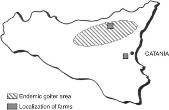
The study was carried out on a total of 72 clinically healthy Sanfratellano horses weighing 585 ± 40 kg ranging in age from 5 to 9 years, which were stabled in two different areas of Sicily (Figure 1). Thirty-six horses (18 males and 18 females) were stabled in farm A (group I) near the city of Catania (150 m above sea level; 37′3°N; 15.4°E) and the other 36 horses (20 males and 16 females) were kept in farm B (group II) situated on the slopes of the Nebrodi mountains (760 m above sea level; 38′5°N; 14.48°E). The last farm is located in an area of severe endemic goitre with the presence of abnormalities in human thyroid function in the population (Squatrito et al., Reference Squatrito, Delange, Trimarchi, Lisi and Vigneri1981; Vigneri, Reference Vigneri1988). The stable management of the two farms varied: the horses on farm A were in individual stables and were not allowed daily access to pasture. They were fed traditional rations, based on a commercial concentrate (approximately 3.5 kg/horse per day), and grass hay (average 8 kg/horse per day), twice daily. The iodine content (anhydrous calcium iodide) was equal to 2 mg. Water was provided ad libitum.
Map of Sicily.

The horses on farm B were kept together in outdoor paddocks, but were kept on pasture most of the time and fed twice a day on a diet of alfalfa hay and oats. Water was available ad libitum. No iodine supplementation was introduced into the diet.
No plant known to be goitrogenic or a member of the Brassica family was present on pastureland. The investigation was conducted in late June.
Measurements
Mean environment temperature was 24°C (22°C to 27°C) and mean relative humidity was 44.60% (38.20% to 51.5%). These were monitored using a Hygrothermograph ST-50 (Sekonic Corporation, Tokyo, Japan).
Blood samples from the jugular (5 ml) were collected using evacuated tubes (Venoject, Terumo®, Leuven, Belgium) at 0900 h in order to minimise the effect of circadian rhythm on hormone measurements. All samples were taken in quiet conditions by the same veterinarian.
No urinary iodine excretion was measured.
Laboratory analysis
Blood samples were kept at 4°C until centrifugation at 1500 × g for 15 min and the plasma was harvested and stored in polystyrene tubes at −20°C until total and free iodothyronines were determined.
Hormone assays were analysed in duplicate using a commercially available immunoenzymatic kit (SEAC-RADIM, Pomezia, Roma). The respective intra- and inter-assay coefficients of variation were as follows: 7.3% and 11.4% for T3; 2.3% and 5.7% for T4; 4.2% and 11.9% for fT3; 6.6% and 9.6% for fT4.
Statistical analysis
All the results are expressed as mean ± s.d. (Table 1 and Figures 2 and 3). Individual results were compared by one-way analysis of variance of repeated measures (RM-ANOVA). Significant differences between the two groups were established using Bonferroni’s multiple comparison test. Significant differences between different gender and age between groups were established using Student’s unpaired t-test. The level of significance was set at <0.05. All calculations were performed using the PRISM package (GraphPad Software Inc., San Diego, CA, USA). The ratios for T3/T4 and fT3/fT4 were also calculated.
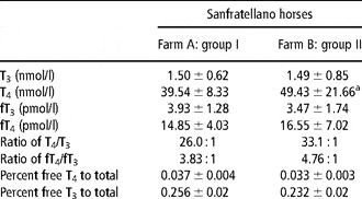
Total and free iodothyronine levels (mean ± s.d.) in Sanfratellano horses from a non-endemic area (farm A: group I) and in horses from an endemic goitre area (farm B: group II)

*Symbol indicates significant (P < 0.02) differences in average hormone concentrations vs group I: a = P < 0.02.
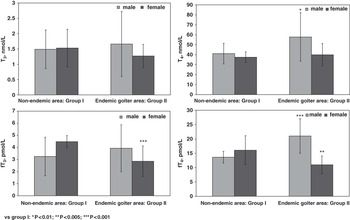
Total and free iodothyronine levels (mean ± s.d.) in male and female horses from a non-endemic and an endemic goitre area.

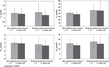
Total and free iodothyronine levels (mean ± s.d.) in young and adult horses from a non-endemic and an endemic goitre area.

Results
The results (Table 1) showed higher thyroxine values (P < 0.02) in group II horses than the values observed in group I horses.
T4 : T3 ratios were lower (26 : 1) in group I than in group II (33.1 : 1); fT4 : fT3 ratios were also lower in group I (3.83 : 1) than in group II (4.76 : 1). The percentage of fT4 and fT3 was similar in the two groups.
On the basis of gender difference, comparison between the two groups showed higher T4 (P < 0.01) and fT4 (P < 0.001) in males, and lower fT3 (P < 0.001) and fT4 (P < 0.005) in females stabled in the endemic goitre area, when compared to the respective values for males and females stabled in the non-endemic area.
On the basis of different age independently from the area of origin (non-endemic or endemic), younger horses (<7 years) showed the highest thyroid hormone levels in both groups. Moreover, all young horses in the endemic area, regardless of age, showed higher (P < 0.01) T4 levels than those horses in the non-endemic area, as previously stated.
Discussion
Thyroid hormone levels were within normal ranges in both groups and the comparison of our results with published data reporting the circulating thyroid hormones of adult healthy horses (Kallfelz and Lowe, Reference Kallfelz and Lowe1970; Reap et al., Reference Reap, Cass and Hightower1978; Anderson et al., Reference Anderson, Nixon and Akasha1988) did not reveal any large discrepancies, although the T4 levels were higher in this study; however, no data regarded the range of adult horse’s thyroid hormones in male and female subjects are available. Furthermore, the results obtained are in agreement with the higher T4 levels reported in growing Thoroughbred foals (Fazio et al., Reference Fazio, Medica, Cravana, Messineo and Ferlazzo2007). Some slight differences may also occur due to different techniques, nutritional factors or geographic environmental variations.
The higher T4 levels recorded in group II (where no clinical symptoms of thyroid disorders were present) compared to physiological ranges reported in the literature show that hypothyroidism found in humans from the same endemic goitre area does not necessarily correlate with lower T4 levels and with evident clinical signs in adult horses. Indeed, like horses stabled in the endemic area, horses stabled in the non-endemic area also showed high T4 levels. Moreover, the higher T4 levels of group II could be explained on the basis of the different altitude of farms (760 m v. 150 m sea level) and environmental temperature (22°C v. 27°C).
In addition, it is possible that different farm management practices might produce different thyroid responses, and sure enough group II horses were kept on pasture most of the time, with plants being the primary source of iodine.
Furthermore, horses in endemic areas had high T4/T3 and fT4/fT3 ratios, while these same horses showed lower percentages for fT4 and fT3 with respect to T4 and T3. These results confirm that changes in fT4 levels generally follow those for T4.
These findings suggest that the horses living in endemic goitre areas (group II) probably have acquired coping strategies to synthesise more T4. These findings could probably be supported by suitable amounts of iodine in the diet for equine thyroid function, or by iodine re-collected in the gastrointestinal tract, as has been reported in humans.
The data obtained confirm that the physiological range of thyroid hormones in horses is wide. In addition, in horses, relatively little research has been done regarding the use of thyroid hormone measurements to evaluate thyroid disease (Sojka et al., Reference Sojka, Johnson and Bottoms1993; Messer et al., Reference Messer, Ganjam, Nachreiner and Krause1995; Breuhaus, Reference Breuhaus2002) and to establish the incidence of thyroid dysfunction in the equine population.
Many intrinsic and extrinsic variables can influence physiological thyroid hormone concentrations: hence, the diagnosis of thyroid diseases in horses, as well as in other species, has usually been difficult to perform. This is because single or isolated measurements of thyroid hormones are hard to interpret, while TRH and TSH stimulation tests, although they are more useful, are not routinely performed in practice (Breuhaus, Reference Breuhaus2002; Long and Voice, Reference Long and Voice2007).
Our data confirm the existence of significant differences between male and female horses in thyroid hormone concentrations, although previous studies performed in growing thoroughbred foals showed higher T4 and fT4 levels in colts than fillies (Fazio et al., Reference Fazio, Medica, Cravana, Messineo and Ferlazzo2007). In addition, our data correlate with the results obtained in humans, with a higher percentage of endocrine thyroid disease in women than in men.
In addition, data obtained confirm that younger horses have higher thyroid hormone levels, and agree with the statement that values decrease slowly over a horse’s life (Malinowski et al., Reference Malinowski, Christensen, Hafs and Scanes1996).
In horses stabled in the same areas of endemic human goitre, with severe hypothyroid cases of goitre, cretinism and deafness, there were no signs recorded of any abnormal clinical symptoms associated with hypothyroidism. Indeed, horses stabled in endemic areas showed paradoxically higher T4 concentrations than physiological and control group values. It would therefore appear that the equine species adapts to an endemic environment through a massive thyroidal response in order to build up a substantial reserve of T4. However, it is not possible to exclude that our subject is affected by exthyroid illness that causes inhibition of the enzyme 5 deidinase, which is responsible for the conversion of T4 to T3. This illness represents an adaptive mechanism to decrease the metabolic rate during periods of illness and stress and the thyroid gland is considered to be normal as reported by Duckett (Reference Duckett1998). In addition, T4 concentrations are similar in male and female horses of the non-endemic area (group I); but in horses from the endemic goitre area (group II), the T4 concentrations are significantly increased in male horses.
It is well known that the fT4 fraction represents the biologically active hormone for tissues; thus, the ratio of fT4 represents a primary factor for determining the fractional turnover of thyroid hormones. Moreover, the binding fraction represents the hormonal store that balances the sudden increase and decrease of hormonal release to tissues. In contrast, T3 is mainly an intracellular hormone and its serum measurement is a less representative value of the total hormonal complex than serum T4 measurements.
These data also showed that the existence of higher concentrations of T4 in horses of all ages was more evident in animals from the endemic area.
In addition, T4 has an intrinsic thyromimetic activity, as protection against hypothyroidism, which could be more pronounced in horses stabled in an endemic goitre area.
The results would seem to suggest a physiological adaptive response of the equine species to a low iodine environment. The possibility that hypothyroidism is present in horses is thus excluded and is supported not only by the lack of clinical signs, but also by the rarity of the cases reported, being limited exclusively to geriatric horses. In conclusion, endemic areas for human goitre are not necessarily so for horses that show an adaptive strategy without presenting any clinical signs of thyroid disorders.
Using the values found in humans (Vigneri, Reference Vigneri1988) as the basis for comparison, our data found the horse to exhibit lower total circulating T4 at 39.54 nmol/l (group I) and 49.43 nmol/l (group II) than the values found in euthyroid humans (95.23 nmol/l) and goitre humans (82.36 nmol/l). However, goitre areas in Sicily still remain a severe problem for humans, although partial beneficial effects of the so called ‘silent iodine prophylaxis’ on iodine deficiency disorders in Sicily endemia were observed (Vermiglio et al., Reference Vermiglio, Finocchiaro, Lo Presti, La Torre, Nucifora and Trimarchi1989). These areas do not appear to be of concern for horses, however, indicating that there is very little understanding of the aetiology of this disease.
Acknowledgements
The authors are grateful to the horse’s owners and veterinarians for their disposition and invaluable contribution to the present study.