The premise
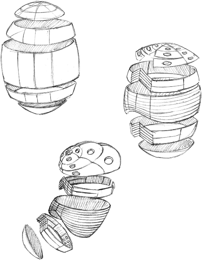
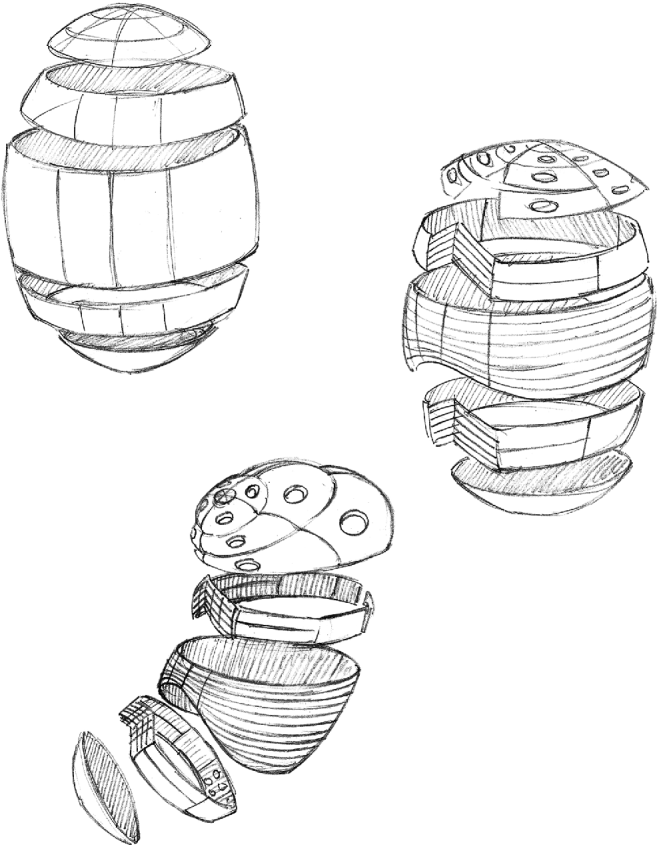
The body is a mosaic enlargement of the pattern of circumferential bands assumed by the columns and rows of molecules that comprise the egg cell membrane.
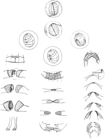
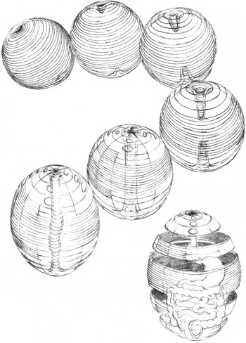
It is well known that cell membranes are built of columns and rows of phospho-lipid molecules that self-organize as underwater double-walled spheres (Oparin, Reference Oparin1952). The membrane has been described as a toroidal surface by Jockusch & Dress (Reference Jockusch and Dress2003) and Kraus (Reference Kraus2006). It is proposed that the expansion of the surfaces within the constraint of the inelastic outer vitelline sphere causes the poles to indent and grow internally towards each other, forming a toroidal bilayer, with the central cable of the outer torus passing through the centre of the inner torus, and that mutual rotation will cause the cable to become buried beneath the dorsal midline as the notochord and nerve chord (Starodubov, Reference Pivar2008, Reference Starodubov, Doronin and Golichenkov2009). The hypothesis presumes that, in response to physical forces, the molecules comprising the egg cell membrane become arranged in circumferential bands separated by polar meridians, subdividing the surface into trapezoidal plates, much like a geodesic globe of Earth. Subject to deformation during subsequent hydrodynamic disruption, each of the plates will curl axially to become a bone of the vertebrate skeleton, the apical cap forming the skull. This topological theorem predicts the form of the bones of the vertebrate skeleton and their configuration in the body with accuracy beyond the possibility of coincidence (Fig. 14).
Egg cells are internally pressurized, inflated spherical membranes that act like bubbles or balloons, combining the familiar properties of both. The model demonstrates that the embryo is formed in the sudden collapse of the equilibrium between the pressurized contents of the cell and the ability of the elastic membrane to contain it. This event is comprehensible in terms of the familiar high school physics experiment where two unequally inflated balloons are conjoined by a tube. Contrary to intuition, the smaller balloon will deflate as it empties its contents into the larger balloon. In the embryological events of fertilization and gastrulation, the thinning of the membrane at the apical cap initiates an aneurismic bulge, resulting in the inflation of the presumptive skull and the sudden collapse of the presumptive rib cage.
Demonstration
The egg is known to result from the primordial germ plasm, a streaming, toroidal, mobile amoeboid cell that crawls through the female body toward the ovaries, swelling and rounding up in size as it goes, finally producing the egg, a thousand times the size of a normal cell (Fig. 18).
The figures describe these predictable, plausible topological events. The egg membrane consists of molecules arranged in circumferential bands. With inflation the bands migrate and stretch, assuming, coincidentally, a configuration not unlike that of the geodesic sphere of Earth, divided into five regions, including a middle equatorial zone, polar caps and two intermediate zones that separate them. The surface of the egg is thereby paved with spherical trapezoidal segments. Each of these will form a bone by curling axially in response to intrinsic tensorial forces (Figs 1 and 2).
Schematic patterning of egg cell membrane
Development of the vertebrate limb

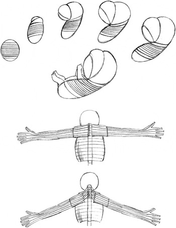
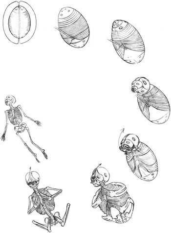
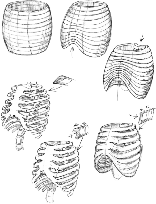
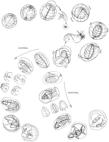
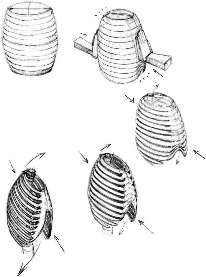
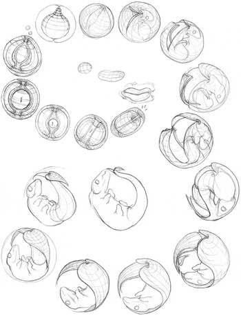
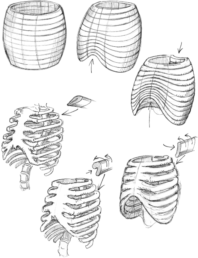
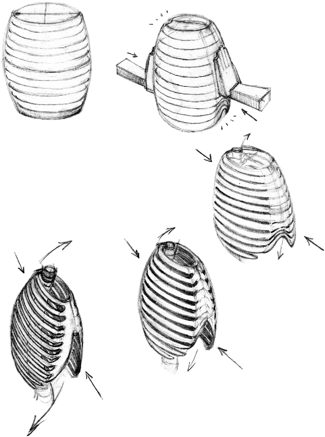
As the egg cell grows larger by inflation the polar caps increase in size and their surfaces become thin. At a certain point the membranes of the polar cap can no longer resist the internal pressure. The segments suddenly swell as an aneurism, precipitating the sudden collapse of the internal pressure equilibrium. The contents of the cell flow into the head region, causing the body region to collapse. The presumptive rib cage compresses from front to back, causing the curvature of the ribs to descend as they curve toward the front, while the front of the rib cage fails by arching upward (Fig. 15). At the same time the zones of the polar cap inflate. The collapse of the front of the rib cage draws the front, or ventral part, of the polar cap beneath the inflated dorsal half, preventing its inflation (Fig. 3).
Hydrostatic transfer

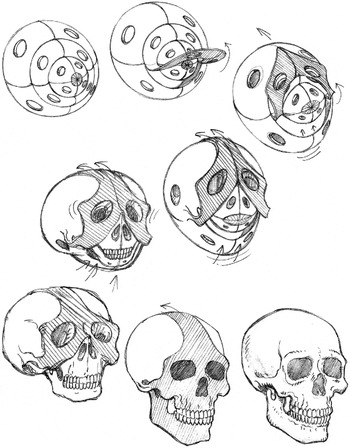
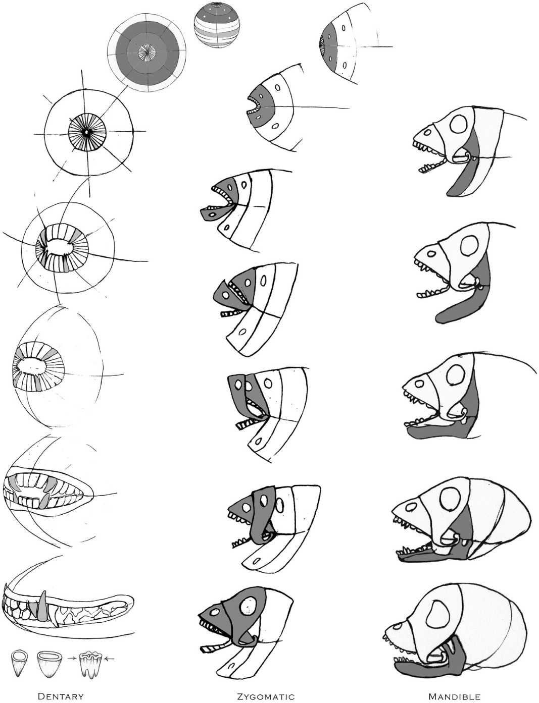
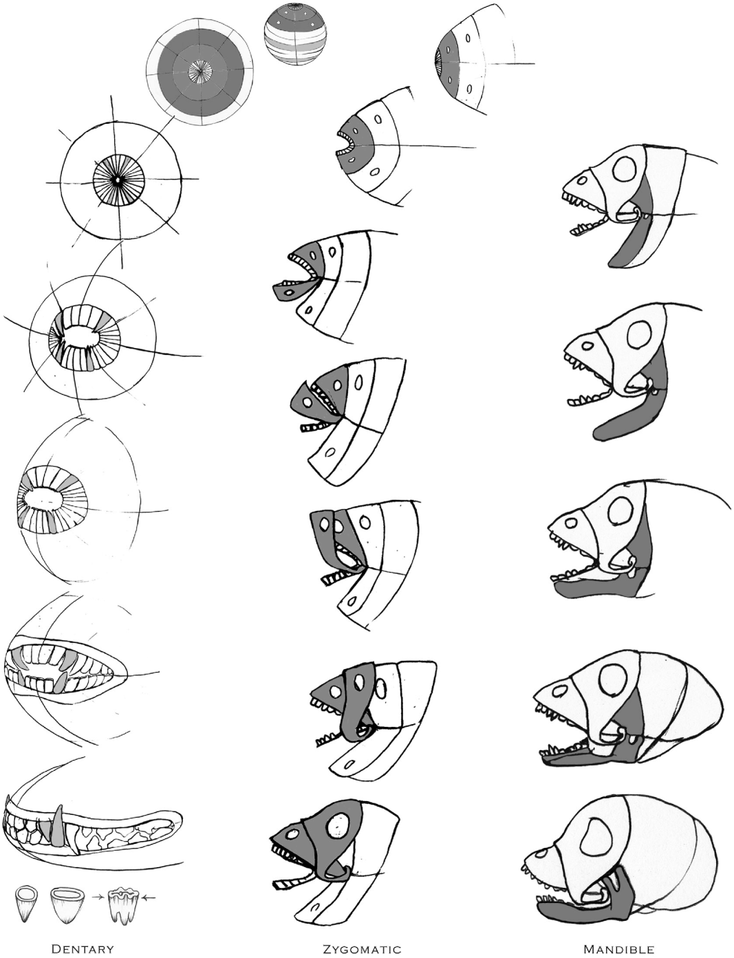
The polar cap comprises two layers of membrane. The top layer of the front, or ventral half, peels backward and comes to lie down on the back, or dorsal half, of the cap, forming the zygomatic arch. The remaining ventral layer is forced into the underside of the dorsal half of the cap (Fig. 4).
Skull morphogenesis

Concurrently, the intermediate zones of the rib cage are displaced as they relocate as the presumptive limbs. The central toroidal cable buries itself in the dorsal midline, installing the notochord and nerve chord (Fig. 5).
Vertebrate embryogenesis

Limb development
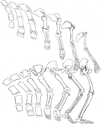
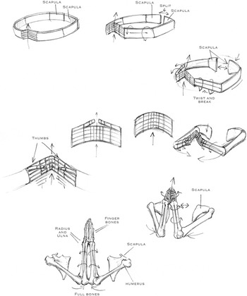
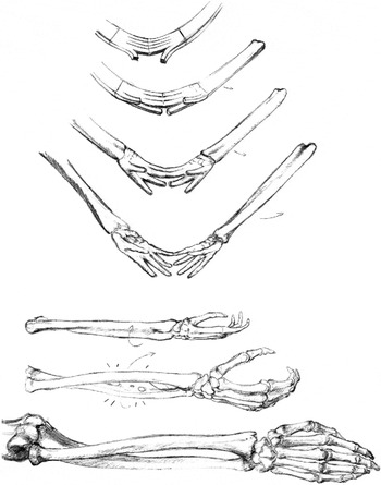
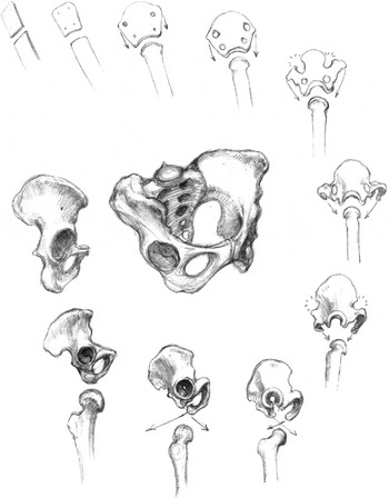
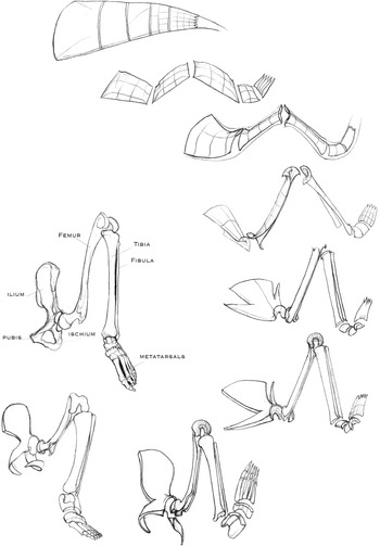
The limbs derive from the band segments located adjacent to the polar caps, apart from the segment crossing the dorsal midline, which is drawn beneath the surface forming the vertebrae. These presumptive limb segments form the scapula, humerus, radius, ulna and carpals of the forelimbs, and the pelvis, femur, tibia, fibula and tarsals of the hind limbs (Gray, Reference Gray and Lewis1930). Axial curling of the scapula and humerus form the shoulder joint. The twisting force in the displacement of the segments separates the radius from the ulna and the tibia from the fibula. The hands derive their characteristic geometry from the sequential subdivision of the carpal bones by halves. The surface features of the bones derive from the deformations experienced as the limbs are drawn together by forces driving the ventral midline toward the anterior (Figs 6–9, 16, 17).
Hands

Hands

Feet

Hands, claws and hooves

Rib cage
The characteristic curvature of the rib cage results from the ventral-dorsal compression of the middle zone of the spherical egg. The ventral side arches anteriorly, and the ribs curve posteriorly as they curve from dorsal to ventral. The contiguous bands of the presumptive ribs each fold in half axially, creating a separating space between them, equal in width of a rib. The hydrostatic failure of the spherical forerunner inflates the skull at the expense of the rib cage (Fig. 10).
Rib cage
Spinal column
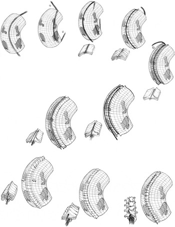
The mutual rotation of the two toroidal spheres through two rotations implants the notochord and the nerve chord beneath the dorsal midline, causing the ribs to fragment. The reassembly of the fragments forms the vertebrae and the somites, the latter forming the interior bands of muscles that encompass the body (Fig. 11).
Vertebrae formation

Skull
The skull takes its form from the apical cap, a two-layer spherical surface divided into four sectors and three zones, each containing a hole at the centre resulting from the thinning of the membrane. Hydrostatic collapse causes the ventral half of the outer layer to fold over upon the dorsal half, forming the zygomatic arch and prefrontal ridges. The remaining ventral segments of the apical cap fold underneath the presumptive mandible (Fig. 19).
Sacrum
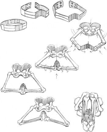
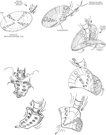
The sacrum is formed from the caudal cap comprising five concentric zones divided by four meridians, with a hole in the centre of each. The coccyx of the spinal column comes to rest in the centre of the caudal cap, covering the dorsal half. The ventral half folds back onto the dorsal half, completing the form of the sacrum (Fig. 12).
Sacrum

Alimentary canal
The presumptive alimentary canal results from the segments of the surface entering the internal canal at the poles that meet interiorly to form the gut tube. An aneurismic inflation forms the stomach. The segmented large intestine is formed from the incursion of the segments of the caudal cap (Fig. 13).
Establishment of toroidal model

The origin of the vertebrate skeleton

Rib cage
Pelvis

Development of the vertebrate limb

Vertebrate development

The origin of the skull
Comparative anatomy
The subject model is of the human species; it is also applicable to the other mammalian species. The fact that reptiles and other predecessor forms, except for birds, do not possess a sternum does not exclude them from the paradigm.
Cell subdivision
For the sake of clarity, the model as described does not take into account cellular subdivision, which occurs concurrently, but is irrelevant to its progress. Each subdivision leaves the resulting cells with an ever-diminishing portion of the original cell membrane. Finally, the blastula is seen as a subdivided replica of the egg cell membrane.
Comments
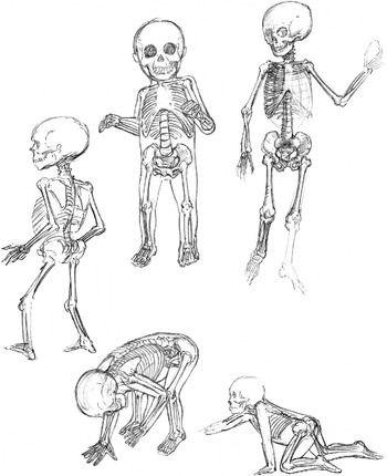
The stages of this hydrostatic model are not contradicted by observed embryology. The well known stages in the laboratory models of chick embryology and Xenopus gastrulation may be reassessed to confirm the predictions of the model. These and other comparisons in embryology offer promise of further confirmation, but are beyond the scope of this investigation. Observed osteogenesis records the gradual calcification of transparent, cartilaginous tissue, which may be presumed to bear the forms of the completed bone, much as an exposed photographic plate bears the image revealed upon chemical development (Fig. 20).
Vertebrate skeleton

The hydrodynamic theory of cell form is a part of the history of morphology. Goethe, who coined the term morphology, dedicated his scientific life to the discovery of a common origin of living form he named the Urform (Steiner, Reference Steiner and Lindeman1988). Wilhelm His, called ‘the father of human embryology’, demonstrated in Unsere Körperform, 1874, the origin of the form of organs with inflated balloons. His' science was denounced by Ernst Haeckel as Gummischlauch wissenschaft (balloon science). It was His who exposed Haeckel's infamous embryo drawing fraud (Haeckel, Reference Haeckel1886). D'Arcy Thompson, in his legendary 1917 tome, On Growth and Form, devotes the hundred pages of Chapter 5 to a definitive mathematical and physical analysis of the behaviour of the cell as a fluid enveloped by an elastic membrane (Thompson, Reference Thompson1917).
Conclusion
The presented model of life sees the human body as a rationally generated structure, independent of the environment, rather than the result of random errors in the genome. Apart from the philosophical implication of the self-organizational origin of life, this understanding can lead to progress in various areas of science (Kauffman, Reference Kauffman1993; Pivar, Reference Pivar2009). In medicine, the mechanically causative theory of disease is a promising field. Self-organization presents the possibilities of embryonic plastic surgery to prevent birth defects, as well as for aesthetic advantages. The technology of tissue engineering may benefit from being informed as to the natural directional vectors of tissue growth, leading to the synthesis of entire organisms.