Introduction
Papillary thyroid microcarcinoma is defined as a focus of papillary thyroid carcinoma that is less than or equal to 10 mm in diameter.Reference Hedinger, Williams and Sobin1 The incidence of thyroid cancer continues to increase, with the vast majority of tumours being less than 2 cm in size.Reference Davies and Welch2 This increase in incidence is believed to be mainly due to a rise in the number of incidental tumours being detected due to widespread use of head and neck imaging (i.e. ultrasound, computed tomography, magnetic resonance imaging and positron emission tomography).
There is considerable controversy in the literature regarding the optimal management of papillary thyroid microcarcinoma, since the majority have an indolent behaviour. However, there is a subset of patients who present with extensive cervical lymph node involvement and/or evidence of metastatic disease, with only small primary tumours in the thyroid gland. In these patients, radical surgery is usually employed, in the form of total thyroidectomy and neck dissection plus adjuvant radioactive iodine and thyroid-stimulating hormone (TSH) suppression.
The aim of this study was to review our institution's experience of the variable disease presentation, management and outcome of patients with papillary thyroid microcarcinoma, and to attempt to identify variables associated with adverse outcomes.
Materials and methods
This study was a retrospective analysis of prospectively collected data from the Sydney Head and Neck Cancer Institute database. Patients included in the study had been treated at our institution between 1987 and 2009.
Institutional ethics approval for the study was granted by the Ethics Review Committee.
Patient demographic details comprised age (at the time of surgery) and sex.
Surgical treatment details were based on the extent of surgery: either hemi- or total thyroidectomy. If a hemi-thyroidectomy was performed then details pertaining to the rate of completion thyroidectomy were obtained. Patients who had a completion thyroidectomy were classified as having undergone total thyroidectomy.
If a neck dissection was performed, the laterality and levels were recorded. Central neck dissection involved removal of lymph nodes in levels six and/or seven, whereas a lateral neck dissection involved removal of lymph nodes in any level between one and five.
Tumours were classified as non-incidental if surgery was performed with a pre-operative diagnosis of (or suspicion of) papillary thyroid cancer. Tumours were classified as incidental if a focus or foci of papillary thyroid microcarcinoma was discovered in the surgical specimen following surgery for other indications.
All thyroid specimens were serially and transversely sectioned at 3–5 mm intervals, after formalin fixation. The dimensions of all obvious abnormalities were recorded, and appropriate sections, along with an alternate slice of normal thyroid tissue, were submitted for paraffin embedding and histopathological evaluation. In cases in which an obvious macroscopic abnormality was not seen, every alternate slice was submitted for histological examination, with more extensive sampling if a microscopic abnormality was found. All specimens were photographed both before and after serial sectioning. No tumours underwent further analysis to assess clonality.
All patients were pathologically staged according to the 2002 American Joint Committee on Cancer staging system.3 The following histopathological details were recorded: maximal tumour diameter; presence of multifocality (and, if present, whether ipsilateral or bilateral); extra-thyroidal extension; and presence of vascular invasion. If a neck dissection was performed, the presence of involved lymph nodes within each level was recorded. When given, the number of treatments and dose of radioactive iodine (in millicurie) were recorded.
Recurrence was defined as local (i.e. within the thyroid bed) or regional (within the lateral neck). Follow up was recorded from the date of the last known review.
Statistical analysis
Data analysis was performed using the SPSS version 11.5 statistical software program (SPSS Ltd, Chicago, Illinois, USA). Disease-free survival was defined as the time between surgery and date of first disease recurrence (locoregional or distant) if the disease recurred, or as the time between surgery and date of death or last follow up if the disease did not recur. A p value of less than 0.05 was considered statistically significant.
Results and analysis
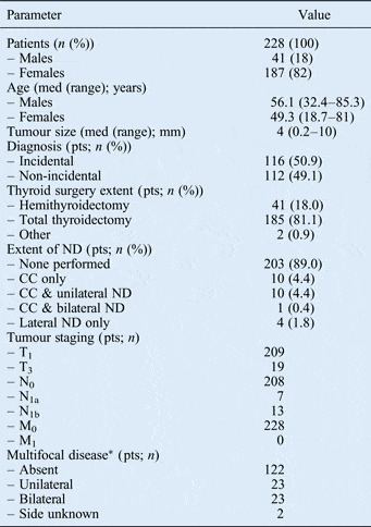
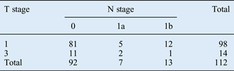
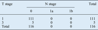
We identified a total of 238 patients with a histopathologically confirmed diagnosis of papillary thyroid microcarcinoma. Ten patients had significant missing data and were thus excluded, leaving 228 patients for analysis (Table I). Pathological tumour and node staging for the non-incidental and incidental tumours is shown in Tables II and III.
Patients' clinicopathological details

*Multifocal tumour information from 170 patients. Med = median; pts = patients; ND = neck dissection; CC = central compartment; T = tumour; N = node; M = metastasis
Tumour and node staging: non-incidental tumours
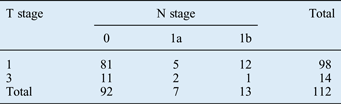
Data represent patient numbers. T = tumour; N = node
Tumour and node staging: incidental tumours
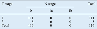
Data represent patient numbers. T = tumour; N = node
The overall median tumour diameter was 4 mm (range, 0.2 to 10 mm). Non-incidental tumours had a greater median diameter (7 mm; range, 0.5–10 mm) than incidentally discovered papillary thyroid microcarcinoma (median diameter, 2.4 mm; range 0.2–10 mm). Non-incidental microcarcinomas were significantly more strongly associated with a younger age (<45 years; p = 0.007) and larger tumour size (≥ 5 mm; p < 0.001), compared with incidentally detected microcarcinomas. The relative risk (incidental group versus non-incidental group) and confidence intervals are shown in Table IV. There was no significant difference with respect to sex, pre-operative thyroid function status or extent of surgery (i.e. hemi- vs total thyroidectomy).
Association between tumour type and patient factors

*Incidental vs non-incidental. †Multifocal tumour information from 170 patients. ‡Size information from 207 patients. RR = relative risk; CI = confidence interval; Inc = incidental; Non-inc = non-incidental; y = years; RAI = radioactive iodine
Regarding the extent of surgery, total thyroidectomy was performed in 185 patients (including 25 patients undergoing initial hemithyroidectomy followed by completion thyroidectomy) and hemithyroidectomy in 41 patients. One patient had only the pyramidal lobe excised, and another had only a thyroglossal remnant excised.
Information regarding multifocal disease status was available in 170 patients. Multifocal disease was found pathologically in 48 (28.2 per cent) patients. Disease was unilateral in 23 (47.9 per cent) patients and bilateral in 23 (47.9 per cent). In two patients, the pathology report did not specify whether the multifocal disease was unilateral or bilateral. In patients who underwent total thyroidectomy (including completion thyroidectomy), multifocal disease was found in 41 (22.2 per cent). These multifocal deposits were unilateral in 17 (41.5 per cent) patients and bilateral in 22 (53.7 per cent). In two (4.8 per cent) patients, the pathology report did not clarify whether the multifocal disease was unilateral or bilateral. Of those patients undergoing hemithyroidectomy only, 5 (12.2 per cent) had multifocal cancers. In patients undergoing initial hemithyroidectomy followed by completion thyroidectomy, the incidence of further foci of carcinoma found in the other thyroid lobe was 41.2 per cent. Multifocality had no association with tumour type (incidental versus non-incidental), sex, age group (<45 versus ≥45 years) or tumour size (<5 versus 5–10 mm).
Overall, the vast majority of patients (215 of 228; 94.3 per cent) presented with localised disease (i.e. T1–3, N0–1a) to the thyroid and/or central compartment. In the non-incidental group, 88.4 per cent of patients presented with disease localised to the thyroid and/or central compartment; thus, 11.6 per cent of patients presented with extensive disease that had spread into the lateral cervical lymph nodes. No metastatic disease was detected during the study period.
Twenty-nine neck dissections were performed in 25 patients. Lymph node metastases were found in 22 (75.9 per cent) neck dissections. The incidence of histopathologically confirmed involvement was 56.5 per cent in the central compartment and 86.7 per cent in the lateral neck. One patient had intra-thyroidal lymph node metastasis. The odds ratio for incidence of one or more pathological features (i.e. extra-capsular spread, extra-thyroidal extension and/or vascular invasion) was 4.67 (95 per cent confidence interval (CI), 1.74–12.49), comparing patients with and without lymph node metastasis (p = 0.001).
We found pathological evidence of minimal extra-thyroidal extension in 19 of 228 (8.3 per cent) patients, of vascular invasion in 10 of 228 (4.4 per cent), and of thyroid extra-capsular spread in 24 of 228 (10.5 per cent).
Ablative radioactive iodine was administered to 41 patients (18 per cent). Six of these patients were given a second dose. The median dose per treatment was 101.4 mCi (range, 100–205 mCi).
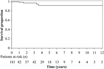
Locoregional recurrence occurred in only 4 of the 228 (1.8 per cent) patients (2 local and 2 regional) (See Figure 1). Two of these patients went on to suffer further recurrence. All four patients were successfully salvaged with further surgery and adjuvant radioactive iodine. Patients with incidentally discovered papillary thyroid microcarcinoma had a shorter median duration of follow up (median, 3.8 months; range, 9 days to 6.8 years) compared with non-incidental patients (median, 16.9 months; range, 3 days to 20 years). No patients died within the study period.
Disease-free survival (locoregional control) for all papillary thyroid microcarcinoma patients.

Discussion
Papillary thyroid microcarcinomas are found both incidentally and non-incidentally. They are also commonly found in thyroids removed for benign indications: up to 22 per cent of thyroid specimens have been reported to harbour foci of papillary thyroid microcarcinoma.Reference Olen and Klinck4, Reference Carlini, Giovannini, Mercadante, Castaldi, Dell'Avanzato and Zazza5 The incidence of detection of papillary thyroid microcarcinoma foci is dependent on how thoroughly the pathologist examines the thyroid histologically.Reference Grodski, Brown, Sidhu, Gill, Robinson and Learoyd6
The biological behaviour of these tumours is highly variable, and the optimal management of patients with papillary thyroid microcarcinoma remains controversial. Although generally regarded as indolent tumours, there is a subset of patients who present with widespread disease that behaves like classical papillary thyroid carcinoma.
The spectrum of treatment ranges from observation alone (advocated by some authors for low-risk tumours) to total thyroidectomy with neck dissection, adjuvant radioactive iodine and TSH-suppressive therapy. The literature generally supports a disease extent based approach. The American Thyroid Association, in their recently revised guidelines, recommend lobectomy alone as sufficient treatment for small (i.e. <1 cm), low-risk, unifocal, intra-thyroidal papillary carcinoma in the absence of prior head and neck irradiation or radiologically or clinically involved cervical lymph nodes (recommendation rating: A).Reference Cooper, Doherty, Haugen, Kloos, Lee and Mandel7 However, a recent meta-analysis failed to demonstrate an evidence base for the management of papillary thyroid microcarcinoma.Reference Roti, Delgli Uberti, Bondanelli and Braverman8
In our cohort, papillary thyroid microcarcinomas were evenly split between incidental (50.9 per cent) and non-incidental (49.1 per cent) tumours. This differs from the findings of Lombardi et al., who reported an incidental rate of 75.5 per cent and a non-incidental rate of 24.5 per cent, and Durante et al., who reported an incidental rate of 77 per cent for papillary thyroid microcarcinoma.Reference Lombardi, Bellantone, De Crea, Paladino, Fadda and Salvatori9, Reference Durante, Attard, Torlontano, Ronga, Monzani and Costante10 Explanations for the variation in reported rates include different geographical populations, with some cohorts arising from areas of endemic goitre.
The vast majority of patients in our cohort presented with localised disease to the thyroid and/or central compartment. However, in the non-incidental group, 11.6 per cent of patients harboured disease which extended beyond the thyroid gland and central neck into the lateral cervical lymph node groups. These patients had advanced disease, despite their small primary tumours, and thus required extensive surgery. Our incidence of lateral neck disease was higher than the 4 per cent reported by Lombardi et al. Reference Lombardi, Bellantone, De Crea, Paladino, Fadda and Salvatori9
Overall, 81.1 per cent of patients were treated with a total thyroidectomy (including 25 patients who had a completion thyroidectomy). Twenty-five (11.0 per cent) patients also had a neck dissection performed. Of those patients undergoing total thyroidectomy, multifocal disease was found in 22.2 per cent. These multifocal tumours were unilateral in 41.5 per cent and bilateral in 53.7 per cent of patients. In the remaining 4.8 per cent of patients, the pathology report did not clarify whether the multifocal disease was unilateral or bilateral. Of those patients undergoing hemithyroidectomy, 12.2 per cent had multifocal disease. However, in patients undergoing initial hemithyroidectomy followed by a completion thyroidectomy, 41.2 per cent also had tumour in the contralateral thyroid lobe. At our institution, hemithyroidectomy is considered adequate treatment for low-risk, small, unifocal tumours without nodules in the contralateral thyroid lobe. Given our study's high incidence of tumours identified in the contralateral lobe, we advocate a completion thyroidectomy in any patient who has multifocal disease in an initial hemithyroid specimen and nodule(s) in the remaining thyroid lobe. It is interesting to note that in Bilimoria and colleagues' study, the extent of surgery (i.e. total versus hemithyroidectomy) did not impact on recurrence or survival.Reference Bilimoria, Bentrem, Ko, Stewart, Winchester and Talamonti11
In our study, multifocal disease was found histopathologically in 48/170 (28.2 per cent) patients. This is higher than the incidence of multifocality (approximately 20 per cent) reported upon routine histopathological examination.Reference Olen and Klinck4, Reference Carlini, Giovannini, Mercadante, Castaldi, Dell'Avanzato and Zazza5 In our incidental group, the incidence of multifocal disease was 22.2 per cent, while in our non-incidental group it was 35.4 per cent. There was no association between multifocality and patient sex or age (<45 versus ≥ 45 years) or the presence of lymph node metastases. Ito et al. and Lombardi et al. have reported multifocal disease rates of 43 and 30.9 per cent, respectively.Reference Lombardi, Bellantone, De Crea, Paladino, Fadda and Salvatori9, Reference Ito, Uruno, Nakano, Takamura, Miya and Kobayashi12 The reported incidence of multifocality depends upon how thoroughly the gland is examined, with rates of up to 80 per cent being found in detailed histological examinations.Reference Mazzaferri and Kloos13 Interestingly, Shattuck et al. have demonstrated that these multifocal tumours have distinctly different clonal origins.Reference Shattuck, Westra, Ladenson and Arnold14
In our cohort, 75.9 per cent of patients undergoing a neck dissection had pathologically confirmed cervical lymph node involvement. The proportion of pathological involvement was 56.5 per cent in the central neck and 86.7 per cent in the lateral neck. This high proportion reflects our practice of performing neck dissections only in those patients with suspicious or confirmed lymph node involvement. Lymph node metastasis was strongly associated with one or more poor pathological features of thyroid carcinoma, with respect to extra-capsular spread, extra-thyroidal extension and vascular invasion (odds ratio, 4.67; 95 per cent CI, 1.74–12.49). Despite the short follow up, and given that none of our patients without central compartment prophylactic neck dissection suffered recurrence, our data do not support routine central compartment neck dissection.
Ito et al. have assessed the probable overall prevalence of papillary thyroid microcarcinoma metastasis.Reference Ito, Uruno, Nakano, Takamura, Miya and Kobayashi12 They performed 594 lymph node dissections in 626 patients, and found a 50.5 per cent prevalence of metastasis. Similarly, Noguchi et al. reported metastatic disease in the central neck in 46.7 per cent of 255 patients.Reference Noguchi, Yamashita, Uchino and Watanabe15 The role of prophylactic neck dissection in this clinical setting is debatable. In their study of clinically node-negative patients, So et al. found central compartment lymph node metastasis in 36.7 per cent.Reference So, Hong, Seo, Baek, Jeong and Chung16 The size threshold risk for metastatic disease is thought to be 5 mm.Reference Machens, Holzhausen and Dralle17 However, Chow et al. and Wada et al. have demonstrated lymph node metastases in 26 and 56 per cent of patients with papillary thyroid microcarcinomas smaller than 5 mm, respectively.Reference Chow, Law, Chan, Au, Yau and Lau18, Reference Wada, Duh, Sugino, Iwasaki, Kameyama and Mimura19 In their study of 259 patients, Wada et al. demonstrated no benefit in performing prophylactic neck dissection in patients with no clinical lymphadenopathy.Reference Wada, Duh, Sugino, Iwasaki, Kameyama and Mimura19 The reported risk factors for metastatic disease are extra-capsular spread, non-incidental diagnosis, multifocal disease and number of removed lymph nodes.Reference Lombardi, Bellantone, De Crea, Paladino, Fadda and Salvatori9 Other reported risk factors are male sex, larger tumours (5–10 mm) and extra-thyroidal extension.Reference So, Hong, Seo, Baek, Jeong and Chung16 At our centre, we do not routinely perform prophylactic neck dissection in the absence of radiologically suspicious lymph node involvement, due to the increased risk of recurrent laryngeal nerve injury and hypoparathyroidism.
Ablative radioactive iodine was administered to 18 per cent of our patients; the median dose was 101.4 mCi, and six patients required a second dose. This incidence of radioactive iodine administration was less than the 36.6 per cent reported by Lombardi et al., and considerably less than the 80.6 per cent rate reported by So et al. Reference Lombardi, Bellantone, De Crea, Paladino, Fadda and Salvatori9, Reference So, Hong, Seo, Baek, Jeong and Chung16 However, it is very difficult to compare studies, as indications for ablative radioactive iodine treatment differ between institutions. Bonnet et al. have advocated radioactive iodine to reduce recurrence in patients with residual lymph node metastasis, invasive tumours or aggressive pathological sub-types.Reference Bonnet, Hartl, Leboulleux, Baudin, Lumbroso and Al Ghuzlan20 So et al. have advocated radioactive iodine for multifocal disease, although this is not supported by the latest American Thyroid Association guidelines.Reference Cooper, Doherty, Haugen, Kloos, Lee and Mandel7, Reference So, Hong, Seo, Baek, Jeong and Chung16 In cases of very low risk papillary thyroid microcarcinoma, post-operative radioactive iodine has been demonstrated to have no benefit.Reference Durante, Attard, Torlontano, Ronga, Monzani and Costante10 In our institution, radioactive iodine is generally administered to patients with poor pathological features (i.e. extra-thyroidal extension, positive margins and/or vascular invasion), evidence of metastatic disease or recurrent disease. As per the American Thyroid Association guidelines, we do not consider multifocal disease an absolute indication for radioactive iodine. In patients with incidental papillary thyroid microcarcinoma discovered after total thyroidectomy, radioactive iodine is generally not given unless there is concern regarding extra-thyroidal extension or a positive surgical margin.
Sugitani and Fujimoto have described three biologically different types of papillary thyroid microcarcinoma.Reference Sugitani and Fujimoto21 Type I papillary thyroid microcarcinoma comprises asymptomatic, incidentally detected tumours; following resection, 6- or 12-monthly ultrasonography is advised. Type II papillary thyroid microcarcinomas are early stage, low-risk tumours that can be treated by lobectomy alone. Type III papillary thyroid microcarcinomas are high-risk, symptomatic tumours that require wide resection followed by ablative radioactive iodine and TSH suppression.
Further information regarding the biological behaviour and risk profile of papillary thyroid microcarcinoma has recently been reported. One promising marker is BRAF mutation status, as BRAF mutagen positive tumours are associated with poorer outcomes.Reference Basolo, Torregrossa, Giannini, Miccoli, Lupi and Sensi22, Reference Xing, Westra, Tufano, Cohen, Rosenbaum and Rhoden23
There may be a role for conservative treatment, as recently reported by Sugitani and Fujimoto.Reference Sugitani and Fujimoto21 In their study, they prospectively followed 230 patients with 300 asymptomatic papillary thyroid microcarcinomas for a mean of 5 years. During this time, 269 (90 per cent) lesions did not change, 22 (7 per cent) increased in size and 9 (3 per cent) decreased in size. Three patients (1 per cent) had surgery for cervical lymph node metastases and nine (4 per cent) had surgery for lesions that had increased in size. No patient developed distant metastases. On multivariate analysis, adverse events were associated with extra-thyroidal extension, extra-capsular lymph node metastasis of 2 cm or more, and poorly differentiated tumours. Surgery was recommended in the following situations: patient preference; backward growth of tumour towards the oesophagus or recurrent laryngeal nerve; development of lymph node or distant metastasis; or increased tumour size. Similarly, in Ito and colleagues' series of 162 patients who elected to have their papillary thyroid microcarcinomas observed, more than 70 per cent of tumours either were stable or decreased in size.Reference Ito, Uruno, Nakano, Takamura, Miya and Kobayashi12 However, in Ito and colleagues' original and follow-up studies, 35 and 32 per cent of patients required surgery, respectively.Reference Ito, Uruno, Nakano, Takamura, Miya and Kobayashi12, Reference Ito, Miyauchi, Inoue, Fukushima, Kihara and Higashiyama24
As expected, follow up in our study was longer in the non-incidental group (median, 16.9 months) than the incidental group (median, 3.8 months). In our series, four patients had locoregional recurrence (recurrence rate, 1.8 per cent). All patients were successfully salvaged surgically. Our findings agree with those of other authors such as Ito et al. (2.7 per cent at 5 years and 5 per cent at 8 years), Lombardi et al. (3.1 per cent) and Bilimoria et al. (4.6 per cent at 10 years).Reference Lombardi, Bellantone, De Crea, Paladino, Fadda and Salvatori9, Reference Bilimoria, Bentrem, Ko, Stewart, Winchester and Talamonti11, Reference Ito, Uruno, Nakano, Takamura, Miya and Kobayashi12 Of note, two of our four patients with locoregional recurrence went on to suffer further recurrence, supporting the need for long-term follow up. Follow-up protocols may be guided by Durante and colleagues' finding that a clear initial 6–12 month ultrasound had a negative predicative value of 100 per cent for persistent or recurrent disease.Reference Durante, Attard, Torlontano, Ronga, Monzani and Costante10 This suggests that yearly neck ultrasounds are not necessary provided the initial ultrasound is negative.
The long-term outlook for patients with papillary thyroid microcarcinoma is generally excellent. Disease-free survival is adversely affected by nodal stage 1b lymph node metastases, male sex, multifocal disease, thyroid extra-capsular spread, number of removed nodes, number of metastatic nodes, larger tumour size (6–10 mm), recurrent laryngeal nerve or oesophageal involvement, older age (>55 years), extra-thyroidal extension, and lymph node extra-capsular spread.Reference Lombardi, Bellantone, De Crea, Paladino, Fadda and Salvatori9, Reference Mazzaferri and Kloos13, Reference Noguchi, Yamashita, Uchino and Watanabe15, Reference Ito, Miyauchi, Inoue, Fukushima, Kihara and Higashiyama24–Reference Yamashita, Noguchi, Murakami, Toda, Uchino and Watanabe26
• The incidence of thyroid carcinoma is increasing due to incidental imaging detection
• This study found equal proportions of incidental and non-incidental tumours
• Although small, some microcarcinomas present with extensive nodal involvement
• In non-incidental cases, cervical nodal involvement is associated with younger age and larger tumours
• Multifocal tumours are common and present more often in both thyroid lobes
• The general prognosis for papillary thyroid microcarcinoma is excellent
It is rare for patients to die from papillary thyroid microcarcinoma. Sugitani and Fujimoto reviewed 178 patients and found no cancer-specific deaths, nor evidence of metastasis in asymptomatic patients without enlarged cervical lymph nodes or recurrent laryngeal nerve palsy.Reference Sugitani and Fujimoto21 Noguchi et al. reported a mortality rate of 0.6 per cent in 2070 patients followed for up to 35 years (mean, 16.5 years), while Bilimoria et al. reported a mortality rate of 2 per cent and a recurrence rate of 4.6 per cent at 10 years' follow up in their large cohort of 52 173 patients.Reference Bilimoria, Bentrem, Ko, Stewart, Winchester and Talamonti11, Reference Noguchi, Yamashita, Uchino and Watanabe15 None of Durante and colleagues' cohort of 946 patients died of disease.Reference Durante, Attard, Torlontano, Ronga, Monzani and Costante10 In our study, no patients died or developed distant metastatic disease, albeit with limited follow up.
Conclusion
The incidence of papillary thyroid microcarcinoma is increasing. Thus, guidelines need to be clarified in order to prevent over-treatment of tumours that in the majority of patients behave indolently. Despite this, a subset of patients with non-incidental tumours will present with advanced disease beyond the confines of the thyroid gland and central compartment of the neck, and will require extensive surgery and adjuvant treatment. Non-incidental tumours occur with equal frequency to incidental tumours, and are associated with younger age (<45 years) and larger tumour size (≥5 mm). We recommend completion thyroidectomy in any patient who has undergone an initial hemithyroidectomy and who is found to have papillary thyroid microcarcinoma and to have any nodules in the contralateral lobe.