Introduction
The recognition of sickness is central to the role of a veterinarian. The disease awareness of both animal owners and veterinarians is based upon the characteristic changes in an animal's behavior associated with sickness. Depression, lethargy, failure to feed, and unexplained weight loss are all common triggers of a visit to a veterinarian. Weight loss due to sickness may have a major economic impact in meat-producing animals. The detection of fever is an essential first step in disease diagnosis. Given their importance however, it is interesting to note that many aspects of sickness behavior remain poorly investigated and incompletely explained. The benefits of some behavioral traits associated with sickness such as loss of appetite are not readily explained by any current theories. In wild animals, for example, sickness behavior may well increase vulnerability to predation. This review examines our current understanding of sickness behavior and explores the links between such diverse subjects as sleep, appetite and body temperature. In effect it will examine the interconnections between the immune system and common behavioral traits of animals.
Sickness is defined as a stereotypical behavioral response to infection or injury. The most obvious signs of sickness include, fever, lethargy, loss of appetite, and weight loss. Less obvious but consistent sickness behaviors include increased slow-wave sleep (SWS), reduced social behavior, reduced exploratory behavior, reduced ability to experience pleasure, increased sensitivity to pain and increased timidity.
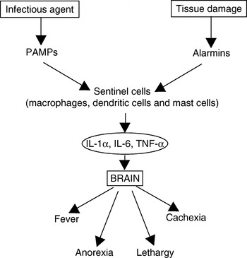
It is now abundantly clear that sickness is an active process and not merely the passive consequences of systemic infection. The behavior of a sick animal serves to promote resistance to infection and ideally improves its chances of recovery. Sickness behavior is not usually caused by the direct actions of pathogens on the brain except in cases of encephalitis or meningitis. Neurons cannot detect the presence of invading bacteria or viruses directly. Conversely, cells of the innate immune system, specifically the major sentinel cells such as dendritic cells, macrophages and mast cells, have receptors that enable them to detect both invading microbes and tissue damage. (Microbes are detected by the recognition of conserved molecular structures called pathogen-associated molecular patterns (PAMPs) while tissue damage is recognized by the presence of products from broken cells (alarmins).) As a result, these sentinel cells will release a mixture of cytokines that inform the brain that microbial invasion is occurring or that tissues are being damaged by pathogens. These cytokines may reach the brain either directly from the bloodstream or indirectly through nerves, especially through the vagus nerve. These cytokines can then bind to receptors on brain cells. As a result, the brain adjusts several homeostatic set-points and triggers the set of metabolic and behavioral changes that we collectively call sickness (Fig. 1). These changes in general, modify physiological processes so as to promote disease resistance and recovery (Johnson, Reference Johnson2002). That said, some of the behaviors associated with sickness might not be obviously beneficial. Likewise, specific sickness behaviors may differ between mammalian species, thus raising additional questions about their precise benefits. It is also important to point out that while cytokines can modulate the activities of neurons, neurotransmitters and neuropeptides also strongly influence immune responses (Szelényi, Reference Szelényi2001).
A simple schematic diagram showing the linkage between innate immune responses and behavior.

Cytokines that affect sickness behavior
While cytokines have classically been regarded as the major signaling molecules of the immune system, they are also, in many cases, potent neuromodulators. Likewise, the cytokine balance within the body is also modulated by the activities of neurotransmitters so that neuroimmune interactions are bidirectional (Del Rey et al., Reference Del Rey, Besedovsky, Sorkin, de Prada and Arrenbrecht1981; Elenkov et al., Reference Elenkov, Wilder, Chrousos and Vizi2000). Although many cytokines can affect brain function and hence behavior, four stand out as being consistently associated with sickness behavior. These include tumor necrosis factor alpha (TNF-α), interleukin-1β (IL-1β), interleukin-6 (IL-6) and high mobility group box protein-1 (HMGB-1). Other cytokines that influence sickness behavior include interleukin-18 (IL-18) and interferon-γ (IFN-γ) (Elenkov et al., Reference Elenkov, Iezzoni, Daly, Harris and Chrousos2005).
TNF-α
TNF-α is a 25 kDa trimeric protein produced by macrophages, mast cells, T cells, endothelial cells, B cells and fibroblasts. Upon detecting invading pathogens, macrophages and mast cells secrete either membrane-associated, or soluble TNF-α. This TNF-α then stimulates both inflammation and sickness behavior. TNF-α triggers the local release of chemokines and cytokines, and promotes the adherence, migration, attraction and activation of leukocytes at sites of microbial invasion. Later, TNF-α facilitates the transition from innate to acquired immunity by enhancing antigen presentation by dendritic cells and T cell costimulation. Its production is stimulated by PAMPs acting through toll-like receptors (TLRs) as well as by molecules secreted by nerves such as the neurotransmitter, substance P (Steinman, Reference Steinman2004). TNF-α is produced very early in inflammation, and this is followed later, by waves of IL-1 and IL-6 (Hawiger, Reference Hawiger2001).
TNF-α in combination with IL-1, triggers changes in vascular endothelial cells. A local increase in TNF-α causes the ‘cardinal signs’ of inflammation, including heat, swelling, pain and redness. Systemic increases in TNF-α depress cardiac output, induce microvascular thrombosis and cause capillary leakage. TNF-α acts on neutrophils to enhance their ability to kill microbes. It attracts neutrophils to sites of tissue damage and increases their adherence to vascular endothelium. It stimulates macrophage phagocytosis. It promotes macrophage synthesis of IL-1, nitric oxide synthase (NOS)-2 and cyclooxygenase (COX)-2. TNF-α also activates mast cells. TNF-α stimulates macrophages to increase its own synthesis together with that of IL-1. In high doses, the physiologic changes triggered by TNF-α cause a systemic inflammatory response syndrome, otherwise called septic shock (Dofferhoff et al., Reference Dofferhoff, Vellenga, Limburg, van Zanten, Mulder and Weits1991).
Interleukin-1β
Activated macrophages synthesize two glycoproteins called IL-1α and IL-1β. IL-1β is produced as a large (31 kDa) pro-protein that is cleaved by caspase-1 to form the 17 kDa active molecule (Barksby et al., Reference Barksby, Lea, Preshaw and Taylor2007). Ten- to 50-fold more IL-1β is produced than IL- and while IL-1β is secreted, IL-1α remains attached to the cell. Thus IL-1α only acts on target cells that come into direct contact with the macrophage. Transcription of IL-1β mRNA occurs within 15 min of ligand binding. It reaches a peak 3–4 h later and levels off for several hours before declining. The stimuli that induce IL-1 production include PAMPs such as bacterial lipopolysaccharides, proinflammatory cytokines such as TNF-α, IFN-α and IFN-β as well as IL-1β itself. IL-1 plays a key role in immune regulation and inflammation because it induces the expression of many effector molecules. These include other cytokines and chemokines, NOS-2 and matrix metalloproteinases. IL-1β acts on vascular endothelial cells to make them adhesive for neutrophils. IL-1 acts on other macrophages to stimulate their synthesis of NOS-2 and COX-2 and so promote more inflammation. Interleukin-1 receptor antagonist (IL-1RA) is an inactive form of IL-1 that binds and blocks IL-1 receptors. As a result, it acts as an inhibitor of IL-1 activities.
IL-18 is another member of the IL-1 family. It originates as a 24 kDa precursor, that is processed by caspase-1 to an active 18 kDa peptide. IL-18 acts through its own receptor complex. Like IL-1β it is produced in precursor form and cleaved on demand by caspase 1. Like IL-1β, IL-18 plays many important roles in sickness and inflammation (Zorilla et al., Reference Zorilla, Sanchez-Alavez, Sugama, Brennan, Fernandez, Bartfai and Conti2007). For example it stimulates neutrophil migration and activation as well as the differentiation of Th1 cells. Recently IL-33 has also been identified as a member of the IL-1 family and probably also plays a role in inflammation (Chen et al., Reference Chen, Carroll and Gadina2006).
Interleukin-6
IL-6 is a 20–30 kDa glycoprotein also produced by macrophages and mast cells. Its production is stimulated by bacterial endotoxins, IL-1 and TNF-α (Harden et al., Reference Harden, du Plessis, Poole and Laburn2006). IL-6 affects both inflammation and acquired immunity. It is a major mediator of the acute-phase reaction and of septic shock. It has been suggested that IL-6 regulates the transition from a neutrophil-dominated process early in inflammation to a macrophage-dominated process later on (Jones, Reference Jones2005). It is of interest to note that IL-6 is produced in large quantities by exercising muscle cells. This may explain why exercise can be pyrogenic in humans (Bradford et al., Reference Bradford, Cotter, Thorburn, Walker and Gerrard2007).
HMGB-1
HMGB-1 is a 28 kDa nuclear architectural protein that binds DNA and ensures the correct folding of DNA molecules within the nucleus (Harris and Andersson, Reference Harris and Andersson2004). It is found in all vertebrate cells and is highly conserved between species. However HMGB-1 has a second function – it is a cytokine with the ability to trigger inflammation and sickness. HMGB-1 is released in two different ways. Thus it is actively secreted from living inflammatory cells such as macrophages and it is passively released from necrotic cells. HMGB-1 is actively secreted by macrophages and monocytes. Its secretion is delayed with a lag of 12–18 h after macrophages are activated by lipopolysaccharides or proinflammatory cytokines such as IFN-γ. It is first acetylated, expelled from the nucleus and migrates to cytoplasmic secretory vesicles. HMGB-1 passively escapes from necrotic cells but it is retained in apoptotic cells. Actively released HMGB-1 and passively released HMGB-1 are molecularly different (Dumitriu et al., Reference Dumitriu, Baruah, Manfredi, Bianchi and Rovere-Querini2005). Acetylated HMGB-1 is readily released from DNA, under-acetylated HMGB-1 is retained. Apoptotic cells under-acetylate their HMGB-1. The actively secreted molecule is highly acetylated (Bonaldi et al., Reference Bonaldi, Talamo, Scaffidi, Ferrera, Porto, Bachi, Rubartelli, Agresi and Bianchi2003). HMGB-1 binds to several different receptors. These include RAGE (receptor for advanced glycation end products), RAGE is expressed on endothelial cells, smooth muscle cells, macrophages, and significantly, on neurons. HMGB-1 also binds to TLR2 and TLR4 and, acting through NF-κB stimulates, cytokine production. HMGB-1 sustains and prolongs inflammation since it induces the secretion of proinflammatory cytokines from macrophages, monocytes, dendritic cells, neutrophils and endothelial cells. HMGB-1 is a chemoattractant for vascular smooth muscle cells; it may also have potent bactericidal activity. Administration of HMGB-1 to normal animals causes fever, weight loss, anorexia, acute lung injury, arthritis and death. Intracerebral administration of HMGB-1 to mice increases TNF-α production and induces anorexia, taste aversion, fever and IL-1 production. So HMGB-1 may either stimulate TNF-α and IL-1 production or it can be produced in response to TNF and IL-1 (Lotze and Tracey, Reference Lotze and Tracey2005). Administration of HMGB-1 in close proximity to the sciatic nerve caused allodynia (the lowering of pain thresholds), as does TNF-α (Hermann et al., Reference Hermann, Holmes and Rogers2005). This may be of relevance to the increased dermal sensitivity experienced in sickness.
Cytokine production in the brain
While most sickness-inducing cytokines are produced at inflammatory sites remote from the brain, it is clear that they are also synthesized within the brain itself (Szeleny, Reference Szelényi2001). For example, intracerebral injection of lipopolysaccharide in rats causes increased production of IL-1β and TNF-α by astrocytes and microglia. The IL-1 activating enzyme, caspase 1, is also produced in the brain (Quan et al., Reference Quan, Zhang, Emery, Bonsall and Weiss1996). Systemic administration of LPS leads to the induction of TNF-α mRNA in perivascular and meningeal cells as well as in neurons (Breder et al., Reference Breder, Tsujimoto, Terano, Scott and Saper1993). Likewise bacterial infections such as meningitis result in increased levels of these two inflammatory cytokines in cerebrospinal fluid. On the other hand, systemic administration of LPS and other PAMPs to rats can lead to increased expression of IL-1β, TNF-α, IL-6 and the IL-1 receptor, IL-1R1, in the brain (Quan et al., Reference Quan, Whiteside and Herkenham1998). Thus within 30 min of intraperitoneal injection of LPS, IL-1β messenger RNA appears in areas surrounding blood vessels. By 8–12 h IL-1 positive cells appear throughout the brain (but not in neurons). Strong staining also develops in both lobes of the pituitary beginning 1–2 h after LPS administration. Cartmell et al. (Reference Cartmell, Luheshi and Rothwell1999) used a different technique. They showed that injection of IL-1RA into the anterior hypothalamus and other nearby regions or the hippocampus prevented the rise in body temperature mediated by intraperitoneal LPS indicating the presence of active IL-1 within that region.
Communications between the immune and nervous systems
While the central nervous system does not have conventional lymphatic drainage and mechanisms exist to exclude potentially harmful lymphocytes from the brain, there are also multiple anatomical connections between the CNS and the immune system (Steinman, Reference Steinman2004). The two systems also share many chemical mediators ranging from small molecules such as nitric oxide to large cytokines and growth factors.
Cytokines exert their effects on the central nervous system through two major routes. Thus cytokines originating in the peripheral lymphoid organs may simply cross the blood-brain barrier. This type of transfer requires that the cytokines reach the CNS by passing through ‘windows’ in the blood-brain barrier (Watkins et al., Reference Watkins, Maier and Goehler1995). These windows are called circumventriclar organs and allow molecules produced by immune cells to be directed at precise sites within the hypothalamus (Sharshar et al., Reference Sharshar, Hopkinson, Orlikowski and Annane2005). Cytokines pass through windows located on the midline of the preoptic nucleus of the hypothalamus. Evidence also exists to suggest that some cytokines such as IL-1 are actively transported into the brain (Banks et al., Reference Banks, Ortiz, Plotkin and Kasin1991).
Some cytokines are produced by neuronal cells within the CNS. They may be produced by glial cells in response to local infections (Breder et al., Reference Breder, Hazuka, Ghayur, Klug, Huginin, Yasuda, Teng and Saper1994; Sternberg, Reference Sternberg1997). Cytokine activation of vascular endothelial cells releases second messengers that can also affect the CNS. These second messengers include NO and prostanoids synthesized by enzymes (NOS and COX-2) whose transcription is induced by pro-inflammatory cytokines such as IL-1.
Afferent fibers of the vagus nerve may also transport pyrogenic cytokines directly into the thermoregulatory centers in the hypothalamus. Thus cytokines signaling trough the vagus stimulate the hypothalamus to secrete corticotrophin-releasing hormone and the pituitary to release adrenocorticotropic hormone.
The brain and immune system also communicate through the autonomic nervous system. Thus parasympathetic pathways innervate lymphoid organs through the neurotransmitter acetylcholine. Likewise sympathetic nerves innervate lymphoid tissues using the neurotransmitter norepinephrine. The norepinephrine can reach target immune cells by diffusion. These nerves communicate directly with the thymus and bone marrow as well as secondary lymphoid organs such as the spleen and lymph nodes (Vizi et al., Reference Vizi, Orso, Osipenko, Hasko and Elenkov1995). Lymphocytes, not only possess receptors for acetylcholine and norepinephrine, but also for other neurotransmitters such as histamine and serotonin (Kin and Sanders, Reference Kin and Sanders2006).
Acetylcholine, the parasympathetic neurotransmitter modulates immune reactions through the vagus nerve (Wrona, Reference Wrona2006). For example, local inflammatory reactions such as arthritis in the paw may be suppressed by vagal nerve stimulation (Tracey, Reference Tracey2007). Vagal nerve stimulation can also suppress the systemic shock response induced by LPS. Activation of the nicotinic acetylcholine receptor expressed on macrophages inhibits their production of TNF-α and IL-1β. As a result, acetylcholine can modulate septic shock and other aspects of sickness behavior (Tracey, Reference Tracey2007).
Complement components may also have a direct effect on the CNS (Francis et al., Reference Francis, Lewis, Akatsu, Monk, Cain, Scanlon, Morgan, Ham and Gasque2003).
Fever
One of the consistent features of infections is the rise in body temperature that we call a fever. Although the temperature rise is normally relatively small, occasionally it can be so high as to cause organ damage, especially to the central nervous system. For example, prior to the availability of antibiotics, pneumococcal pneumonia caused fevers reaching 40°C within 12 h. It has long been regarded as a beneficial process. Thus Thomas Sydenham, the 17th century English physician said ‘Fever is a mighty engine which nature brings into the world for the conquest of her enemies’. It is fair to say however that the benefits of a fever remain unclear to this day.
Temperature homeostasis in mammals is regulated by neurons located in the preoptic area of the hypothalamus, in a region that contains the medial and lateral parts of the preoptic nucleus, the anterior hypothalamus, and nearby regions of the septum. This region contains a central thermostat that keeps core body temperature within a very narrow range despite fluctuations in ambient temperature (Biddle, Reference Biddle2006).
The mechanisms by which a fever is induced involve two major pathways. Thus cytokines such as IL-1β, IL-6, TNF-α and HMGB-1 act directly on the brain. These cytokines bind to brain receptors and effectively reduce the firing rate of warm-sensitive neurons in the temperature-regulating region (Krueger, Reference Krueger1991). This alters the thermoregulatory set-point so that body temperature is stabilized at a higher than normal temperature. This leads to increased heat production and reduced heat loss. Sick animals seek warmer environments and assume a posture minimizing heat loss.
The second pathway involves cytokine induction of the production of prostaglandin E2 in either cerebral endothelial cells or in microglia. The small prostaglandin molecules may then enter the brain to affect the temperature sensitive neurons (Roth et al., Reference Roth, Rummel, Barth, Gerstberger and Hubschle2006).
Fever is almost certainly beneficial for most, but not all species of vertebrates. Increased body temperature enhances several different immune mechanisms and increases resistance to infection. Thus in mice, artificially elevated body temperatures increase resistance to herpes simplex virus, poliovirus, Coxsackie B virus, rabies virus, and Cryptococcus neoformans. However fever decreases resistance to Streptococcus pneumoniae (Eiseman et al., Reference Eiseman, Mallette, Wotkyns, Summers and Tong1956). (It is of interest to note that mice, paradoxically reduce their body temperature in response to endotoxin (Conti et al., Reference Conti, Sanchez-Alavez, Winsky-Sommerer, Morale, Lucero, Brownell, Fabre, Huitron-Resendiz, Henriksen, Zorrilla, de Lecea and Bartfai2006).) An artificially increased body temperature increases resistance of rabbits to S. pneumoniae and C. neoformans, of dogs to herpesvirus (Carmichael and Barnes, Reference Carmichael and Barnes1969), of pigs to transmissible gastroenteritis (Furuuchi and Shimizu, Reference Furuuchi and Shimmizu1976) and of ferrets to influenza A (Mackowiak, Reference Mackowiak1994). A high body temperature also inhibits the growth of pathogens such as Treponema pallidum. It is notable that Julius Wagner von Jauregg won the Nobel Prize for Medicine, 1927 when he showed that artificially induced high fevers led to a cure for syphilitic dementia presumably by killing intracerebral T. pallidum.
Multiple mechanisms have been investigated to determine how an elevated body temperature enhances immune function. Thus the production of nitric oxide and inducible nitric oxide synthase is susceptible to changes in temperature (Pritchard et al., Reference Pritchard, Li and Repasky2005). At normal body temperature, stimulation by both IFN-γ and LPS were required to induce macrophage NO production. At 39.5°C however LPS alone was sufficient. Furthermore, significantly more NO and iNOS were generated at the higher temperature. Thus changes in body temperature may well lower the threshold required to produce effector molecules. Fever-range temperatures also promote lymphocyte trafficking across high endothelial venules (Chen et al., Reference Chen, Carroll and Gadina2006).
The central importance of fevers is emphasized by the fact that elevation of body temperature in response to infection occurs even in cold-blooded animals (ectotherms) and insects. These animals raise their body temperature by behavioral changes.
For example, infected lizards (Dipsosaurus dorsalis) kept in a constant temperature environment cannot change their body temperature. Lizards kept in warm environments survive better than those in a colder environment (Krueger et al., Reference Krueger, Ringler and Anver1975). If provided with a choice of two environments, one warm and the other cold, healthy lizards maintain their body temperature within a fairly narrow range by alternating between the two environments. Lizards challenged with killed bacteria changed their behavior and spent more time in the warmer environment. As a result, their mean body temperature was increased (Vaugn et al., Reference Vaugn, Bernheim and Kluger1974). This is known as a behavioral fever.
The maintenance of body temperature comes at a cost in energy. Some small mammals such as mice cannot afford to expend this energy and appear to benefit from a reduced body temperature. For example, transgenic mice with a reduced core body temperature have an increased life-span (Conti et al., Reference Conti, Sanchez-Alavez, Winsky-Sommerer, Morale, Lucero, Brownell, Fabre, Huitron-Resendiz, Henriksen, Zorrilla, de Lecea and Bartfai2006).
The persistence of a fever is controlled by regulatory cytokines, most especially interleukin-10. Thus fevers induced in rats by either LPS or Staphylococcus aureus are reduced in severity and duration by simultaneous administration of IL-10 (Cartmell et al., Reference Cartmell, Ball, Bristow, Mitchell and Poole2003). This appears to be a result of a decline in inflammation-induced IL-6 levels in blood. Conversely, neutralization of IL-10 increases the amplitude and duration of toxin-induced fever.
Anorexia
A fully-grown animal tends to maintain a relatively constant weight despite short-term fluctuations in food intake and energy expenditure. This weight stability is a result of mechanisms that maintain energy reserves at a relatively constant level and resist changes in this value. This homeostasis is regulated by the central nervous system which attempts to hold body weight at a set-point. If body weight changes, control mechanisms induce behavioral responses that re-establish the original value. Thus changes in body weight during sickness represent alterations in the body weight ‘set-point’. The control of this set-point is also a function of the hypothalamus. Under normal circumstances, the arcuate nucleus of the hypothalamus receives signals from numerous sources that inform it of such factors as temperature, metabolic signals, body weight, and food intake. Meal-related inputs such as smell, texture, taste and gastric distention are communicated to the hypothalamus by the vagal nerve. These signals are integrated and evoke a response that alters appetite (energy intake) and expenditure (van den Top and Spanswick, Reference van den Top and Spanswick2006). The central control of eating behavior appears to be aimed at appetite suppression. Thus animals have a perpetual drive to eat and the CNS counters this with multiple, overlapping appetite suppressing pathways. Excessive activity in these pathways will therefore result in anorexia.
Anorexia is a consistent feature of sickness and, like fever, appears to be mediated by inflammatory cytokines. Cytokines produced during inflammation suppress appetite through multiple pathways. Thus they may induce aversion to specific foods, they may induce nausea through vagal pathways, or they may act on the arcuate nucleus and the melanocortin pathway to suppress appetite. For example, injected IL-1β suppresses appetite while blockade of the IL-1 receptors on brain cells prevents this anorexia (Rothwell and Lueheshi, Reference Rothwell and Luheshi2000). This infection-induced anorexia is apparently beneficial insofar as it enhances survival in the face of infection, For example, Murray and Murray (Reference Murray and Murray1979) force-fed infected mice and 100% died while ad-lib fed infected mice – 50% died. The mice permitted to eat at will, consumed only 58% of the feed of uninfected control animals. Their survival was positively correlated with their degree of anorexia and their weight loss.
During infections, TNF-α causes gastric stasis. Thus acting on receptors in the brainstem it stimulates sensory areas and triggers efferent signals that act through the gastric enteric plexus to cause gastroinhibition. TNF-α thus completely disrupts autonomic control of the gut resulting in stasis associated with nausea and vomiting (Hermann and Rogers, Reference Hermann and Rogers2008).
Analysis of the time-course of lipopolysaccharide-induced cytokine release and anorexia in rats suggests that different cytokines and hormones may act at different times during the process (Kim et al., Reference Kim, Kim, Ahn, Kim, Kim, Lee and Park2007). Thus food intake is depressed by 2 h after a single injection of lipopolysaccharide and remains decreased for 24 h. However TNF-α levels peaked before 2 h, IL-6 levels peaked between 2 and 4 h, and leptin levels peaked between 8 and 16 h. Insulin levels were high between 2 and 16 h. Hypothalamic cytokines were elevated by 30 min after LPS injection and remained high for 16 h.
Fighting infection requires significant energy consumption. Medieval physicians considered a fever to result from some sort of fire within the body. They also believed that food served to fuel that fire. Consequently it was believed, as pointed out in Withal's Dictionary, in 1574. ‘Fasting is a great remedie of feuer’. Modern science has not come up with a much better hypothesis. It has been hypothesized however that filling of the intestine by food triggers a cytokine response that promotes a Th1 bias in the immune system (Bazar et al., Reference Bazar, Yun and Lee2005). The theory further suggests that this bias may help antiviral immunity but not antibacterial immunity. According to this highly speculative theory, anorexia ‘may’ promote a Th2 response that enhances antibacterial resistance while feeding ‘may’ help antiviral resistance.
There appears to be a solid link between some eating disorders and cytokine production. For example, anorexia nervosa patients show a significant increase in blood levels of TNF-α and IL-6. ‘Recovered’ anorexia patients have normal IL-6 levels but their TNF levels tend to remain elevated (Kahl et al., Reference Kahl, Kruse, Rieckmann and Schmidt2004). The significance of this is unclear since in mice, IL-6 does not affect appetite (Dunnn and Swiergiel, Reference Dunn and Swiergiel1998).
IL-18 is a potent appetite suppressant and it appears to act as a homeostatic regulator of appetite to suppress excessive food intake. Thus IL-18 levels decrease in individuals undergoing weight loss. Administration of IL-18 suppresses appetite, feed conversion efficiency and weight regain in food-deprived mice (Zorilla et al., Reference Zorilla, Sanchez-Alavez, Sugama, Brennan, Fernandez, Bartfai and Conti2007). It is possible therefore that IL-18 produced in inflammatory foci also contributes to appetite suppression.
Th2 cells secrete multiple cytokines such as IL-4, and IL-13 and in general promote antibody-mediated immune responses such as allergies. Activated Th2 cells also express a gene for a protohormone, promelanin-concentrating hormone. This is proteolytically processed to produce melanin-concentrating hormone, a potent appetite stimulant (Sandig et al., Reference Sandig, McDonald, Gilmour, Arno, Lee and Cousins2007). Besides providing a link between our current epidemics of allergies and obesity, it is tempting to suggest that alterations in the Th1:Th2 balance may play a key role in controlling appetite. From the point of view of animal production, have we, by selecting animals for high growth rate and feed conversion, selected animals with a Th2 bias? Limited studies have been undertaken in this area. The case of commercial broilers however, suggests the opposite, that modern birds may have enhanced Th1 responses (Cheema et al., Reference Cheema, Qureshi and Havenstein2003).
While both LPS and IL-1 reduce food intake in rodents, it is well recognized that this effect can be blocked by COX inhibitors. More specifically, COX-1 appears to mediate early effects while COX-2 causes anorexia later in the process suggesting that the cytokines act through prostanoids (Dunn and Swiergiel, Reference Dunn and Swiergiel2000; Swiergiel and Dunn, Reference Swiergiel and Dunn2002).
Fatigue and sleep
Sleep is a complex phenomenon consisting of several distinct forms of sleep. These forms are reflected in the electrical activity of the brain as measured on an electroencephalogram (EEG). In small mammals such as rodents, sleep is their major opportunity for physical rest, confines the animal to a thermally insulated nest, allows metabolic rate to drop and is a very effective way of conserving energy. However it is clear that sleep is distinctly different from hibernation, so it must serve a function in addition to energy conservation (Heller and Ruby, Reference Heller and Ruby2004).
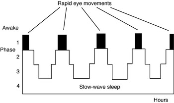
Normal sleep occurs in defined cycles (Fig. 2). Thus an individual cycles between ‘deep’ and shallow sleep throughout the night. Phase 1 sleep is very shallow being a transition from wakefulness to ‘real sleep’. Phase 2 sleep is slightly deeper and occupies about 45% of human sleep time. The deeper sleep phases (phases 3 and 4) are characterized by generation of low frequency waves on the EEG and so are called slow wave sleep (SWS). SWS accounts for about 20% of nightly sleep. Between these periods of SWS, sleep lightens and there are periods of non-SWS (NSWS). NSWS is characterized by significant brain activity, dreaming and rapid eye movements (REM). REM sleep occurs about every 90 min in sleeping humans and constitutes about 20–25% of sleep.
A simple schematic diagram showing the normal phases of sleep.

Sleep, inflammation and immunity are intimately linked since they share regulatory molecules. Sleep influences immunity through the production of certain cytokines while immunity influences sleep patterns through production of these same cytokines. For example, cancer patients being treated with TNF-α exhibit dose-dependent lethargy and fatigue (Spriggs et al., Reference Spriggs, Sherman, Michie, Arthur, Imamura, Wilmore, Frei and Kufe1988). Conversely, inhibition of TNF-α by administration of soluble TNF-receptor p75 improves severe fatigue in rheumatoid arthritis patients (Pollard et al., Reference Pollard, Choy, Gonzalez, Khoshaba and Scott2006). TNF-α also has direct effects on spontaneous sleep in experimental animals. Thus intracerebroventricular injections of TNF-α or IL-1β enhance non-REM sleep (Krueger and Majde, Reference Krueger and Majde2003).
For many years, the lethargy associated with sickness behavior was considered to be a result of physical weakness brought about by the diversion of energy towards fighting infections. Thus it was seen as a passive process of little benefit to the sufferer. Studies however have shown however that sickness behavior is motivational. That is, the animal is motivated to behave in a certain manner rather than simply a consequence of infection-induced weakness. Psychological studies showed for example, that endotoxin-treated rats were motivated to become inactive (Szelenyi and Szekely, Reference Szelenyi and Szekely2004). The occurrence of sickness-induced lethargy points to a relationship between the immune system and sleep that is considerably more than a simple energy-conserving process. For example, it is accepted that sleep deprivation tends to make humans more susceptible to infections, especially minor respiratory infections (Bryant et al., Reference Bryant, Trinder and Curtis2004). Conversely sleep is a prominent feature of sickness behavior. The mechanisms by which sleep and sleep deprivation affect and are affected by the immune system are unclear. It is abundantly clear however that infections cause increased sleep or changes in sleep patterns. In general animals show an increase in SWS following infection. This sleep is also more intense than normal. It is commonly followed by a period of less intense sleep (Toth and Krueger, Reference Toth and Krueger1988). Increased sleep can also be induced by microbial PAMPS such as muramyl peptides and lipopolysaccharides. There are dose-dependent increases in SWS duration and decreased REM sleep. These effects can be separated from the pyrogenic response induced by these molecules (Shohem et al., Reference Shohem, Ahokas, Blettele and Krueger1987). In humans, low doses of LPS induce detectable increases in blood cytokine levels and SWS time and intensity but are insufficient to cause a fever (Mullington et al., Reference Mullington, Hinze-Selch and Pollmacher2001).
Is the increased sleep associated with infections coincidental or has it a beneficial effect on immune function? Toth et al. (Reference Toth, Tolley and Krueger1993) classified sleep patterns in rabbits inoculated with E. coli, S. aureus, or C. albicans on the basis of the duration of the period of enhanced sleep. Patterns characterized by prolonged sleep were associated with a more favorable prognosis and less severe clinical signs than were patterns characterized by relatively short periods of enhanced sleep followed by prolonged sleep suppression. Animals that eventually died had demonstrated reduced sleep when compared to rabbits that survived the infection (Toth et al., Reference Toth, Tolley and Krueger1993). Likewise in elderly humans, poor sleep quality, including difficulty in falling asleep, is significantly associated with mortality.
Many molecules affect the duration and intensity of sleep. Two cytokines, IL-1β and TNF-α both regulate normal sleep (Takehashi et al., Reference Takehashi, Kepes, Fang and Krueger1995), especially the duration of SWS. This increase in SWS occurs within an hour of injection and, depending on the dose, may persist for 4–10 h. The effect is especially marked with low doses of cytokines. Conversely, high doses of IL-1β and TNF-α may decrease sleep (Opp et al., Reference Opp, Obal and Krueger1991). Administration of exogenous IL-1β and TNF-α increases the duration of NREM sleep in many different species of mammal (Krueger et al., Reference Krueger, Fang, Taishi, Chen, Kushikata and Gardi1998). These cytokines also induce increases in the amplitude of slow EEG waves during NREM sleep. It is significant that minor increases in ambient temperature or low doses of microbial products also increase the amplitude of these slow waves (Krueger and Madje, Reference Krueger and Madje1994). It is also of interest to note that intraperitoneal administration of IL-1β to rats or mice increases the duration of their NREM sleep but reduces the amplitude of the EEG slow waves (Hansen and Krueger, Reference Hansen and JM1997). Conversely, a deficiency of IL-1β or TNF-α reduces spontaneous sleep as does inhibition of the IL-1β or TNF-α receptors. Mice that lack the 55 kDa TNF receptor or the IL-1 type I receptor sleep less than do controls (Feng et al., Reference Feng, Wang and Krueger1997, Reference Feng, Wang and Krueger1998). There is a diurnal rhythm in brain cytokines and the highest levels of TNF-α and IL-1β in brain occur during peak sleep periods. Nitric oxide also promotes sleep. IL-10, a cytokine that suppresses the activities of the pro-inflammatory cytokines, reduces sleep (Opp et al., Reference Opp, Smith and Hughes1995). The response to intracerebral inoculation of pro-inflammatory cytokines has different effects on sleep and fever indicating that these responses are induced independently (Welter et al., Reference Welter, Meyers and Krueger1989). It appears that these sleep-inducing cytokines act through the transcription factor NF-κB, and NF-κB inhibitors such as IL-10, IL-4 and IkB reduce sleep (Kubota et al., Reference Kubota, Kushikata, Feng and Krueger2000).
The role of other cytokines in sleep induction is less clear. Thus IL-2, IL-15, IL-18, IFN-α and IFN-γ increase sleep duration while IL-4, IL-10 and IL-13, the classical Th2 cytokines, reduce spontaneous sleep. The role of IL-6 is less clear with contradictory results being obtained (Bryant et al., Reference Bryant, Trinder and Curtis2004). It is likely that the Th2 cytokines act by inhibiting the actions of the pro-inflammatory cytokines.
Infections affect sleep patterns. In general, animals show increased duration of SWS during infections but this may be followed by a period of less sleep but the effects may be quite complex. Episodic diseases such as Trypanosomiasis may be associated with episodic periods of somnolence that relate primarily to disturbances in circadian rhythm (Darsaud et al., Reference Darsaud, Bourdon, Mercier, Chapotot, Bouteille, Cespuglio and Buguet2004). Humans with influenza show decreased sleep during the incubation period and increased sleep when symptomatic (Bryant et al., Reference Bryant, Trinder and Curtis2004). Asymptomatic HIV infection increases duration of SWS in the latter half of the night (Darko et al., Reference Darko, Miller, Gallen, White, Koziol, Brown, Hayduk, Atkinson, Assmus, Munnell, Naitoh, McCutchan and Mitler1995).
The body temperature changes during sleep and ambient temperature influences sleepiness. The mechanisms involved are probably not the same as those involved in a fever (Krauchi, Reference Krauchi2007). After high doses of IL-1, antipyretics reduce a fever but do not influence sleep. The time-course of a fever is usually much shorter than the period of sleepiness. Small mammals drop their temperature during sleep and require a nest or other form of thermal insulation to prevent chilling. In these species, sleep conserves significant energy. Human temperature drops only slightly while sleeping.
It is of interest to note that there appears to be a link between insomnia and an individual's Th1:Th2 balance (Sakami et al., Reference Sakami, Ishikawa, Kawakami, Haratani, Fukui, Kobayashi, Fujita, Araki and Kawamura2002–Reference Sakami, Ishikawa, Kawakami, Haratani, Fukui, Kobayashi, Fujita, Araki and Kawamura2003). Thus NK cell activity, serum IFN-γ and IL-4 were measured in the blood of healthy adult men. Of these, 9.2% reported insomnia based on a standardized questionnaire. These insomniac men had a significantly lower IFN-γ level and a lower IFN/IL-4 ratio than men who reported sleeping normally. Insomniac men had a significantly higher blood IL-4 level than noninsomiacs. These results suggested that a link might exist between the blood level of these cytokines, relative Th1 and Th2 activity, and sleep quality. A similar situation may occur in women. Thus women with lower levels of IL-6 in their blood slept better (Friedman et al., Reference Friedman, Hayney, Love, Urry, Rosenkranz, Davidson, Singer and Ryff2005). Women with the highest IL-6 levels slept less well and had poor relationships with others.
It is also of interest to note that exposure of hamsters to short day lengths decreases the production of proinflammatory cytokines and consequently the symptoms of infection (Wen and Prendergast, Reference Wen and Prendergast2007). Administration of standardized doses of IL-1β to hamsters exposed to short (9 h) photoperiods demonstrated that the effect on sickness behavior was, at least in part, due to a reduced response to the cytokines suggesting that the hypothalamus was refractory to their stimuli. The regulation of sleep is controlled by circadian clock mechanisms (Cavadini et al., Reference Cavadini, Petrzilka, Kohler, Jud, Tobler, Birchler and Fontana2007), and the circadian rhythm is driven by a pacemaker within the suprachiasmatic nuclei in the hypothalamus. These cells possess a molecular clockwork mechanism whereby the transcription factors induce the production of the transcriptional repressor genes Per and Cry. However their gene products, PER and CRY, suppress their own expression and so generate oscillations that result in the rhythmic expression of these genes on a circadian basis. Both IL-1β and TNF-α suppress the expression of certain of these clock genes. As a result of this impairment of circadian function, infected animals will exhibit altered sleep–wake behavior and increased fatigue (Cavadini et al., Reference Cavadini, Petrzilka, Kohler, Jud, Tobler, Birchler and Fontana2007). Thus sick animals will appear somnolent and sleep at times when they would normally be awake and active. Likewise normally active animals may take longer rest periods.
The effects of IL-6 on sleep are different from IL-1 and TNF-α. Thus TNF-α and IL-1 both increase NREM sleep and suppress REM sleep. IL-6 however acts only on NREM sleep (Hogan et al., Reference Hogan, Morrow, Smith and Opp2003). Thus central administration of IL-6 increased NREM sleep in rats while administration of anti-IL-6 antibodies reduced NREM sleep. The doses required to induce sleep were higher than those required to induce a fever.
In wild animals, sleep represents a period of vulnerability where predators may approach without being detected. As a result, many wild mammals, especially prey species sleep only shallowly and for short periods. Conventional sickness behavior, by causing fatigue, may result in deeper or longer sleep, and hence enhance vulnerability. This appears to be less of an issue in carnivores, especially if domesticated, where sleep is unthreatened and hence can be indulged in frequently.
One unique feature of human sleep is that it involves fasting for a significant fraction of the day. Few other mammals fast for so long. Thus herbivores continue digesting food while sleeping. Carnivores classically gorge themselves on food and then sleep while it digests. Rodents wake up periodically to snack. Sleeping humans show a surge in growth hormone input during the early stages of sleep and this may serve to protect against the adverse effects of this sleep-associated fasting (Horne, Reference Horne1988). The fasting may of course be sustained by high levels of TNF-α and IL-1 while sleeping.
Mood changes
Anhedonia is the inability to experience pleasure and it is well recognized that sickness makes us miserable. Why should this happen? Some evidence comes from depressed human patients who have high levels of inflammatory cytokines in their bloodstream (Irwin and Miller, Reference Irwin and Miller2007). Likewise, administration of some cytokines can cause clinical depression. Patients with major depression show elevated IL-6, IL-1β, and TNF-α as well as other inflammation-associated proteins. A positive correlation exists between these cytokine levels and the severity of depression. Likewise, interferon-α induces depression. IFN-α has been used to treat infectious diseases and cancer. IFN-α induces IL-1β, TNF-α and IL-6. IFN-α treated patients develop major depression. They show alterations in serotonin metabolism. They respond to anti-depressant therapy. Correlation does not however prove causation and not all cases of depression are linked to cytokine levels. Whether similar mood changes affect other mammals is not known but if they do, could contribute to the subtle signs of sickness noted by experienced pet owners and veterinarians.
Cytokines also induce alterations in the metabolism of neurotransmitters including serotonin, norepinephrine, and dopamine. All of these molecules are associated with the regulation of emotion. As a result, humans receiving cytokine therapy show altered brain function relevant to depression. Unresponsiveness to antidepressants is associated with high IL-6 and acute-phase protein levels. These treatments may improve the patient's mood but leave other symptoms such as fatigue unaffected. IL-1RA inhibits cytokine-induced sickness and has an anti-depressant affect in animals. In humans the anti-TNF-α drugs used to treat inflammatory diseases can significantly improve a patient's mood. High levels of blood IL-6 in older women are associated with poor sleep quality and poor social relationships. Individuals with good relationships but poor sleep quality have low IL-6 levels! The role of IL-6 may however differ between species since infection-related behaviors, such as feeding, exploratory, anxiety and depression are unaffected in IL-6-deficient mice (Swiergiel and Dunn, Reference Swiergiel and Dunn2006).
Weight loss
While prolonged anorexia will, of course lead to weight loss, a much more significant loss of body mass occurs in sick animals in response to the mobilization of adipose tissue reserves and subsequent loss of fat and muscle cells. In fact, the metabolic changes that occur with cancer and infection cachexia are very different from those induced by starvation (Melstrom et al., Reference Melstrom, Melstrom, Ding and Adrian2007). Cachexic patients show a disproportionately greater loss of muscle mass than starved patients and both fat and muscle stores are significantly depleted. This results in a preferred loss of skeletal muscle as compared to smooth muscle (Mitch and Goldberg, Reference Mitch and Goldberg1996). Historically chronic infections such as tuberculosis cause a major weight loss called ‘consumption’. Likewise AIDS in sub-Saharan Africa is known as ‘Slim disease’. The severe wasting of respiratory muscles may eventually cause these patients to succumb to pneumonia.
Chronic inflammation can also inhibit the overall growth of an animal. Historically, reductions in infections and inflammation have therefore lead to increased height in humans and increased longevity (Crimmins and Finch, Reference Crimmins and Finch2006). Recent increases in human life-expectancy have been associated with a significant decrease in chronic inflammation. Infections (and vaccines) are known to reduce weight gain in cattle, pigs and chickens and hence may have a significant economic impact.
The weight loss associated with inflammation is mediated by TNF-α. It was originally called cachectin since it mediated the cachexia of inflammation (Beutler and Cerami, Reference Beutler and Cerami1986). The majority of skeletal protein catabolism in chronic inflammatory states is mediated by an ATP-Ubiquitin-dependent proteasome pathway. This pathway involves the attachment of multiple molecules of the small peptide ubiquitin to proteins that are to be degraded. This attachment is an energy-requiring process involving ATP. These ubiquitin-labeled proteins are then degraded into small oligopeptides by the 26S proteasome – a highly effective proteolytic complex. The small oligopeptides containing 6–9 amino acids are further degraded into single amino acids. The normal function of this proteasome pathway is to degrade any defective proteins produced in error. The accelerated muscle degradation seen in chronic inflammation appears to be due to activation of this ATP-dependent pathway (Melstrom et al., Reference Melstrom, Melstrom, Ding and Adrian2007).
Multiple cytokines, including TNF-α, IL-1β, IL-6 and IFN-γ stimulate protein degradation via the proteasome pathway. TNF-α seems especially effective in this respect (Reid and Li, Reference Reid and Li2001). Thus nude mice injected with TNF-α-transfected cells undergo severe cachexia. Passive administration of anti-TNF-α to mice leads to a reduction in protein and weight loss caused by cancer. TNF-α administration causes a significant increase in the production of ubiquitin leading to enhanced proteolysis. Similarly, anti-IL-6 treatment decreased muscle atrophy in tumor-bearing mice. IL-6 up-regulates an ubiquitin ligase E2α-II. This enzyme controls polyubiquination, the rate-limiting step in the ubiquitin–proteasome system. The role of IL-1β in cachexia is less clear. Thus chronic administration of IL-1β causes a decrease in muscle mass in rats but IL-1β is also a major appetite suppressant. In rats with methylcholanthrene sarcomas, anorexia is associated with up-regulation of IL-1β and its receptor in the brain. IFN-γ produced by activated Th1 cells has similar activities to TNF-α in that it up-regulates expression of the ubiquitin genes.
TNF-α also affects lipid metabolism since it directly interferes with the metabolism of triglycerides and cholesterol. Thus patients suffering from acute inflammation with increased circulating TNF-α have increased triglyceride concentrations early in the course of the process (Gabay and Kushner, Reference Gabay and Kushner1999). TNF-α acts on adipose tissue cells through its receptor TNFR1 to increase both MAP kinases and c-jun. This leads to activation of protein kinase A that phosphorylates hormone-sensitive lipase (HSL) and perilipins. Perilipins are proteins located on the surface of the lipid droplet in the adipocytes (Popa et al., Reference Popa, Netea, van Riel, van der Meer and Stalenhoef2007) and they regulate substrate accessibility for HSL. Activated perilipins translocate away from the lipid droplet and so permit the HSL to hydrolyze the triglycerides inside. TNF-α also down-regulates the production of perilipins so permitting further lipolysis. As a result of these activities, free fatty acids are released into the circulation, enter the liver and provide the substrate for triglyceride synthesis. Triglyceride synthesis is further enhanced by TNF-α, IL-1β and IL-6 since these cytokines increase hepatic citrate which in turn activates acetyl-CoA carboxylase that is critical for fatty acid synthesis. TNF-α also decreases lipoprotein lipase activity further increasing plasma triglyceride concentrations. The result of all these changes is that adipose tissue shrinks as a response to a decrease in the size of the lipid droplets.
Clearly this mobilization will make energy-rich lipids available for immune function activity. It may also have an effect in regulating inflammatory responses. For example, adipocytes and other cells located within adipose tissue now appear to be a major source of many inflammatory cytokines (Faina, Reference Faina2006). As adipose tissue grows it results in a significant increase in macrophage numbers. These macrophages in adipose tissue are a major source of both TNF-α and IL-6 (Weisberg et al., Reference Weisberg, McCann, Desai, Rosenbaum, Leibel and Ferrante2003). Thus the blood levels of inflammatory cytokines increase in the obese. Obesity markedly increases the release of TNF-α, IL-6 and IL-8 by adipose tissue. Visceral adipose tissue is a much more significant source of these cytokines than subcutaneous adipose tissue (Faina, Reference Faina2006). Human adipocytes secrete significant amounts of IL-6 and IL-8 but the non-fat cells in adipose tissue secrete even more. Leptin on the other hand is a cytokine produced only by adipocytes so its level is determined by the size of an individual's fat stores. Leptin inhibits appetite. Thus appetite is enhanced as fat stores are depleted. Leptin is a potent stimulator of Th1 responses. Leptin is structurally related to other cytokines including IL-6.
Disadvantages of sickness behavior
While it is well accepted that sickness behavior is designed to improve resistance to infections, it is pertinent to ask whether this is always the case. Thus it may not always pay an animal to express its sickness behavior. When an animal acts ‘sick’, it sends multiple messages to observers. These may include, for example, ‘I may be infectious – avoid me! I am easy to catch – eat me! I cannot maintain social dominance – now is your chance to overtake me!’ Veterinarians are not the only ones that can recognize and respond to sickness behavior.
Because of these messages of weakness, hiding sickness behavior may be critical to survival in wild animals. It may be essential to avoid overt demonstrations of vulnerability. An excellent example of this effect is seen in ‘Sick bird syndrome’. This is the name given to the manner in which wild birds successfully hide the fact that they are sick until it is too late to treat them. They are especially effective in hiding their sickness in the presence of a perceived threat such as a human. Another relevant example is observed in cheetah hunting tactics. Thus it is not uncommon for a hunting cheetah to expose itself to a herd of prey. The minor stampede triggered by this enables a cheetah to identify target animals that are sluggish, slow or otherwise present as worthwhile prey. Likewise, keeping up with the herd is vital if an animal is to take advantage of its defensive benefits. Hiding deep within a herd may help sick and susceptible individuals to avoid being targeted although predators such as wolves appear to have little difficulty in identifying susceptible targets.
Conversely, it may be asked, ‘Does it ever pay to act sick?’ Is it possible that the expression of sickness behavior is in part, socially conditioned and its expression may depend on an animal's survival strategies? For example, K strategists are slowly reproducing large animals that put a lot of care into the raising of their young. Examples include elephants, musk oxen and humans. The loss of an ‘infant’ represents a major loss in these species. As a result, some investment in the care (protection) of the sick may therefore be worthwhile. Sickness behavior may trigger such care. Some mammals unquestionably protect their sick or injured? Elephants are an excellent example of a species that expresses concern for the sick and injured and make attempts to ‘encourage and support’ family members. Human societies also provide care for the ‘sick’ and only abandon their sick and injured when either the disease is frightful, and/or, the patient is seen as beyond saving. On the other hand, in r strategists, such as rapidly breeding small animals such as rodents, disease losses are easily tolerated because there are large numbers of offspring. Nothing is gained by investing in the care of the sick or slow. Sickness behavior in these species will attract nothing but predators. Malingering could have some short-term benefits in K species, but as in humans, it is likely that such behavior would be recognized as such.
Sickness behavior therefore, while enhancing resistance to ‘micropredators’ such as bacteria and viruses, may simultaneously increases susceptibility to ‘macropredators’ such as carnivorous birds and mammals. The net effect of these offsetting advantages and disadvantages will depend on the particular circumstances of an animal. Prey species in general may find it advantageous to hide their increased susceptibility. Predator species may not be so constrained, especially if they belong to a pack. Domestication itself, of course, provides a measure of protection to animals and thus may permit an animal the luxury of exhibiting gross sickness behavior.
Given the central role of sickness behavior in triggering the attention of owners and veterinarians, it is surprising that it has been so little investigated in the veterinary literature. Behavioral studies may well provide information of significant usefulness in disease diagnosis.