Aortic coarctation is defined as congenital narrowing of the aorta, which typically occurs at the aortic isthmus.Reference Goudar, Shah and Shirali 1 Severe aortic coarctation induces haemodynamic compromise and necessitates ductal opening. Hence, severe aortic coarctation requires early surgery. Although spontaneous regression of mild-to-moderate aortic coarctation has been reported,Reference Chehab, Abdel-Massih, Smayra and Saliba 2 that of severe aortic coarctation has not been reported. We report a case of trisomy 18 with spontaneous regression of severe aortic coarctation.
Case report
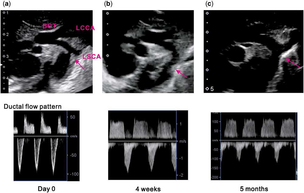
A female infant weighing 1592 g was born, via vaginal delivery, at gestational age 35 weeks and 1 day. She had Apgar scores of 4, 5, 6 at birth. Fetal ultrasound indicated trisomy 18 and detected persistent left superior caval vein, aortic coarctation, and ventricular septal defect. Physical findings such as overlapping fingers, rocker bottom feet, and micrognathia were compatible with trisomy 18, which was diagnosed based on postnatal chromosome analysis. A postnatal echocardiogram revealed persistent left superior caval vein, a large perimembranous ventricular septal defect (5.2 mm), patent ductus arteriosus (4.7 mm) with dominant right-to-left shunt, and severe aortic coarctation (1.2 mm, Fig 1a). This patient had a left aortic arch with the left common carotid artery originating from the brachiocephalic trunk. Although the diameters of the aortic valve (5.5 mm) and ascending aorta (4.9 mm) were relatively maintained, the transverse arch was narrow (2.3 mm). The distance between the brachiocephalic trunk and left subclavian artery was as long as 10.0 mm. Alignment between the isthmus and descending aorta was not straight (Fig 1a and b). These findings suggested severe aortic coarctation requiring ductal opening.
Echocardiographic changes in the aortic arch and isthmus. Upper panel shows aortic isthmus shape and lower panel shows ductus arteriosus flow pattern on day 0, at 4 weeks, and 5 months. ( a ) The diameter of the aortic isthmus at day 0 was as narrow as 1.2 mm, with a body weight of 1.59 kg. Red arrow indicates posterior shelf. Ductus flow pattern was bidirectional with right–left shunt dominance. These findings suggested aortic coarctation. The left common carotid artery (LCCA) originates from the brachiocephalic trunk (BCT). The distance between the brachiocephalic trunk and left subclavian artery (LSCA) was as long as 10.0 mm. ( b ) The diameter of the aortic isthmus at 4 weeks was 2.5 mm, with a body weight of 1.67 kg. Despite a tendency towards dilation, the isthmus angle still showed a figure 3 sign. Ductus flow pattern was bidirectional. ( c ) The diameter of the aortic isthmus at 5 months increased to 4.5 mm, with a body weight of 3.2 kg. The shape of the isthmus was fully normalised. Ductus flow pattern was bidirectional with left-to-right shunt segment dominance. Single red arrows indicate the aortic isthmus.

However, we carefully monitored the patency of the ductus arteriosus and the haemodynamics without initiating prostaglandin E1 for the following reasons: there is a natural tendency for patent ductus arteriosus in trisomy 18 patients,Reference Kosho, Nakamura and Kawame 3 the use of prostaglandin E1 could induce increased pulmonary flow, and stabilisation without the use of prostaglandin E1 would enable smooth transition to home care. As in a previous report,Reference Kosho, Nakamura and Kawame 3 the ductus arteriosus remained open. Cardiopulmonary status stabilised, and extubation was performed on day 4. Hospital discharge was on day 142. Thereafter, respiratory status gradually worsened, partly owing to increased pulmonary flow, as shown on echocardiography and chest radiography. Contrary to our expectation, the aortic isthmus spontaneously dilated (2.5 mm at 4 weeks, and 4.5 mm at 5 months, Fig 1b and c, respectively), and aortic coarctation had almost fully regressed. Alignment between the isthmus and descending aorta normalised and patent ductus arteriosus flow was bidirectional, with a dominant left-to-right shunt (Fig 1c). Therefore, the patient was no longer duct-dependent. She underwent pulmonary artery banding and patent ductus arteriosus ligation at 6 months. Her postoperative course was favourable, with no pressure gradient between the upper and lower extremities.
Discussion
To our knowledge, this is the first case of severe aortic coarctation with spontaneous regression. Our conservative strategy for management of the severe aortic coarctation and ventricular septal defect/patent ductus arteriosus avoided neonatal surgery and enabled a smooth transition to home care. We finally performed pulmonary artery banding and patent ductus arteriosus ligation at 6 months, and the postoperative cardiopulmonary status and general condition were stable.
Spontaneous regression of aortic coarctation has rarely been reported. Chehab et al reported three cases of spontaneous regression of mild-to-moderate aortic coarctation without ductus-dependent circulation. These cases were diagnosed after the neonatal period. The infants presented at 5 weeks to 2 months of age.Reference Chehab, Abdel-Massih, Smayra and Saliba 2 Our report differs from these cases because severe aortic coarctation was prenatally diagnosed, the aortic coarctation was apparently duct-dependent after birth according to its shape (Fig 1a), and the aortic isthmus had been evaluated throughout the perinatal period with clear echocardiographic images (Fig 1a and b).
In contrast to a complex aortic coarctation case necessitating surgery during the neonatal period,Reference Kaneko, Kobayashi and Achiwa 4 there was no apparent heart failure during the neonatal period in this case, suggesting gradual reduction of pulmonary resistance; this might have helped maintain lower body blood flow, enabling observation for isthmus development. Prostaglandin E1 was not used in this case, possibly contributing to a gradual adaptation process by avoiding rapid reduction of pulmonary resistance. Clinical decision-making for a trisomy 18 patient requires careful consideration of all benefits and risks of intervention. Given the natural tendency for a patent ductus arteriosus in trisomy 18,Reference Kosho, Nakamura and Kawame 3 spontaneous regression of severe aortic coarctation can occur, as in our case. Therefore, cautious waiting and careful observation during the neonatal period are options in the management of aortic coarctation/large patent ductus arteriosus in trisomy 18. Because the mechanism remains unknown, evaluation of similar cases is needed to clarify the basis for spontaneous regression of aortic coarctation.
Acknowledgements
The authors thank the cardiovascular surgeons and medical staff of Saitama Medical University International Medical Center who were involved in the treatment of this patient.
Financial Support
This research received no specific grant from any funding agency, commercial, or not-for-profit sectors.
Conflicts of Interest
None.
Supplementary materials
To view supplementary material for this article, please visit https://doi.org/10.1017/S1047951118000161